
1. Introduction
During locomotion, the distal limbs of the horse behave like a spring-mass system that stores energy during the damping phase when the hoof is down on the ground and restores it during the propulsion phase. (Wilson et al. Citation2003). The anatomical structures that constitute the limb of the horse are subjected to strong mechanical stresses during the locomotion, depending on several parameters including the nature of the ground (Crevier-Denoix et al. 2013) or the movement performed (Murray et al. Citation2006). These mechanical stresses repeated during the sporting exercise can lead to micro-lesions possibly leading to more serious attacks with the appearance of a lameness (Singer et al. Citation2008). The tracking of the locomotion of the horse, especially for the detection of lameness (Pfau et al. Citation2013; Bosch et al. Citation2018), is at the core of studies in equine science. In addition, the adaptation of inertial measurement unit (IMU) systems sized for human biomechanical studies to equine science is challenging.
The objective of this preliminary study is to determine, using motion capture, the limits of the use of IMU designed for human biomechanics for the analysis of the biomechanics of the distal limb of the horse. This study will also serve to characterize the specific material requirements for the measurement of horse locomotive parameters in order to develop an “onboard” tool for preventing locomotor lesions in horses.
2. Methods
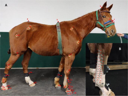
A motion capture system (MOCAP) (Vicon, Oxford Metrics Ltd, Oxford, UK), gold standard in motion biomechanics, was used to track horse limbs’ 3D displacements. 18 cameras (T160 Vicon) were set up on both sides of a high-speed treadmill. 5 horses were equipped with 35 reflective kinematic markers on specific anatomical points (). A layer of 37 small markers (half-spheres of 3 mm) was also placed on the surface of the flexor tendons and the suspensory ligament of the right forelimb (), areas of predisposition to locomotor lesions. In this paper we will focus on the markers of the right forelimb. Two IMUs (ProMove-mini, Inertia Technology BV, Enschede, The Netherlands) were also positioned on the distal limb of the horse, one in the center of the third metacarpal bone on the dorsal side and the second in the center of the first phalanx also on the dorsal side of the limb. Three speeds were predefined (walk at 1.5 m/s, trot at 4 m/s and trot at 6 m/s) and each horse made three trials at each speed. An average of 25 strides were recorded at steady speed for each trial. For each stride, stance/stride ratio (SSR) and protraction/retraction (PR) amplitude of the limb during stance phase are calculated from MOCAP and IMU. Lateral and longitudinal tendon deformities are also measured at each stride from the markers layer.
Figure 1. Placement of the kinematic markers according to the anatomical points of interest (A) and kinematic markers map on the surface of the flexor tendons and the suspensory ligament (B).
3. Results and discussion
The accelerometer's measurement of stride events requires a wide measurement range to avoid saturation (>16 g) of the sensors while accurately measuring small changes.
The use of the map of small markers (half-spheres of 3 mm) allowed to calculate the surface deformations in the area of the flexor tendons and the suspensory ligament. The small size of markers limited the noise generated by the inertia of the markers. Measurements showed lateral deformities of up to 6 cm on average at trot (6 m/s) on the surface of tendons and ligament in their distal part. This result must be taken into account for the fixing system of the ‘onboard’ measurement device.
The horse is a massive animal and measurements of locomotor parameters on his distal limb have many constraints. IMUs have been used for several years in human biomechanical analysis and calibration systems have been developed to overcome their limits for human movement (Lepetit et al. Citation2018). Studies have also been conducted in the equine model, especially for the detection of gait asymmetry, but few have focused on the biomechanical specificities of the distal limbs using IMUs. This requires specific technical adaptations due to the quadruped specific gait patterns and high impacts at landing. The direct comparison of the MOCAP with the IMUs allowed to tune a system of biomechanical analysis of the distal limb with a single IMU positioned on the canon bone. Locomotor parameters were calculated from the MOCAP, but also from the accelerometer's and gyroscope's signals of the IMU positioned on the canon bone according to different methods. Our preliminary results demonstrated correspondences between MOCAP and IMU methods. Nevertheless, the development of a specific method for calculating the amplitude of distal limb range of motion is necessary to obtain accurate results. The comparison of IMU’s calculation methods adapted to the specificities of the equine limb movements would be the next methodological step.
4. Conclusions
The use of IMUs for equine science is promising but challenging. Our findings demonstrate a lack of precision in the measurements of the movements of the distal limb. The parameters measurement range and the surface deformations of the tendons require specific developments and cannot be a simple translational approach of the methods already existing for humans.
Table 1. Results (mean ± sd) of the SSR measurements and the PR amplitude of the limb during stance phase from MOCAP and IMU on three horses trotting at a speed of 4 m/s.
Acknowledgements
The authors thank the Centre d’Imagerie et de Recherche sur les Affections Locomotrices Equines (CIRALE), the Association Nationale de la Recherche et de la Technologie, the Agence Nationale de la Recherche, the Pôle Hippolia and the Région Nouvelle Aquitaine for their financial and logistical support.
References
- Bosch S, Serra Bragança F, Marin-Perianu M, Marin-Perianu R, van der Zwaag B, Voskamp J, Back W, Van Weeren R, Havinga P. 2018. EquiMoves: a wireless networked inertial measurement system for objective examination of horse gait. Sensors. 18(3):850.
- Crevier-Denoix N, Falala S, Holden-Douilly L, Camus M, Martino J, Ravary-Plumioen B, Vergari C, Desquilbet L, Denoix JM, Chateau H, et al. 2013. Comparative kinematic analysis of the leading and trailing forelimbs of horses cantering on a turf and a synthetic surface. Equine Vet J. 45:54–61.
- Lepetit K, Ben Mansour K, Boudaoud S, Kinugawa-Bourron K, Marin F. 2018. Evaluation of the kinetic energy of the torso by magnetoinertial measurement unit during the sit-to-stand movement. J Biomech. 67:172–176.
- Murray RC, Dyson SJ, Tranquille C, Adams V. 2006. Association of type of sport and performance level with anatomical site of orthopaedic injury diagnosis. Equine Vet J. 38(S36):411–416.
- Pfau T, Starke S. D, Tröster S, Roepstorff L. 2013. Estimation of vertical tuber coxae movement in the horse from a single inertial measurement unit. Vet J. 198(2):498–503.
- Singer E. R, Barnes J, Saxby F, Murray J. K. 2008. Injuries in the event horse: training versus competition. Vet J. 175(1):76–81.
- Wilson AM, Watson JC, Lichtwark GA. 2003. Biomechanics: a catapult action for rapid limb protraction. Nature. 421(6918):35–36.