
1. Introduction
The rapid amplitude change of the cerebral systolic arterial input flow increases the brain volume. Then cerebrospinal fluid (CSF) is quickly displaced out of the cranium toward the spinal canal; intracranial pressure (ICP) increase is therefore limited. Nevertheless, this first CSF response is also limited and has to be quickly replaced by cerebral blood venous outflow. The venous contribution is slower than the CSF but at the end drains out the cranium all the blood volume inputed previously. Due to the narrow aqueduct of Sylvius, only a small CSF volume from the ventricles is useful in the cerebral dynamics (Balédent et al. Citation2001). Cerebral hydrodynamics knowledge has benefited considerably from the introduction of phase contrast magnetic resonance imaging (PCMRI), the unique technique to investigate the small but rapid CSF oscillations (Feinberg and Mark Citation1987). Recently a new EPI PCMRI acquisition was introduced (Chen et al. Citation2015) to quantify CSF and blood flow in real time. Only few post processing software exist and are not completely dedicated to extract all the cerebral information present in PCMRI. The objective of this work is to present PCMRI and our dedicated software used to investigate cranio spinal dynamics. We also aim to describe power and limit of MRI to investigate cerebral hydrodynamic.
2. Methods
Using a 3 T MRI and the last PCMRI sequences all the main CSF and blood flows of the cranio spinal system can be quantified in ten minutes. Healthy and patients’ populations were investigated for the last 20 years. CSF flow was quantified in the spinal subarachnoid spaces, the pontine cistern, the foramens of Magendie and the aqueduct of Sylvius. Blood flow is quantified in the internal carotid and the vertebral arteries, straight and sagittal sinus, jugular and epidural veins.
A post processing software was developed (Interactive data language) to visualize, segment and calculate key parameters of flow obtain by conventional and EPI PCMRI. For all the anatomic levels CSF and blood flows are calculated along cardiac cycle. From EPI PCMRI breathing impact in the flows is now investigated.
3. Results and discussion
These flow data are functional information’s complementary to the morphological imaging. Cerebral blood flow and CSF oscillations are easily accessible. We have shown that exist a temporal organisation and amplitude of these flows during the cardiac cycle. Because blood viscosity is not negligible, cerebral venous outflow is delayed and CSF flow out the cranium is important to limit intracranial pressure increase during systolic period. By using EPI PCMRI real time flow investigation is now possible. Our results have shown that breathing impact CSF and cerebral blood flows (Daouk et al. Citation2016). Arterial and venous blood flows during inspiration were respectively higher than during expiration (10% and 8%). All these flows quantifications are used to better investigate the cranio spinal system. In case of patients presented alterations of their cranio spinal dynamics. We have shown how CSF is function of arterial and venous cerebral flows. Post processing is very important to extract quantitative and interpretable information from the large number of images and no commercial dedicated tool still exists. These physiological functional data are useful to better understand the fluid mechanics of the brain. These new flow investigations help to diagnose pathologies as hydrocephalus or idiopathic intracranial hypertension. All the CSF and blood flows measured by PCMRI are potential data that could be used as input for numerical simulation models to access unmeasurable parameters as compliance, flow resistance or pressure of the system.
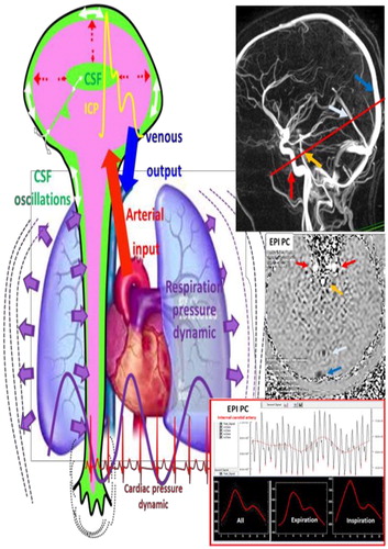
CSF and blood flows circulate in the cranio spinal compartments. The main “motor” of these flows is the heart but respiratory modulate the amplitude of the flows. We can see an MRI cerebral angiography used to select (red line) the level of quantitative velocity acquisition (EPI PCMRI). EPI PC shows arterial flows in white intensities and venous flows in black intensities. Our home-made post processing software shows an example of an internal carotid artery flow change during cardiac cycle and breathing ().
Figure 1. Cranio spinal dynamics investigated by MRI.
4. Conclusion
PCMRI sequence is useful to investigate CSF and cerebral blood flow. Convivial and accurate post processing tool are necessary to optimize use of PCMRI and to be easily used in clinical practice. Such new flow investigations combined with advanced numerical flow simulations open new ways to understand cerebral pathologies as idiopathic Hydrocephalus, idiopathic hypertension, Chiari malformation and syringomyelia where cerebral fluids dynamics seems to be altered.
References
- Balédent O, Henry-Feugeas MC, Idy-Peretti I. 2001. Cerebrospinal fluid dynamics and relation with blood flow: a magnetic resonance study with semiautomated cerebrospinal fluid segmentation. Invest Radiol. 36(7):368–377.
- Feinberg D.A, Mark A.S. 1987. Human brain motion and cerebrospinal fluid circulation demonstrated with MR velocity imaging. Radiology. 163(3):793–779.
- Chen L, Beckett A, Verma A, Feinberg D. 2015. Dynamics of respiratory and cardiac CSF motion revealed with real-time simultaneous multi-slice EPI velocity phase contrast imaging. NeuroImage. 122(122):281–287.
- Daouk J, Bouzerar R, Balédent O. 2016. Heart rate and respiration influence on macroscopic blood and CSF flows. Acta Radiol. 58(8):977–982.