
 ?Mathematical formulae have been encoded as MathML and are displayed in this HTML version using MathJax in order to improve their display. Uncheck the box to turn MathJax off. This feature requires Javascript. Click on a formula to zoom.
1. Introduction
Collagen is the most prevalent protein constituting the intervertebral disc (IVD) extracellular matrix (ECM). Annular collagen constitutes a structured network formed by two fiber families oriented about ±30° with respect to the transverse plane. Local measurements have shown that the orientation angle decreases from the inner AF to the outer AF (Holzapfel et al. Citation2005). The collagen network shape enables the mechanical strength of the AF and therefore the tensile properties of the IVD. With age the ECM of the IVD undergoes several modifications affecting its ability to to absorb and to redistribute the applied mechanical charges. Among these alterations, collagen network looses its organized shape which leads to a loss in IVD mechanical properties (Whatley and Wen Citation2012).
At the cervical level, IVDs are mostly exposed to flexion-extension motion (Mikuckyte and Ostasevisius 2017). Although lateral bending and axial rotation are less frequent, they are always exercised as secondary motions during spine flexion-extension. For these complex loads, the collagen fiber network seems to be the main load bearing in the AF (Ayturk et al. Citation2010).
In order to investigate the importance of the collagen network structure in the cervical IVD mechanics, we propose to perform a parametric study using an hyperelastic fiber reinforced finite element (FE) model of cervical IVD. The parametric study is carried out by varying the collagen fiber orientation.
This study highlights that the cervical IVD behavior is sensitive to collagen network organisation. It also shows that axial stress is more affected by modifying the fiber orientation than shear stress in flexion motion.
2. Methods
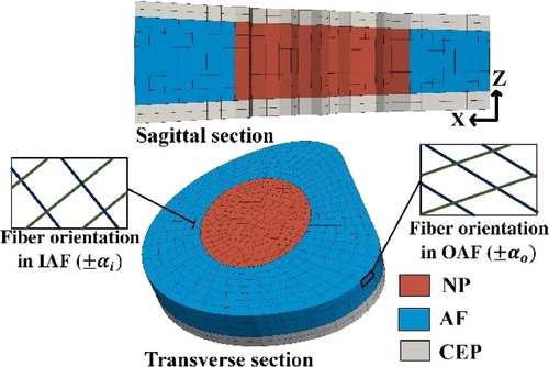
A 3 D geometry of a C6-C7 IVD was constructed to carry out the study (). A biphasic swelling model was used to take into account the internal osmotic pressure role in the IVD behavior. The permeability of the tissue was taken strain dependent.
Figure 1. Geometry of C6-C7 IVD.
The IVD is assumed to be a saturated porous media described by a superposition of two intrinsically incompressible phases: an hyperelastic solid for the ECM and a fluid phase for the water saturating the pores space. The strain energy density of the solid phase is composed of an isotropic compressible NeoHookean part for the non-fibrillary matrix and an anisotropic part for the fiber network.
where for each fiber family i,
denotes the director vector of the fiber orientation and Wfi their anisotropic strain energy density (Gasser et al. Citation2006). In the current model, Wfi was null in the NP and in the cartilaginous endplates (CEP). Two families of fibers with the same mechanical properties were defined in the AF.
In order to study the effect of the collagen network orientation under physiological spine flexion, two parameters were varied to construct 5 cases. The two parameters are the fiber orientation angles in the inner AF (IAF) and the outer AF (OAF) respectively αi and (). Cases 1, 2 and 3 permit to study the effect of varying the fiber direction when αi = αo. Cases 3, 4 and 5 permit to investigate the effect of using a local distribution of the fiber orientation (αi ≠ αo) with a fixed mean angle αm = 35°. The remaining model parameters were taken from Chetoui et al. Citation2019.
Table 1. Parameters sets used in the parametric study.
The range of motion of the flexion and the secondary lateral bending and axial rotation at C6-C7 level were taken from in vivo measurement (Anderst et al. Citation2015).
The simulations were performed using the open source software LMGC90. Firstly, a preconditioning step was performed to set the initial internal fields and swelling followed by a 100 N creep-compressive load step to model the head weight. Finally, to simulate the flexion, a displacement-driven load was applied on the upper CEP for 10 seconds. The used mesh, obtained after a mesh sensitivity analysis, is constituted of 5940 hexahedral elements (26106 nodes).
3. Results and discussion
Each case took about 16 hours of computing time. The stress field did not vary qualitatively with the fiber orientation. Only the value of the stress was affected. With constant fiber orientation, the AF stress increases with α. The AF maximal shear stress is obtained at the end of the flexion load step. For α = 35°, the maximal shear stress σxz (z being the axial direction of the IVD) reaches 0.954 MPa. A decrease of 10° in the angle α induces a diminution of 1.7% in σxz (). The compressive stress σzz drops by a greater rate equal to 11.7% when α decreases from 35° to 25°.
Figure 2. Maximal AF shear stress with respect to α.
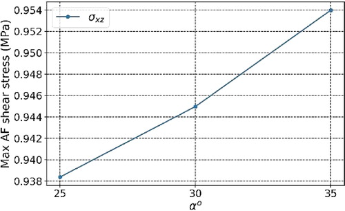
The AF stress is directy proportional to the value of the fiber orientation angle with respect to the transverse plane. When collagen fibers direction get closer to the axial direction, they become more resistant to the disc radial swelling and the axial strain.
In the case where αi ≠ αo, varying the difference between the outer AF and the inner AF fiber orientation has no significant effect on the maximal shear stress. A relative difference of 0.6% is found between σxz in cases 3 and 5. The difference between the maximal compressive stresses σzz found with the same range of angles is more relevant. σzz increases by 10.7% when αi −αo drops from 30° to 0 ().
Figure 3. Maximal AF compressive stress with respect to the distribution of α.
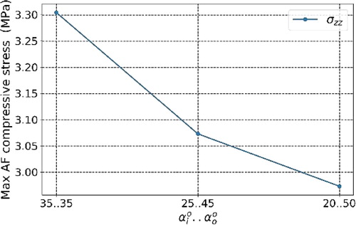
Varying the fiber orientation spatial distribution affects noticeably the value of the axial stress. This result highlights the relevance of defining local fiber orientations at the AF level in IVD FE analysis.
In flexion load, the contribution of the collagen fiber in the AF shear stress does not seem relevant compared to its contribution in the axial stress. By analogy, similar results will be found under lateral bending. We anticipate that the shear stress will be more affected by changing the fiber orientation under axial rotation.
4. Conclusions
The present work investigated the contribution of the collagen network orientation in the cervical IVD mechanics under flexion load. The obtained results did not only show the importance of the collagen network structure for the IVD behavior, but also confirmed that simulating the IVD mechanics under non axial loads strongly requires the use of a fiber reinforced model.
Additional information
Funding
References
- Anderst W, Donaldson III W, Lee J, Kang J. 2015. Three-dimensional intervertebral kinematics in the healthy young adult cervcal spine during dynamic functional loading. J Biomech. 48:1286–1293.
- Ayturk UM, Garcia JJ, Puttlitz CM. 2010. The Micromechanical role of the annulus fibrosus components under physiological loading of the lumber spine. J Biomech Eng. 132:061007.
- Chetoui M, Boiron O, Ghiss M, Dogui A, Deplano V. 2019. Assessment of IVD degeneration related properties using FE models based on ρH weighted MRI data. Biomech Model Mechanobiol. 18(1):17–28.
- Gasser T, Ogden R, Holzapfel G. 2006. Hyperelastic modelling of arterial layers with distributed collagen fibre orientation. J Roy Soc Inter. 3:15–35.
- Holzapfel G, Schulze-Bauer C, Feigl G, Regitnig P. 2005. Single lamellar mechanics of the human lumbar annulus fibrosus. Biomech Model Mech. 3:125–140.
- Mikuckyte S, Ostasevicius V. 2017. Numerical study of lateral bending influence on lumbar intervertebral disc. JVE, Vibroeng Proc. 15:71–76.
- Whatley RB, Wen X. 2012. Intervertebral disc (IVD): Structure, degeneration, repair and regeneration. Mater Sci Eng. 32(2):61–77.