
1. Introduction
Tibial eminence fracture is a bone avulsion of the tibial insertion of the anterior cruciate ligament. These fractures are classified as type 1: no displacement, type 2: partially displaced, type 3: completely displaced, and Zaricznyj added type 4 for comminuted fractures. Type 3 and 4 fractures and orthopedic treatment failures require surgical management however, there is no gold standard on the fixation method. The two most common methods are screwing and suturing. The use of suspensory fixation has many advantages over the screw and is easier to use than sutures. Obtaining stable osteosynthesis is necessary to fight against laxity and allow early mobilization. The objective of this study is the biomechanical comparison of two systems of fixation of tibial eminence fracture type III: screwing and a Pullup® suspension system. Our hypothesis is that the Pullup® has a structural strength equivalent to the screw.
2. Methods
We used fourteen frozen knees from seven anatomical subjects. The muscles and tissues were removed leaving only the femur-anterior cruciate ligament-tibia set. We performed a standardized type III fracture of 15 × 15 × 5 mm as described in the literature (Griffith et al. Citation2004). The matched pairs were randomized into the group A fixed by screw or B fixed by Pullup®.
The tests were performed on a traction machine with tracking markers using cameras (). These were pull-out tests with continuous femoral traction at 5 mm/minute until failure. The main criterion of evaluation was the load at break: maximum load observed before release. The secondary criterion was the load during the displacement of the fracture pad.
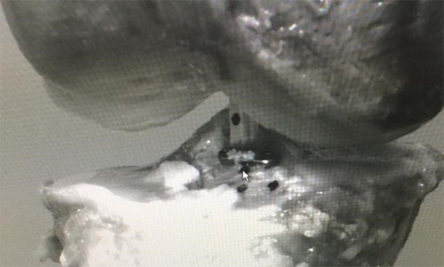
Figure 1. Photograph of tracking markers.
3. Results and discussion
All tests were performed until failure (). Pullout forces on average were higher for Pullup® 98.14 N (±35.26) than for screws 82.04 N (±32.7) without significant difference: p = 0 37. Likewise, for the forces necessary for a displacement of the pad ().
Figure 2. Displacement of the pad mm as a function of the forces N (highest point = load at break).
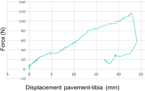
Table 1. Synthesis of the forces (N) according to the displacement of the pad (mm).
There does not seem to be any significant difference between the Pullup® and the screw.
The Pullup® has several advantages over the screw. It requires a smaller diameter tunnel which allows it to be used for smaller fracture fragment and carries a lower damage risk. Sekiya has shown that the suspensory fixation with a larger interface is more applicable to the small fragment (Sekiya et al. Citation2016) and with less risk of fragmentation when tightening. Moreover, it does not require removal of the material in clinical practice unlike the screw which can cause a risk of conflict. Finally, it is technically more reliable because the fracture reduction is done using a tibial guide. We decided to compare the suspensory fixation to the screw because it addresses the same types of fractures (excluding fractures of type IV) and are both technically less complex than the suture as shown by (Seon et al. Citation2009) and (Senekovic & Balazic Citation2014). The comparison with the suture is also difficult in the face of the multitude of different techniques in the literature. Furthermore, no superiority of the suture on the screw has been clearly established on the biomechanical level as shown by (Li et al. Citation2018) and (Mahar et al. Citation2008) clinical level as shown in the literature review published in 2014 by (Coyle et al. Citation2014). There is only one biomechanical study in the literature that focuses on the suspensory fixation in this type of fracture. This study of (Hapa et al. Citation2012) showed a superiority of Endobutton on the different suture systems and the anchors. However, it was performed on anatomical pieces of sheep. The distinct anatomy of the femur-anterior cruciate ligamenthuman tibia complex can hardly be reflected using the knees of a quadruped. Regarding the clinic, the decline in suspensory fixation is low, there is only one series of (Loriaut et al. Citation2017) with two years of decline. In this study the results are favorable in terms of both stability and functional scores, but it is a small non-comparative series with only 5 patients.
4. Conclusions
The Pullup® suspensory fixation appears to be as mechanically as reliable as the osteosynthesis screw for the type III tibial eminence fracture and offers a greater choice of technique to surgeons.
References
- Coyle C, Jagernauth S, Ramachandran M. 2014. Tibial eminence fractures in the paediatric population: a systematic review. J Child Orthop. 8(2):149–159.
- Griffith JF, Antonio GE, Tong CWC, Ming CK. 2004. Cruciate ligament avulsion fractures. Arthrosc J Arthrosc Relat Surg. 20(8):803–812.
- Hapa O, Barber FA, Süner G, Özden R, Davul S, Bozdağ E, Sünbüloğlu E. 2012. Biomechanical Comparison of tibial eminence fracture fixation with high-strength suture, endobutton, and suture anchor. Arthrosc J Arthrosc Relat Surg. 28(5):681–687.
- Li J, Yu Y, Liu C, Su X, Liao W, Li Z. 2018. Arthroscopic fixation of tibial eminence fractures: a biomechanical comparative study of screw, suture, and suture anchor. Arthrosc J Arthrosc Relat Surg. 34(5):1608–1616.
- Loriaut P, Moreau P-E, Loriaut P, Boyer P. 2017. Arthroscopic treatment of displaced tibial eminence fractures using a suspensory fixation. Indian J Orthop. 51(2):187–191.
- Mahar AT, Duncan D, Oka R, Lowry A, Gillingham B, Chambers H. 2008. Biomechanical comparison of four different fixation techniques for pediatric tibial eminence avulsion fractures. J Pediatr Orthop. 28(2):159–162.
- Sekiya H, Takatoku K, Kimura A, Kanaya Y, Fukushima T, Takeshita K. 2016. Arthroscopic fixation with endobutton for tibial eminence fractures visualised through a proximal superomedial portal: a surgical technique. J Orthop Surg. 24(3):417–420.
- Senekovic V, Balazic M. 2014. Bioabsorbable sutures versus screw fixation of displaced tibial eminence fractures: a biomechanical study. Eur J Orthop Surg Traumatol. 24(2):209–216.
- Seon JK, Park SJ, Lee KB, Gadikota HR, Kozanek M, Oh LS, Hariri S, Song EK. 2009. A clinical comparison of screw and suture fixation of anterior cruciate ligament tibial avulsion fractures. Am J Sports Med. 37(12):2334–2339.