
1. Introduction
For several decades, Total Hip Arthroplasty (THA) has become the treatment of choice for advanced hip osteoarthritis. However, this intervention is still prone to complications. Dislocation is the second most common complication after aseptic loosening and is very influenced by the acetabular cup orientation. This last decade, more and more authors mentioned that the safe zone proposed by Lewinnek (Lewinnek et al. Citation1978) is not suitable for every patient because of the patient specific spine-hip kinematics (Rivière et al. Citation2017).
Several solutions have been developed in order to take into account this parameter for THA (Lazennec et al. Citation2015; Pierrepont et al. Citation2016; Kochman et al. Citation2017). All of them are either invasive, difficult to use or expensive. A non-invasive ultrasound (US) based device has been recently proposed which allows the acquisition of the pelvic tilt, i.e., the angle between the Anterior Pelvic Plane (APP) and the vertical, in several daily positions: in the supine, standing and sitting positions (Dardenne et al. Citation2018). The accuracy of this system has been analysed according to a mechanical test bench. This accuracy was very satisfying with an error of 1.15 ± 0.82° whatever the tilt and the soft tissue thickness, when a clinical precision of 2° is expected.
The goal of this paper is therefore to evaluate in-vivo the intra and inter-observer precision of this device.
2. Methods
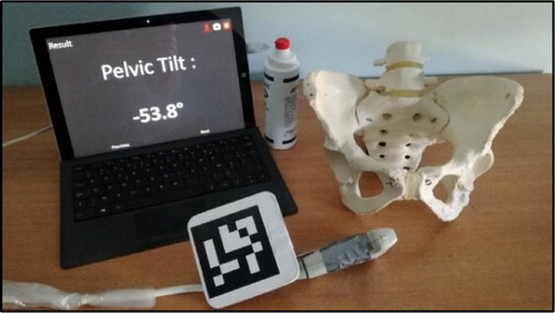
The device is composed of a two-dimensional US probe (L12-5L60N, Telemed®) connected to a tab (Surface Pro 3, Microsoft®) which is equipped with an accelerometer and a front-facing RGB camera (). The three-dimensional (3D) position and orientation of the US probe is determined thanks to an ARUCO marker rigidly fixed to the US probe and localized by the front-facing camera. Both the US probe and the RGB camera have been beforehand calibrated. The APP is determined by acquiring with the US probe the right anterior iliac spines, the left anterior iliac spines and the pubic symphysis. Landmarks are then manually placed by the user on the three US images. The pelvic tilt is finally estimated by computing the angle between the APP, defined by the three landmarks, and the vertical provided by the embedded tab accelerometer.
Figure 1. Device including the tab and the US probe equipped with an ARUCO marker.
To evaluate the precision, pelvic tilt measurements were realized with the device on three healthy subjects and by three physicians. These subjects had a low, medium and high Body Mass Index (BMI), i.e., respectively with a BMI less than 20, a BMI between 24 and 26, and a BMI greater than 30. Among the three physicians, there were an expert who used the device on ten patients before the study, an intermediate who never used the device before but is experienced in osteoarticular US, and a novice who never used the device before and is not experienced in US. For each subject, the pelvic tilt was measured ten times by the three physicians in the supine, standing and sitting positions, resulting in 90 measurements per subject. The inter and intra-observer precisions have been analysed according to the BMI and the positions using the intraclass correlation coefficient (ICC). The criteria defined by Cohen were used: low precision (ICC < 0.40), medium precision (0.40 < ICC < 0.59), good precision (0.60 < ICC < 0.74) and excellent precision (0.75 < ICC < 1.0).
3. Results and discussion
For the inter observer precision, the ICC was 0.998, 0.998 and 0.996 for respectively a low, moderate and high BMI. The ICC was 0.983, 0.925 and 0.69 for respectively the supine, sitting and standing positions.
For the intra observer precision, the ICC was 0.999 for low and medium BMI, whatever the user’s expertise. For high BMI, the ICC was 0.992, 0.998 and 0.996 for respectively the expert, intermediate and novice users. Regarding the positions, the ICC was 0.984, 0.981 and 0.977 for the supine position, 0.874, 0.942 and 0.787 for the standing position, and 0.868, 0.955 and 0.986 for the sitting position, for respectively expert, intermediate and novice users.
4. Conclusions
The in-vivo inter-observer precision was therefore excellent for all measurements and excellent according to the BMI. The inter-observer precision was also excellent regarding the supine and the sitting positions and good concerning the standing position. The intra-observer precision was excellent for all measurements, whatever the user’s expertise, the BMI and the positions.
This study shows therefore that besides the accuracy, which has been studied last year (Dardenne et al. Citation2018), the inter and intra-observer precision of our system meets the clinical requirement.
References
- Dardenne G, Pluchon J-P, Guezou-Philippe A, Letissier H, Hamitouche C, Lefèvre C, Stindel E. 2018. An easy-to-use portable ultrasound based device for the measurement of the pelvic tilt. Epic Ser Heal Sci. 2(EasyChair):39–42.
- Kochman A, Goral A, Martin T, Marek W, Kozak J, Morawska-Kochman M, Synder M. 2017. Application of navigated ultrasound for assessment of the anterior pelvic plane in patients with degenerative hip diseases. J Ultrasound Med. 36(7):1373–1380.
- Lazennec J-Y, Rousseau M-A, Brusson A, Folinais D, Amel M, Clarke I, Pour AE. 2015. Total hip prostheses in standing, sitting and squatting positions: an overview of our 8 years practice using the EOS imaging technology. Open Orthop J. 9(1):26–44.
- Lewinnek GE, Lewis JL, Tarr R, Compere CL, Zimmerman JR. 1978. Dislocations after total hip-replacement arthroplasties. J Bone Jt Surg. 60(2):217–220.
- Pierrepont JW, Stambouzou CZ, Miles BP, O’Connor PB, Walter L, Ellis A, Molnar R, Baré JV, Solomon M, McMahon S, et al. 2016. Patient-specific component alignment in total hip arthroplasty. Reconstr Rev. 6.
- Rivière C, Lazennec J-Y, Van Der Straeten C, Auvinet E, Cobb J, Muirhead-Allwood S. 2017. The influence of spine-hip relations on total hip replacement: a systematic review. Orthop Traumatol Surg Res. 103(4):559–568.