
1. Introduction
Peripheral Venous Catheter Insertion (PVCI) is the most common procedure performed by health professionals, but still remains technically difficult. Helm et al. (Citation2015) observed that PVCI failure occurred 35% to 50% of the time leading to premature removal. A more realistic part-task simulator is currently in development to reduce this failure rate. Such a simulator should help healthcare students to train their gesture, and increase their skills before performing PVCI on patients. It is therefore crucial that such simulators behave like in vivo human skin when introducing the catheter into the vein.
Now, it is well known that force needed to puncture human tissues highly depends on several parameters such as geometry of the penetrator, loading conditions (velocity and angle insertion), as well as the degree of pre-stretch of soft tissues (Shergold and Fleck Citation2004). In order to select the best artificial material to mimic the mechanical response of skin and vein for a PVCI procedure, it is planned to develop an experimental setup, with fixed conditions close to the physiological ones for the medical procedure of interest here. Therefore, the present study is intended to analyse the gesture of a health professional during in vivo PVCIs, in order to extract the loading conditions of interest, as well as the rupture force of skin and vein. An experimental protocol is then set up with fixed conditions coming from the in vivo analysis, and tested with an ex vivo arm. A comparison between the rupture forces obtained in vivo and ex vivo is finally proposed.
2. Methods
2.1. In-vivo protocol
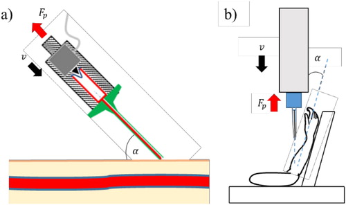
PVCI was performed on the ventral forearm of five healthy volunteers aged 39.2 ± 15.3 years old by a nurse instructor. The medical gesture was recorded by two high-speed cameras (FASTCAM SA3, Photron, Japan) at 125 Hz. A tracking of targets located on the side of the catheter was performed with the help of an image correlation software (VIC 3 D®-Correlated Solution) in order to get its insertion angle, and its speed via its measured displacement (see ). The parameter was obtained by the projection of the needle axis on an orthonormal plane located in the neighbouring of the perforation area. The catheter includes a miniature 5 N load sensor (LSB 200-5 N; Andilog, France) to measure the puncture load (). The authorization for the study was granted by the ethic committee CPP of Ile-de-France V and the National Agency of Medicines Security under the reference number 2018-A02895-50.
Figure 1. (a) In vivo test. (b) Ex vivo setup.
2.2. Ex-vivo protocol
Puncture was performed on the forearm of an 87 years old fresh cadaver with a tensile machine testing (INSTRON 8802–High Wycombe, England), on which was mounted a 5 N load (SMT1-5N; PM Instrumentation, France). The same catheter as for in vivo tests was used. The displacement of the catheter is measured optically in the same way than for the in vivo protocol.
The average loading parameters extracted from in vivo tests were used to determine the experimental conditions for the ex vivo tests (see ).
3. Results and discussion
shows the typical mechanical response of skin for a successful in vivo PVCI.
Figure 2. Typical curve for a successful PVCI, with remarkable steps.
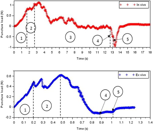
First, the load is increased due to skin stiffness until rupture (step 1) then cutting and friction forces come in (step 2) (Okamura et al. Citation2004) as the catheter progressed until touching the vein where a sudden load increase is visible right before its rupture. The following increase is due to friction forces as the practitioner is progressing into the vein’s lumen until stopping its gesture to put a cotton on the punctured area (step 3). Finally, the catheter is withdrawn from the vein (step 4) and the skin (step 5). The analysis of the gesture of the practitioner in vivo gives a mean puncture velocity and a mean insertion angle of 11.38 ± 0.9 mm s−1 and 13.9 ± 4°, respectively. shows the mean peak load measured to puncture the skin and the vein as well as friction forces isolated during the withdrawal of the catheter. The in vivo results are consistent with the only other study performed under similar conditions (Okuno et al. Citation1998) for which a peak load of 0.64 ± 0.23 was obtained when puncturing the vena mediana cubiti of 10 volunteers.
Table 1. Comparison between ex vivo and in vivo puncture loads.
Overall, less force was needed to puncture ex vivo tissues and to withdraw the catheter. However, it is not yet possible to conclude on whether these differences are due to tissue state or age.
4. Conclusions
An in vivo protocol was developed to extract the loading conditions during puncture of the forearm as well as an ex vivo protocol reproducing said loading conditions. Further analysis over more volunteers is currently performed to confirm the results. An attempt to check the influence of age, sex and BMI over puncture loads will be tackled.
Acknowledgements
The authors would like to thank Patrick Attali, nurse instructor, for performing the PVCIs and the project SAMSEI supported by ANR 11 IDFI 0034.
References
- Helm RE, Klausner JD, Klemperer JD, Flint LM, Huang E. 2015. Accepted but unacceptable: peripheral IV catheter failure. J Infus Nurs. 38(3):189–203.
- Okamura A.M, Simone C, O'Leary M.D. 2004. Force modeling for needle insertion into soft tissue. IEEE Trans Biomed Eng. 51(10):1707–1716.
- Okuno D, Togawa T, Saito H, Tsuchiya K. 1998. Development of an automatic blood sampling system: control of the puncturing needle by measuring forces. Proc IEEE Eng Med Biol Soc. vol. 20. p. 1811–1812.
- Shergold OA, Fleck NA. 2004. Mechanisms of deep penetration of soft solids, with application to the injection and wounding of skin. Proc R Soc Lond A. 460(2050):3037–3058.