
1. Introduction
Training for triathlon requires athletes to spend multiple hours of training in swimming, cycling and running. This training load increases the risk of injuries in lower back and the lower limb (Gosling et al. Citation2008). Despite an abundant literature, the evaluation of the risk of low back pain remains complex as it lacks evidence-based recommendations and reliable functional test. Several functional movement tools are suggested to evaluate this risk in assessing mobility and joint stability (Cook et al. Citation2014). However, it does not specifically investigate the range of motion of low back. The one-sided tilt test (also know as ‘hip drop test’) is an active voluntary movements used by osteopaths to analyse the one-side range of motions at the right and left side from a static position in individuals with nonspecific back pain (Chila Citation2011). The challenge is the clinical interpretation of this test though three-dimensional kinematic adaptations and musculoskeletal strategies of the movement performed on the test side and on the opposite side simultaneously on several regions (lumbar, pelvis, hip and knee). The aim of this pilot study was to use a musculoskeletal modelling approach to propose a functional screening approach of lumbar-pelvic-femoral complex range of motion during the one-sided tilt test.
2. Methods
2.1. Participants
Twenty-two well-trained and asymptomatic triathletes (age: 38.8 ± 12; years’ experience: 8.3 ± 9; training hours: 10.1 ± 3, male: 19; female: 3) were recruited for this study after completing a consent form and a questionnaire to exclude medical pathology. All components of the study were designed by the Research Department of the Institute of Osteopathy in collaboration with the M2S lab (#2018-277) according to the principles of the Declaration of Helsinki.
2.2. Protocol
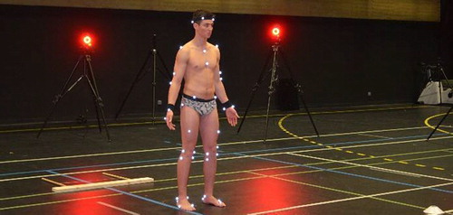
The one-sided tilt test is used to examine the range of motions of the low back when tilting the pelvis to the right and to left side. To do this, the participant had to bend his knee allowing the pelvis to tilt to the same side. The test was explained to the participants and repeated several times before the recordings (). After a static trial, participants were instructed to perform the right and left one-sided tilt test in an alternate sequence.
Figure 1. Experimental set up.
2.3. Musculoskeletal modelling
Three-dimensional kinematics were obtained from a 24-camera motion analysis system (Vicon, Oxford, UK). Markers data served as an input of a full musculoskeletal model, developed by Raabe and Chaudhari (Citation2016), to compute lumbar and lower limb joint angles from the recommended OpenSim calculation steps (Delp et al. Citation2007). The model was scaled to match the participants’ anthropometry using anatomical landmarks (segment lengths) and joint angles were calculated with a global optimisation-based inverse kinematics procedure. All joint angles were estimated at the peak of the right knee flexion (right hand side) and for the left knee flexion (left hand side) for standardising the procedure.
2.4. Statistical analysis
Latent class analysis was then used to identify different classes of movement combination. Significance level was not corrected for multiple testing and was set at p < 0.05.
3. Results and discussion
All participants performed the test on both side. Forty-four trials were analysed.
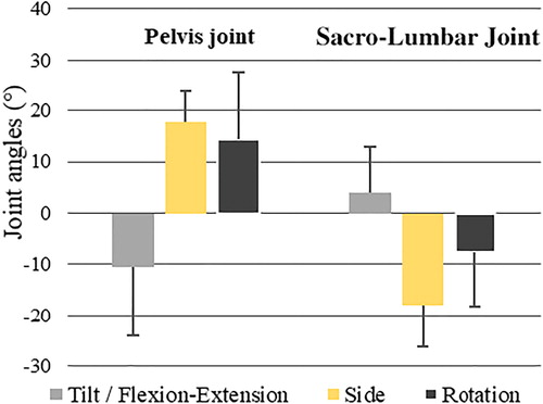
We observed an opposite kinematics between the pelvis (anterior tilt, ispsilateral side, controlateral rotation) and the sacro-lumbar joint (extension, contralateral side, ispsilateral rotation) ().
Figure 2. Sacro-lumbar and pelvis joint angles during the one-sided tilt test (mean of all trials).
Based on its results, four classes of possible movement combinations were identified (from Class 1 with the lowest range of motion to Class 4 with the highest range of motion) and characterised by an increase in knee flexion. In addition, highest ranges of motion (Class 4) occurred when the ipsilateral knee was more flexed and when the controlateral knee is close to full extension ().
Table 1. Mean of all degree of freedom joint angles of each class of participants.
Knee flexion was most limited with reduced range of motion of pelvis tilt, then rotation and finally list. These patterns were different between the left and right side in 31.8% of the studied population.
4. Conclusions
The main finding of this pilot study is that biomechanical analysis allowed to better understand musculoskeletal strategies during the one-sided tilt test. Polyarticular functional dynamics could help understand different strategies and kinematic adaptations linked to over or under mechanical load of specific joints. In addition, this approach permitted to identify athletes with limited range of motion on lumbar-pelvic-femoral complex. The use of a musculoskeletal approach allows the possibility in future studies of accessing data that are difficult to measure (muscle lengths and joint forces). A better understanding of how anatomical structures function and interact during functional movement is fundamental to prevent low back pain and to objectify the impact of osteopathic treatment for example.
Acknowledgements
We acknowledge all participants and especially Thomas Bagory, Ilona Bieda and Victorien Grosset for our help during data acquisition.
References
- Chila AG. 2011. Foundations of osteopathic medicine. 3rd ed. Philaldephia, PA: Lippincott Williams & Wilkins.
- Cook G, Burton L, Hoogenboom BJ, Voight M. 2014. Functional movement screening : the use of fundamental movements as an assessment of function - part 1. Int J Sports Phys Ther. 9(3):396–409.
- Delp SL, Anderson FC, Arnold AS, Loan P, Habib A, John CT, Guendelman E, Thelen DG. 2007. OpenSim: open-source software to create and analyze dynamic simulations of movement. IEEE Trans Biomed Eng. 54(11):1940–1950.
- Gosling CM, Gabbe BJ, Forbes AB. 2008. Triathlon related musculoskeletal injuries: the status of injury prevention knowledge. J Sci Med Sport. 11(4):396–406.
- Raabe ME, Chaudhari AMW. 2016. An investigation of jogging biomechanics using the full-body lumbar spine model: model development and validation. J Biomech. 49(7):1238–1243.