
1. Introduction
Since the end of the XIXth century, motion capture (Mocap) technologies do continuous innovation. Today, several technologies are enabled, and the most common are optical systems. These optical motion capture systems are sold at different prices. In the context of an open call for tenders for the choose of one system to another, the question if the accuracy will increase with the price of the system is still open.
In this study, we will propose to compare knee joint flexion/extension angle during treadmill practice recorded with a Vicon system (Oxford Metric, Yarnton, UK); a Mokam (Kinestesia, Verton, FR), and the freeware Kinovea.
The aim of this study is to compare the accuracy of these three motion capture systems during gait and run for 2D knee joint flexion/extension angle estimation.
2. Methods
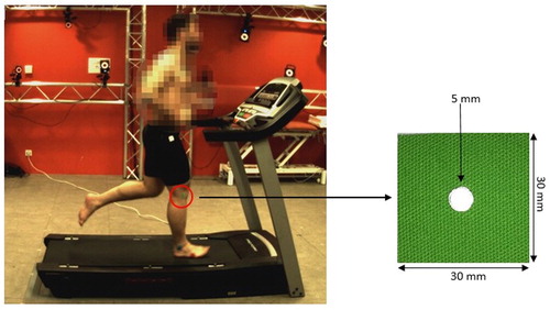
6 subjects (3 men and 3 women) aged 26 (±4) years old realised walk and run at 2; 3; 4; 6; 8; 10 and 12 km/h on a treadmill. The subjects were equipped with three 30 mm square coloured Mokam markers with at their centre a 5 mm Vicon passive marker (). To record these movements a 37 Vicon camera system (100 Hz), a Mokam (100 Hz) and a Bonita Video Camera (33 Hz) with Kinovea freeware were used (Pfister et al. Citation2014; Hisham et al. Citation2017; Hanley et al. Citation2018). Records were made simultaneously.
Figure 1. Greater trochanter, lateral condyle of femur and lateral malleolus markers.
After labelling of the point of greater trochanter (hip), lateral condyle of femur (knee) and lateral malleolus (ankle), two continuous gait cycle were used for data analysis. The first step was to calculate the 2D flexion/extension angle after orthogonal projection in the sagittal plane using raw coordinates given by the systems (Perry Citation1992). As the three systems do not record at the same frequency, we normalized the data from 0 to 100% of the two gait cycles. Multiple Correlation Coefficient (CMC) and Root Mean Square (RMS) with a two-way ANOVA analysis were computed to compare angles obtained by the three systems (Rezgui et al. Citation2013).
3. Results and discussion
shows a representative data of the 2D flexion/extension knee joint angle during treadmill locomotion at 4 km/h. As the subjects are non-pathologic, knee joint flexion/extension pattern corresponds to the normal gait pattern as found in literature (Perry Citation1992; Novacheck Citation1998). At standing position, the knee was set up at 0°. The negative values are due to a knee recurvatum.
Figure 2. Example of knee joint flexion/extension angle variation at 4 km/h with the 3 systems.
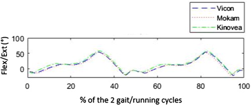
In the , the MCC values are all over 0.86 with a mean of 0.95. RMS values are 29,14 for Vicon, 28,54 for Mokam and 30,35 for Kinovea. The two-way ANOVA analysis showed non-significative difference (F = 0.96).
Table 1. MCC between the three systems for all subject at all speeds.
Our results demonstrated equivalent ability to measure the 2D knee flexion/extension angle during treadmill exercise. Our finding supports previous observations (Damsted et al. Citation2015). This similarity could be explained by the fact that knee motion of the treadmill is quasi-planar with limited range of rotations in the frontal and transverse planes (Riley et al. Citation2008). Consequently, the benefit to occur 3D analysis is limited in this case.
4. Conclusions
Based on the choice of a motion capture system only by the price of the system could be inadequate. Each system has a field of applications. Consequently, the specification of the motion capture system has to be in correspondence with the field of applications. In the present case, 2D knee flexion-extension angle investigation on treadmill exercises with low cost system is equivalent accurate. To conclude, we recommend to writers of open call for tenders for motion capture system to carefully specify applications and the specifications in relation to applications.
Acknowledgements
Thanks to Kinestesia for the technic assistance on the Mokam.
Additional information
Funding
References
- Damsted C, Nielsen RO, Larsen LH. 2015. Reliability of video-based quantification of the knee and hip angle at foot strike during running. Int J Sports Phys Ther. 10(2):147–154.
- Hanley B, Tucker CB, Bissas A. 2018. Differences between motion capture and video analysis systems in calculating knee angles in elite-standard race walking. J Sports Sci. 36(11):1250–1255.
- Hisham NAH, Nazri AFA, Madete J, Herawati L, Mahmud J. 2017. Measuring ankle angle and analysis of walking gait using kinovea. Int Med Dev Technol Conf. 2017:247–251.
- Novacheck TF. 1998. The biomechanics of running. Gait Posture. 7(1):77–95.
- Perry J. 1992. Gait analysis: normal and pathological function. Thorofare, NJ: Slack.
- Pfister A, West AM, Bronner S, Noah JA. 2014. Comparative abilities of Microsoft Kinect and Vicon 3D motion capture for gait analysis. J Med Eng Technol. 38:274–280.
- Rezgui T, Megrot F, Fradet L, Marin F. 2013. On the imitation of CP gait patterns by healthy subjects. Gait Posture. 38:576–581.
- Riley PO, Dicharry J, Franz J, Croce UD, Wilder RP, Kerrigan DC. 2008. A kinematics and kinetic comparison of overground and treadmill running. Med Sci Sport Exerc. 40:1093–1100.