
 ?Mathematical formulae have been encoded as MathML and are displayed in this HTML version using MathJax in order to improve their display. Uncheck the box to turn MathJax off. This feature requires Javascript. Click on a formula to zoom.
1. Introduction
Identification of the wave reflection and mainly its delay time on the incident wave has significant clinical value in detecting cardiovascular or cerebrovascular diseases. Indeed, an increase in arterial stiffness induces an early return of the reflected wave which results in an increase of the amplitude of the arterial wave. We can classify the methods of determining the delay time t0, according two modes, the first mode would concern the direct determination of t0 from a waveform analysis of a single signal like presure or velocity (Sugawara et al. Citation2010) and the second mode, which is indirect, which consists of the separation of the incident and reflected waves and which requires the recording of several hemodynamic data (Feng and Khir Citation2010). Usually, the direct methods defined the delay time by the point of inflection when the rise of the wave changes curvature due to the superposition of the incident wave and the reflected wave (Sugawara et al. Citation2010). The aim of this study was to implement a direct and objective method for noninvasive determination of the delay time in the internal carotid artery based on cepstral analysis. To show the precision of the proposed method we specially focused on the effect of age. Our method is based on the acquisition of instantaneous blood velocity at a single site in the center of the internal carotid.
2. Methods
2.1. Materials
18 healthy subjects were enrolled in this study whose 9 young volunteers having an average age between years 29 ± 7, and 9 middle-old having average age 64 ± 22 years. Blood axial velocity was measured in internal carotid artery at level C2-C3, by using phase contrast magnetic resonance using 1.5 T and 3 T machines. The velocity was acquired with a 2 D sequence cine phase-contrast and was synchronized with the cardiac cycle with a temporal resolution of the order of 20–31 ms. The length L of the internal carotid is estimated by using Philips DICOM software during the acquisition with a spatial resolution of 0.5 mm. This estimation is based on the three-dimensional flight time of the cerebral angiogram.
2.2. Physical and mathematical models
The geometric model adopted in this study was limited by an effective reflection area in zr, located downstream of the measurement site which is in z (Nichols et al. Citation2011; Abdessalem et al. Citation2007; Schubert et al. Citation2015). We assumed that the blood velocity wave propagates in elastic, linear media with a frequency f and a speed wave c and the measured velocity v(t) at the time t was the resultant of an incident vi(t) and a reflected component vr (t) (Abdessalem et al. Citation2007), where:
(1)
(1)
And, if K and a are the reflection and attenuation coefficient, we put and vr (t) can be expressed as follows:
(2)
(2)
The delay time being t0 = 2(zr –z)/c, thus in its impulse form, v(t) is a convolution product (*):
(3)
(3)
A cepstral analysis of the measured signal was made, to determine t0.
2.3. Cepstral analyse of the velocity signal
The cepstrum was defined as the inverse Fourier transform (TF) of the logarithm of the Fourier transform (Peube Citation2010). The TFV of v(t) is:
(4)
(4)
Then the logarithm of the square of the modulusallows to separate the contribution of the reflection wave from the incident wave and we get a periodic function with frequency t0:
(5)
(5)
Finally, the cepstrum CV of v is calculated by applying the inverse Fourier transform of Equation (5) to return to the temporal space and by analogy by the Fourier transform t0 is called quefrency:
(6)
(6)
where, CVi is the cepstrum of the incident wave. A script Matlab was developed to perform the calculation of CV. To extract the representative peaks of the reflections given by f (t0) of high quefrency, a high-pass filter has been applied to CV to eliminate CVi which generally has a low quefrency (Grabisch and Balluet Citation1985).
3. Results and discussions
3.1. Determination of the delay time t0
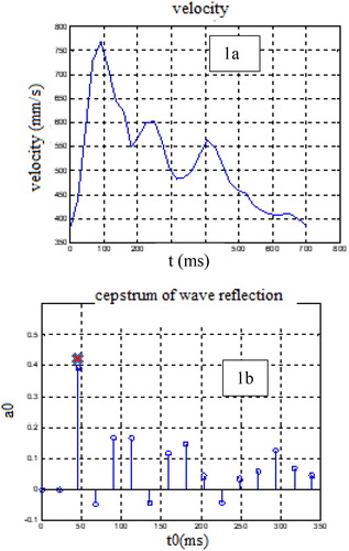
The first result of this study, related to blood velocity in the internal carotid artery and its cepstrum of young healthy subjects can be seen in . Every reflection has an amplitude ai and its delay time ti. The first two peaks always represent the peaks which correspond to the incident wave. Thus, the peak, which comes after the two first ones with the highest amplitude is used as a threshold to distinguish incident and reflected wave peaks. The peak used for the calculation of the time delay is the third peak (t0,a0) with the highest amplitude lower than 1 because the reflection wave is always lower than the incident wave.
Figure 1. Blood velocity signal and its Cepstral analysis: (a). Blood velocity signal. (b). Cepstral analysis of the blood velocity, the red cross indicates the selected peak corresponding to the delay time.
3.2. Determination of t0 as a function of age and length of internal carotid
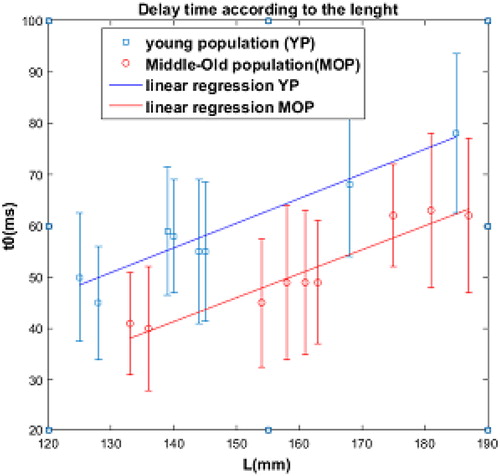
In we have represented t0 according to the length L of the artery for the young and the middle –old subjects. A linear regression of the values obtained shows a substantially parallel evolution. On the other hand, as expected, the values of t0 of the middle-old subjects are lower than those of the young subjects. The results of the linear regression show an average difference of 12.5 ms. it can be concluded here that the proposed method is sensitive to the increase in arterial stiffness with age.
Figure 2. Delay time t0 according to the length of the internal carotid artery and the age.
4. Conclusions
In this study, we presented a new technique using cepstral analysis that provides a convenient way to determine non-invasively the delay time of the reflected wave on the forward wave. The proposed method is a simple, easy and accurate technique that can be used to locally identify the reflection waves and eventually the wave speed. Moreover, it has the advantage of examining short vessels that are difficult to access under normal and pathological conditions. Although the temporal resolution of MRI is less than t0 for the cases studied, this method would gain in power with better temporal resolution as is the case with the Echo-Doppler.
References
- Abdessalem KB, Sahtout W, Flaud P, Gazah H, Fakhfakh Z. 2007. Numerical simulation of non-invasive determination of the propagation coefficient in arterial system using two measurements sites. Eur Phys J Appl Phys. 40(2):211–219.
- Feng J, Khir AW. 2010. Determination of wave speed and wave separation in the arteries using diameter and velocity. J Biomech. 43(3):455–462.
- Grabisch M, Balluet JC. 1985. Identification en présence d’échos par le Cepstre. Trait Signal. 2(1):11–27.
- Nichols WW, O’Rourke MF, Vlachopoulos C. 2011. MCDonald’s blood flow in arteries. 6th ed. Hodder Arnold.
- Peube JL. 2010. Cepstral analysis: establishment of fluid mechanics and transport phenomena. London: UK: ISTE; 483, p. 485.
- Schubert T, Pansini M, Bieri O, Stippich C, Wetzel S, Schaedelin S, von Hessling A, Santini F. 2015. Attenuation of blood flow pulsatility along the Atlas slope: a physiologic property of the distal vertebral artery. AJNR Am J Neuroradiol. 36(3):562–567.
- Sugawara J, Hayashi K, Tanaka H. 2010. Distal shift of arterial pressure wave reflection sites with aging. Hypertension. 56:920–925.