
1. Introduction
Assessment of scapular kinematics has been a challenging topic for several decades. While invasive procedures, such as biplanar fluoroscopy or intra cortical pins, have showed high accuracy to measure scapular kinematics, non-invasive methods, such as stereophotogrammetry, are still not satisfying because of large soft tissue artifacts. In this context, correction procedures have to be developed to minimize the negative effect of soft tissue artefacts on scapular kinematic estimates.
A first approach consists of using an acromion-marker-cluster method coupled with a single calibration to identify scapulo-thoracic kinematics. This method has been extended with double (Brochard et al. Citation2011) calibrations to improve scapular kinematics accuracy. Although these approaches are easy to use, they may lead to joint dislocation when considering the whole kinematic chain. Multibody kinematic optimization has then been developed to address this issue. Especially, closed loop kinematic chain has gained interest for the last decade in order to improve scapulo-thoracic kinematic estimates (Duprey et al. Citation2017). This approach consists of optimizing joint angles by minimizing the distance between the positions of experimental markers and the virtual markers of the kinematic model, with the latter being obtained from a single calibration usually performed while the participant hold an anatomic pose. The closed loop is ensured by constraining the scapula to slide along the thorax. Nevertheless, scapular misorientation are still observed especially for high degrees of arm elevation (Michaud et al. Citation2017), which are far from the calibration pose. Combining double calibrations with multibody kinematic optimization may be innovative and a promising approach to address current issues when computing scapular kinematics.
The purpose of this study was to develop and assess a new procedure to estimate scapular kinematics when combining closed kinematic chain algorithm and double calibration.
2. Methods
2.1. Participants
After having signed an informed consent, four healthy participants (age [22–34], height [1.61–1.85], mass [65–75]) were involved in this study, approved by the local ethical committee.
2.2. Procedure and data collection
Participants were fitted with 18 reflective markers stuck on the thorax and the right clavicle, scapula and arm. For the scapula, three markers were located on the acromion. Marker trajectories were recorded with a 10 camera optoelectronic system (Qualysis, Sweden).
Participants were asked to perform five movements of interest and five static poses (25 in total) representing intermediate instants of each motion. For the three analytical movements (i.e., arm elevations in the coronal, scapular and sagittal plan), poses were considered at 0°, 45°, 90°, 135° and 170° of arm elevation. For the two functional movements (mimic a box lifting and a unilateral shot put-test [USPT]), the five poses split the movements equally from its start t its end. For each pose, an adjustable scapula locator was placed according to three bony landmarks of the scapula after palpation, namely the acromion process, trigonum spinae and inferior angle. To assess the variability of the measurements with the scapula locator, scapula locator configuration was repeated three times, and two measurements per pose for the analytical movements were performed.
2.3. Kinematics
The kinematic model was composed of four segments (thorax, clavicle, scapula, humerus) and 14 degrees of freedom (thorax = 6, sterno-clavicular joint = 2 rotations, acromio-clavicular = 3 rotations, gleno-humeral joint = 3 rotations). Then, four multibody kinematic optimization procedures were implanted to compute scapulo-thoracic joint angles. First, the reference method (REF) consisted of tracking scapular marker trajectories of the scapula locator. High weightings (100 vs 1 for skin markers) were allocated to the scapula markers to obtain its true location. Second, a multibody kinematic optimization was implemented using a closed loop chain enforcing the scapula to keep two contact points with an ellipsoid representing the thorax (ELL). The initial calibration was performed while the participant maintained a static anatomical pose. For this procedure, thorax and sterno-clavicular generalized coordinates obtained with REF were used, while acromio-clavicular joint angles were obtained by tracking the three skin markers located on the scapula.
Two methods using double calibration procedures were used to compute scapulo-thoracic angles, based either on the amplitude-proportional (DC1) or on a time-proportional deformation (DC2) (Zhang Citation2002).
The misorientation of the scapula computed with ELL, DC1 and DC2 was assessed by computing the helical axis angle between scapula orientation measured with these three methods and REF values.
2.4. Statistics
Given the small sample size, for each misorientation angle, the effect size (Cohen’s d) was computed to assess the difference between the three methods relative to REF procedure. A strong effect was associated to an absolute value superior to 0.80.
3. Results and discussion
The scapula locator calibration presented variability of 6.1 ± 4.3%. When two measures were performed with the scapula locator, 2.7 ± 0.7° difference was observed.
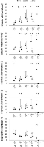
As expected, double calibration methods improved scapular estimates of the last pose in all movements since Zhang’s equations (2002) ensure a full correction of the joint angles at the pose of the second calibration (). Although the double calibration procedure requires an additional measurement at the end of each movement, it counteracts the high scapular misorientation observed for high degrees of arm elevation (up to 48° for analytical movements). For analytical movements, DC2 seemed also to improve scapular kinematic estimates and decrease its variability from 90° to the full arm elevation with on average 14° of misorientation (for P3 and P4) against 28° for ELL. Nevertheless, for low arm elevations, near to the pose of the single calibration, DC2 yielded to greater misorientation than ELL since the initial pose is influenced by the misorientation of the last pose.
Figure 1. Mean and standard deviation of scapular misorientation for abduction, scaption, flexion, box lifting, and USPT (up to bottom) during the five intermediate poses (P1–P5). *, † and § for strong effects between ELL and DC1, ELL and DC2, DC1 and DC2 respectively.
4. Conclusions
Double calibration methods associated with closed loop multibody kinematic optimization improved scapular kinematic estimates, especially at high arm elevation. Nevertheless, scapular misorientation at low arm elevation were not fully corrected, requiring further studies with more participants to completely address scapular kinematic estimate issues.
References
- Brochard S, Lempereur M, Remy-Neris O. 2011. Double calibration: an accurate, reliable and easy-to-use method for 3D scapular motion analysis. J Biomech. 44(4):751–754.
- Duprey S, Naaim A, Moissenet F, Begon M, Cheze L. 2017. Kinematic models of the upper limb joints for multibody kinematics optimisation: An overview. J Biomech. 62:87–94.
- Michaud B, Duprey S, Begon M. 2017. Scapular kinematic reconstruction–segmental optimization, multibody optimization with open-loop or closed-loop chains: which one should be preferred? Int Biomech. 4(2):86–94.
- Zhang X. 2002. Deformation of angle profiles in forward kinematics for nullifying end-point offset while preserving movement properties. J Biomech Eng. 124(5):490–495.