
 ?Mathematical formulae have been encoded as MathML and are displayed in this HTML version using MathJax in order to improve their display. Uncheck the box to turn MathJax off. This feature requires Javascript. Click on a formula to zoom.
1. Introduction
Balloon Kyphoplasty is a minimally invasive surgery, which has acquired great importance recently for treating compressive bone fractures by balloon inflation and PMMA cement injection (Achatz et al. Citation2017). The PMMA cement is subjected to challenging body environment once it is injected into the cancellous bone. The PMMA and porous cancellous bone interface experiences complex in vivo 3 D loading which causes compressive and fracture configuration. Many researchers have worked intensively on the mechanical analysis of the bone and PMMA cement. However, works reported on mechanical analysis of bone and PMMA cement interface were limited and none when it comes to fracture analysis.
Therefore, the work reported here focusses mainly on understanding and evaluating the fracture properties (specific fracture energy (Gf) of the cancellous bone, PMMA cement and Bone/PMMA interface using a recently developed integrated methodology (wedge splitting test (WST) and Heaviside based digital image correlation (H-DIC)) (Bokam.P et al., Citation2020).
2. Methods
2.1. Materials
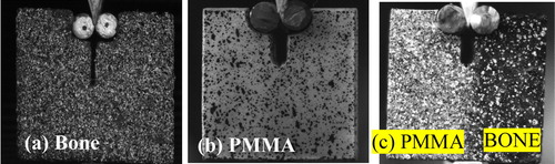
Three groups of specimens (4 samples per each group) were prepared for present study as shown in . (a) cancellous bone extracted from bovine shinbone; (b) PMMA (Kyphon® Xpede™ Bone Cement) prepared by mixing the powder (Methylmethacrylate-styrene-copolymer 69.1% w/w, Barium sulfate 30%w/w) and liquid (Methylmethacrylate (monomer) 99.4% w/w). (c) cancellous bone extracted from proximal epiphysis region (bovine shine bone) is placed in a mold and in the other half of the mold, the PMMA cement is poured and let it cure. The bonded (Cancellous bone + PMMA) sample was then removed and machined to prepare wedge-splitting samples.
Figure 1. Top: Speckle pattern on WST specimen for (a) cancellous bone, (b) PMMA cement, (c) PMMA + bone interface sample.
2.2. Method
The principle of the wedge splitting method and specimen shapes considered similar as described by E.K. Tschegg Citation1986. The wedge unit transforms the vertical load (Fv) from the compression test machine (under displacement control) into a higher magnitude of the horizontal force (Fh). This horizontal force Fh (EquationEquation 1(1)
(1) ) leads to the crack initiation at the notch allowing the crack propagating through the specimen.
(1)
(1)
where Fv is the vertical load applied by the test machine, 2α is the wedge angle (here α = 15 degrees) and µ is the coefficient of friction for the roller bearing.
The entire experimentation was carried in the presence of two CMOS camera (1280 × 1024 pixels with a video acquisition frequency of 0.5 Hz) which are used to record images for the optical full field measurements using Heaviside Digital Image Correlation (H-DIC) method (Valle et al. Citation2015) to retrieve the crack opening displacement and corresponding crack length. The recorded images were analyzed using Digital Image Correlation H-DIC (Valery et al. 2015) to retrieve the crack opening displacement (COD) and the corresponding crack length.
From the principle of WST, to evaluate the specific fracture energy (Gf) required to open a crack of unit area, the total dissipated energy calculated from load –COD curve was divided with the fractured area (EquationEquation 2(2)
(2) ).
(2)
(2)
where ‘A’ is the ligament area, δ is the COD, and F is the splitting force (two times (Fh)).
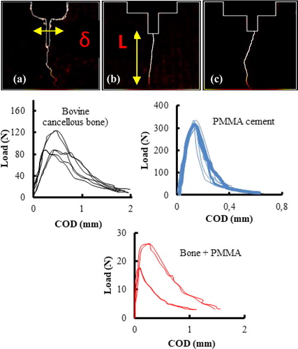
The ligament area (A) is obtained by multiplying the crack length (L) by the thickness of the sample. The values of the crack length (L) and COD (δ) were obtained from the H-DIC analysis as shown in (top)).
Figure 2. Top: Von mises equivalent strain maps with crack propagation for (a) cancellous bone, (b) PMMA cement, (c) PMMA-bone samples obtained using the H-DIC. Bottom: Load-COD curves obtained for the three groups of samples.
3. Results and discussion
shows the von mises equivalent strain maps and Load- COD curves obtained from the 2D H-DIC analysis of the images which are recorded during the test.
The maximum force (Fmax) at which the crack initiated for the three groups are 99 ± 15 N, 308 ± 12 N, and 22 ± 4 N, respectively. From the load-COD curves for the cancellous bone and PMMA + bone interface samples, we observe variation in the Fmax. This can be directly linked to the porosity of the cancellous bone. In addition, the presence of large pores near the crack tip will lower the values of the Fmax. The specific fracture energy (Gf) values for the three groups of samples are reported in the .
Table 1. Specific fracture energy (Gf) values obtained using WST and H-DIC.
The evaluated values are compared with available literature for cancellous bone and PMMA cement specimens. In addition, the study presents the first results of specific fracture energy values for the PMMA + bone interface samples. However, we observe the obtained values for the samples are lower compared to the PMMA cement. This can be due to the variation of porosity in the cancellous bone and the weak bonding between the cancellous bone and PMMA cement.
4. Conclusions
The present findings contribute the first reports on the specific fracture energy (Gf) values of cancellous bone and PMMA cement interface. The bonding between the cancellous bone and PMMA cement gives an idea to the interfacial fractures between bone and PMMA cement and to understand the re-fracture complications that arouse post kyphoplasty operation.
References
- Achatz G, Riesner HJ, Friemert B, Lechner R, Graf N, Wilke HJ. 2017. Biomechanical in vitro comparison of radiofrequency kyphoplasty and balloon kyphoplasty. Eur Spine J. 26(12):3225–3234.
- Bokam P, Germaneau A, Rigoard P, Vendeuvre T, Valle V. 2020. Evaluation of fracture properties of cancellous bone tissues using digital image correlation/wedge splitting test method. J Mech Behav Biomed Mater. 102:103469.
- Merta I, Berger L, Heidfogel G, Kuhn KD, Lewis G, Tschegg EK. 2017. Size and boundary effects on notch tensile strength and fracture properties of PMMA bone cement. Polym Test. 59:441–448.
- Tschegg EK, Celarek A, Fischerauer SF, Stanzl-Tschegg S, Weinberg AM. 2012. Fracture properties of growth plate cartilage compared to cortical and trabecular bone in ovine femora. J Mech Behav Biomed Mater. 14:119–129.
- Tschegg EK. 1986. Equipment and appropriate specimen shapes for tests to measure fracture values. AT No. 390328. Vienna, Austria: Austrian Patent Office.
- Valle V, Hedan S, Cosenza P, Fauchille AL, Berdjane M. 2015. Digital image correlation development for the study of materials including multiple crossing cracks. Exp. Mech. 55:389–391.