
 ?Mathematical formulae have been encoded as MathML and are displayed in this HTML version using MathJax in order to improve their display. Uncheck the box to turn MathJax off. This feature requires Javascript. Click on a formula to zoom.
1. Introduction
Physiotherapy is a health care discipline based on movement assessment and treatment. Mechanical Diagnosis and Therapy (MDT) is a reliable and efficient approach using repeated end-range movements to reverse symptoms among patients presenting with musculoskeletal disorders (May et al. Citation2018). The rationale behind this approach in regard to low back pain is based on the dynamic nature of the pathological intervertebral disc biomechanics. When the Annulus Fibrosus (AF) is centrifugally fissured (radial fissure), the Nucleus Pulposus (NP) could spread into the fissure under patient movement influence. For example, an extension movement can push the NP into an anterior fissure and increase patient pain while a flexion movement would purge the fissure of the NP and reduce patient pain.
The mechanical behaviour of NP in a healthy disc has already been fully described. However, evidence for disc pathological condition remains conflicting, inconsistent and suffers from methodological weaknesses due to imaging technical limitations (Kolber and Hanney Citation2009).
MRI technologies have significantly improved over the last decades, increasing the accuracy of data acquisition and processing. For example, quantitative MRI with T1 and T2 mapping returns relaxation times pixel-wise, allowing objective characterization of the disc (Galley et al. Citation2017). Nevertheless, such techniques have never been used to assess the nucleus dynamic properties.
We aimed to characterize the mechanical behavior of intervertebral disc by using MRI relaxometry (T1/T2 mapping) as a quantitative marker to assess, quantify and compare the NP displacement and strain of a cadaveric lamb vertebral segment with intact and fissured disc according to sagittal loads.
2. Methods
2.1. Specimen preparation
We used a fresh lamb spine segment comprising 3 vertebrae and 2 intervertebral discs, wrapped in a saline soak gauge and placed under creeped load 1.5 h before the experiments with an axial compressive load. The same disc was characterized before and after inducing an anterior tear.
2.2. MRI acquisition and processing
We used a 3.0-Tesla imager (Siemens, Munich, Germany) to run 3 different types of sequences for each mechanical state:
multi-slice 2D T2-weighted Turbo Spin Echo
multi-echo spin echo sequence for T2 mapping
Inversion Recovery TSE for T1 mapping
Images were processed using scripts and functions developed with MATLAB (MathWorks, Natick, MA). The maps were obtained after normalizing the data and fitting them pixel wise according to:
S is the normalized signal intensity in arbitrary units, t the time obtained from the echo train or inversion times, and a/b constants used for fitting purposes.
2.3. Mechanical loading
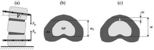
We controlled the boundary conditions by fixing the lower vertebra and by using two different elastic systems to impose displacements with an arm level on the upper vertebra providing an angular variation and combined axial/bending loads (). We imposed 3 loading states (neutral, flexion and extension) on both disc conditions: healthy vs fissured.
Figure 1. Measurement of the nucleus sagittal boundary displacement (w), the nucleus pulposus (NP) migration (m) within the annulus fibrosus (AF) and nucleus sagittal strain according to compression (Fc) and bending (Fb) force: (a) setup; (b) reference mechanical loading state (neutral, non-fissured disc); (c) mechanical state with partial migration of the nucleus.
2.4. Measurements
We used a mark tracking system to measure the bending angle and the axial strain. We measured the global nucleus displacement by evaluating: 1- the centroid position of NP relative to the centroid position of the spinal canal, 2- the displacement on sagittal boundaries of the NP (w-w0), and 3- the NP strain in the sagittal direction ((w – w0)/w0). We also estimated the degree of nucleus migration into the fissure by calculating the ratio of the nucleus displacement (m) and the thickness of the annulus ().
3. Results and discussion
We observed a comparable biomechanical behavior of the disc as was previously published (Kolber and Hanney Citation2009), showing an anterior migration of the NP in extension and a posterior migration in flexion.
The radial fissure was visible using all MRI sequences. On T2-weighted images, its morphology changed depending on the bending load with a grade 2 fissure on the Dallas classification when the disc is in extension and a grade 1 when it is in flexion (Sachs et al. Citation1987).
The quantitative mapping confirmed the NP migration into the fissure with respect to the applied movement. In extension, the T1 relaxation time of the tissue inside the fissure is superior to 800 ms, similar to the one measured in the nucleus. This characteristic disappeared in flexion (see ). Our results show that the nucleus elongated by 21.1 mm in extension while it contracted by 3.1 mm in flexion. The anterior boundary of the nucleus moved forward by 5.0 mm in extension while it moved backward by 0.6 mm in flexion (see ).
Figure 2. T1 map of the fissured disc positioned in extension (left) and flexion (right).
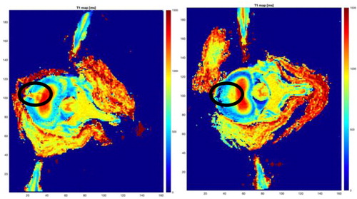
Table 1. Mechanical measurement of the disc angle variation, NP strain and displacement (mean values).
The AF fissure modified the biomechanics of the NP by increasing its strain and its displacement in all our measurements (see ).
These results revealed a relationship between disc abnormality and displacement possibility with a pattern similar to the one hypothesized by MDT therapists.
These results are encouraging but need to be validated in larger sample sizes, with human specimen at different stages of disc degeneration and by using different fissure orientations.
4. Conclusions
Quantitative MRI mapping is an effective way of assessing disc radial fissure mechanical behaviour. The NP moves in a consistent pattern according to bending load with either healthy or fissured disc. However, the fissure increases the displacement preference which appears consistent with the clinical hypothesis raised by MDT practitioners.
Disclosure statement
No potential conflict of interest was reported by the authors.
Additional information
Funding
References
- Galley J, Maestretti G, Koch G, Hoogewoud H-M. 2017. Real T1 relaxation time measurement and diurnal variation analysis of intervertebral discs in a healthy population of 50 of 50 volunteers. Eur J Radiol. 87:13–19.
- Kolber MJ, Hanney WJ. 2009. The dynamic disc model: a systematic review of the literature. Phys Ther Rev. 14(3):181–189.
- May S, Runge N, Aina A. 2018. Centralization and directional preference: An updated systematic review with synthesis of previous evidence. Musculoskelet Sci Pract. 38:53–62.
- Sachs BL, Vanharanta H, Spivey MA, Guyer RD, Videman T, Rashbaum RF, Johnson RG, Hochschuler SH, Mooney V. 1987. Dallas discogram description. A new classification of CT/discography in low-back disorders. Spine. 12(3):287–294.