
1. Introduction
Nordic hamstring exercise (NHE) is one of the most common exercise used in soccer, rugby or sprinting in order to prevent hamstring injury. It is well established in the literature that implementation of NHE into the training process can significantly reduce the incidence of hamstring injuries in football (Petersen et al. Citation2011; Al Attar et al. Citation2017). In fact, several studies reported numerous positive adaptations following an implementation of several weeks of NHE training such as a significant improvement in eccentric hamstring strength (Mjolsnes et al. Citation2004; Mendiguchia et al. Citation2020). However, few football players (beginners or experts) are able to fully perform the movement in its entire range of motion and thus take full advantage of this exercise. Previous studies have shown that changes in the knee angle starting positions can affect exercise effectiveness and the activation of particular muscles. In fact, Park and Yoo (Citation2014) reported that changes in support angle during the Roman chair exercise differentially activated muscle groups in the lower extremities. In this way, it is possible to strengthen the muscle on all its length since most of the hamstring muscle injuries occur in the final part of the swing phase (Šarabon et al. Citation2019). To the best of our knowledge, there are a few studies considering parameter changes in the NHE. Šarabonet al. (2019) reported that muscle activity decreased or remained similar in all modified knee angle starting positions compared to the standard NHE for all measured muscles (i.e., biceps femoris, semitendinosus). However, the effects of knee angle starting position on the spontaneous biomechanical organization used to perform the NHE remain unclear.
Thus, the aim of the present study was to analyse the effect of knee angle starting position on the kinetic and kinematic adaptations during the NHE. More precisely, we studied the spontaneous motricity used in order to lowering their upper body towards a prone position, as slowly as possible until participant can no longer control the lowering.
2. Methods
2.1. Participants
Nine healthy male with previous experience with NHE (24 ± 7 years, 180 ± 7 cm, 79.4 ± 8.2 kg) participated in the study. All participants were free of lower limb injury in the twelve months prior to data collection. Informed written consent was obtained from all participants prior to data collection.
2.2. Experimental protocol
After a 10 min warm-up consisting of light cycling aerobic activity, each participant performed two series of 10 repetitions of manual resistance eccentric contractions of the hamstrings
After the warm-up, on both leg, two EMG surface electrodes (TrignoWireless EMG Systems; Delsys, Boston, MA, USA) were placed on semitendinosus and biceps femoris and kinematic markers and clusters were attached on the different segments ().
Figure 1. Set up for NHE variations evaluation.
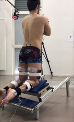
Two force sensors were fixed on both ankles to measure contact forces normal to the lower leg at 200 Hz and synchronized with 12 cameras of an optoelectronic motion capture system (Motion analysis corporation, Raptor 4, Santa Rosa, USA) in order to track 32 reflective markers at 200 Hz to record kinematics of every trial. Surface EMG signals were sampled at 2000 Hz.
The NHE were tested in randomized order for three conditions of different knee angle starting position (0˚ (i.e. leg support align with the floor), 20˚ and 30˚ relative to the ground respectively) (). For each slope condition, participants were asked to perform three repetitions of the NHE. Each participant performed a familiarization session before the study. The rest between each trials of the same knee angle starting position was 2 min and 5 min between each slope.
2.3. Data analysis and statistics
Knee angles (defined as rotation between the distal and proximal segment using Euler angles with a rotation sequence Z (F-E), Y (A-A), X (P-S), negative values correspond to knee flexion), knee angular velocity and maximal forces for both legs were computed with Visual3D software (C-Motion, Germantown, USA). Standard statistical methods were used to compute means and standard deviation of the parameters studied for each participant and each condition. The peak EMG activity for all muscles, which was determined as highest mean value on 250 ms window length during the movement, was computed.
Three repeated measures ANOVA were used to identify the effect of slope condition. Newman Keuls post-hoc test was used when significant level (p < 0.05) was reached.
(MathWorks, Natick, MA).
3. Results and discussion
The average maximal force for both legs were 255.2 ± 68.2, 262.7 ± 34.7 and 278.2 ± 42.0 N for the 0˚, 20˚and 30˚ slopes conditions, respectively. The repeated-measures ANOVA revealed no significant effect of knee angle starting position on the maximal force. Moreover, the statistical analysis showed no significant asymmetry between both legs.
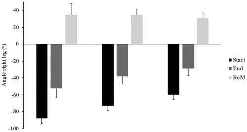
shows the average knee angle at the beginning and at the end of the movement as well as the range of motion for each condition. The statistical analysis showed a significant decrease of knee angle at the beginning and at the end of the movement with an increase of the slope condition. However, no significant effect was observed on the knee range of motion. Consequently, by combining the three knee angle starting positions, the participants are enabled to perform the exercise through a larger range of motion compare to the standard NHE and achieve similar maximal forces.
Figure 2. Effect of the slope condition on the start position (Start), end position (End) and knee range of motion (RoM).
Average knee angular velocity values were 40.8 ± 23.5, 39.1 ± 20.6 and 28.1 ± 16.5 N for the 0˚, 20˚and 30˚ slopes conditions, respectively. Our results show a trend (p = 0.07) towards a decrease in the angular velocity of the knee with an increase in slope, suggesting better control of the movement. Finally, for all measured muscles the muscle activity remained similar whatever the slope condition. The present results concerning the force are in agreement with a recent previous (Šarabon et al. 2019).
4. Conclusions
The results of the present study suggest that using different start positions when performing NHE might be of interest to adapt the difficulty and thus obtain a better control of the movement and work on a greater range of motion without the help of external equipment.
Disclosure statement
No potential conflict of interest was reported by the authors.
References
- Al Attar WSA, Soomro N, Sinclair PJ, Pappas E, Sanders RH. 2017. Effect of injury prevention programs that include the nordic hamstring exercise on hamstring injury rates in soccer players: a systematic review and meta-analysis. Sports Med. 47(5):907–916.
- Mendiguchia J, Conceição F, Edouard P, Fonseca M, Pereira R, Lopes H, Morin J-B, Jiménez-Reyes P. 2020. Sprint versus isolated eccentric training: comparative effects on hamstring architecture and performance in soccer players. PLoS One. 15(2):e0228283Feb 11
- Mjolsnes R, Arnason A, Osthagen T, Raastad T, Bahr R. 2004. A 10-week randomized trial comparing eccentric vs. concentric hamstring strength training in well-trained soccer players. Scand J Med Sci Sports. 14(5):311–317.
- Park SY, Yoo WG. 2014. Effects of hand and knee positions on muscular activity during trunk extension exercise with the Roman chair. J Electromyogr Kinesiol. 24(6):972–976.
- Petersen J, Thorborg K, Nielsen MB, Budtz-Jørgensen E, Hölmich P. 2011. Preventive effect of eccentric training on acute hamstring injuries in men’s soccer: a cluster-randomized controlled trial. Am J Sports Med. 39(11):2296–2303.
- Šarabon N, Marušič J, Marković G, Kozinc Z. 2019. Kinematic and electromyographic analysis of variations in nordic hamstring exercise. PLoS One.;14((10)):e0223437