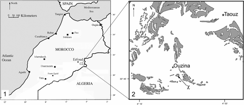
FIGURE 1 Locality maps. 1, Map of Morocco and surrounding area (modified from Chatterton et al., 2006; McKellar and Chatterton, 2009). 2, Ordovician (shaded regions) locality map for trilobite/trace locality (marked with χ) within the Tafilalt basin (modified from Fetah et al., 1986).
In Volume 17, Number 4 of Ichnos: An International Journal for Plant and Animal Traces, all figures in the article entitled “Rusophycus carleyi (James, 1885), Trace Fossils from the Lower Ordovician of Southern Morocco, and the Trilobites that Made Them”, by S. Gibb, B. D. E. Chatterton, and M. K. Gingras, pages 271–283, appeared in incorrect resolution. The figures in correct resolution appear below.
FIGURE 2 Microbial mat “cracking” and preferential iron staining along the crack lines from the Upper Fezouata Formation, Ouzina, southern Morocco. 1, Asaphellus aff. fezouataensis in bottom right corner (UA13664). 2. Asaphellus aff. fezouataensis in middle (UA13667n). Scale bar is 1 cm. (See Color Plate VI.)
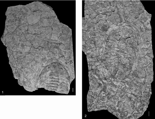
FIGURE 3 The proximity of Asaphellus aff. fezouataensis to Rusophycus carleyi. 1, A. fezouataensis with white arrow directed at a lobe of R. carleyi under the right pleural region of the trilobite (UA13664). The trilobite would have become trapped within the sediment. The left lobe is not visible because perhaps the trilobite had dug alongside another R. carleyi and only the right lobe of the most recent behavior is recorded. 2, R. carleyi, convex hyporelief, with white arrow pointing to the right lateral section of the cephalon of a trilobite (UA13657). 3, R. carleyi, convex hyporelief, with white arrow directed towards the left genal spine of a trilobite (UA13662). 4, R. carleyi, convex hyporelief, with the white arrow at the right ventral lateral section of the cephalon and the black arrow pointing at the transected portion of the hypostome of the trilobite, within the trace (UA13661). Specimens UA13662 & UA13661 were submerged in ethyl alcohol to enhance the exoskeleton within the trace, while UA13664 & UA13657 were coated in ammonium chloride. Specimens are from the Upper Fezouata Formation, Ouzina, southern Morocco. Scale bar is 1 cm.
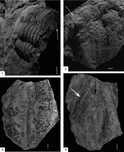
FIGURE 4 Rusophycus carleyi (UA13658), convex hyporelief with left lateral border of pygidium (white arrow), and thoracic segment impressions (white arrows outlined by black), from the Upper Fezouata Formation, Ouzina, southern Morocco. Scale bar is 1 cm.
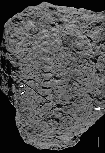
FIGURE 5 Scatter plot (length vs. width) of Rusophycus carleyi and the trilobites from the Ouzina locality. The length and width of the trilobite, when only fragments were available were inferred by extrapolating proportions from parts, with ratios taken from four Asaphellus species from the Ordovician of Morocco. The black diamonds are the trilobites and the grey squares are Rusophycus carleyi. All specimens are from the Upper Fezouata Formation, Ouzina, southern Morocco.
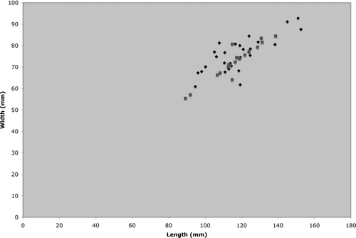
FIGURE 6 Rusophycus carleyi (UA13655), convex hyporelief, from the Upper Fezouata Formation, Ouzina, southern Morocco. 1, Lateral oblique close-up of hypostome impression (black arrow). 2, Oblique lateral view of trace. 3, Lower posterior view of endopodal spine scratches within the medial opening. 4, Close-up of coxal impressions, with the left being the anterior end of the trace. Scale bar is 1 cm.

FIGURE 7 Rusophycus carleyi. 1, UA13656 displaying endopodal spine scratches within the posterior lateral medial opening. 2, UA13660 displaying endopodal spine scratches within the lateral medial opening. 3,4, UA13659. 3, Entire view of convex hyporelief of trace. 4, Close-up of thoracic segment impressions on trace. All specimens are from the Upper Fezouata Formation, Ouzina, southern Morocco. Scale bar is 1 cm.
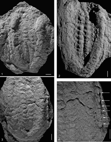
FIGURE 8 Schematic of the tracemaker (trilobite), Asaphellus aff. fezouataensis, removing itself from the substrate, resulting in the trace Rusophycus carleyi. From the striations/ridges within the mesial opening, it is assumed that A. aff. fezouataensis would have podomeral spines. Sketch created by Darrin Molinaro.
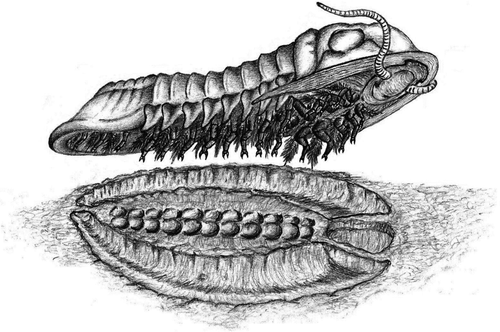
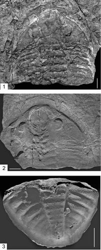
FIGURE 9 Asaphellus aff. fezouataensis, all dorsal views. 1, UA13664, cephalon and majority of thorax (submerged in ethyl alcohol to enhance trilobite morphology). 2, UA13665, cephalon, minority of thorax and taphonomically altered hypostome (coated with ammonium chloride). Important note, with regard to this specimen is the pits observed on the posterior end of the cephalon, associated with the alimentary structures (diverticulae). The pits are similar in outline to trilobite alimentary diverticulae illustrated by Chatterton, Johanson, and Sutherland (1994, Figs. 1.1–1.3, 2.1, 2.4 and 3.4). 3, UA13663, pygidium (coated with ammonium chloride). All specimens are from the Upper Fezouata Formation, Ouzina, southern Morocco. Scale bar is 1 cm.