

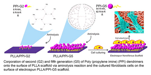
Abstract
The aim of this comparative study was focused on the surface chemistry, in vitro biodegradation and biocompatibility of poly(L-lactic acid) (PLLA) scaffolds incorporated with poly(amidoamine) (PAMAM) and poly(propylene imine) (PPI) dendrimers. The functionality of these two types of dendrimers from second, third and fifth generations (G2, G3 and G5) onto the surface of PLLA film and electrospun scaffolds was investigated and characterized by a series of analyses including X-ray photoelectron spectroscopy, atomic force microscope, Fourier transform infrared spectroscopy, contact angle, scanning electron microscope, weight loss, water uptake and pH changes after degradation in different media. The successful incorporation of dendrimers into the surface of PLLA scaffolds was observed. PPI-G5 could further increase the hydrophilicity and surface roughness of PLLA scaffolds compared to PPI-G3 and PPI-G2. Further improvement on PLLA surface chemistry was observed when PAMAM-G5 was applied compared to PPI-G5. The pH value of Roswell Park Memorial Institute Medium (RPMI) media containing raw electrospun PLLA scaffold decreased to 5.2 from its original value of 7.2. However, the local acidity of RPMI medium caused by PLLA hydrolysis was greatly reduced on the surface of PLLA/PAMAM-G5 and PLLA/PPI-G5 with pH values of 7.8 and 7.5, respectively. Moreover, the viability of fibroblast cells on the surface of PLLA/dendrimer scaffolds was considerably increased compared to raw PLLA. Fibroblasts showed higher attachment and homogeneous spread morphology on the surface of PLLA/PAMAM-G5 and PLLA/PPI-G5. These observations underline the favorable surface properties of PLLA scaffolds incorporated with dendrimers, to be used as a biodegradable biocompatible scaffold tailored for biomedical applications.
Graphical Abstract
Acknowledgments
We acknowledge and are grateful to Dr. Majid Mohammadi-Mossahebi for his dedicated help with cell culture and MTT assay test. AFM and XPS tests were performed at UNSW Mark Wainwright Analytical Centre’s Electron Microscopy Unit (EMU) and Solid State and Elemental Analysis Unit (SSEAU), respectively. We also would like to thank Dr. Majid Ebrahimi Warkiani for his valuable comments regarding AFM analysis. The authors wish to acknowledge two anonymous reviewers for their critical comments and suggestions, which helped to significantly improve this article. We greatly appreciate Dr. Emilio Echevarria for his immense help and thoughtful comments for editing.