

Abstract
In this study, epithelial cell adhesion molecule (EpCAM) aptamer-activated nanoparticles (Ap-NPs) were synthesised to enhance treatment efficiency in colorectal cancer (CRC). PLGA [poly(d, l-lactide-co-glycolide)] copolymer was fabricated by conjugation of COOH-PEG-NH2 to PLGA-COOH through an EDC/NHS-mediated chemistry. Afterwards, 5-fluorouracil-loaded (FU) nanoparticles were prepared using the water/oil/water double emulsion solvent evaporation method. The in vitro cytotoxicity of formulations was evaluated using the MTT assay in HCT-116, CT-26 and HEK-293 cell lines. For in vivo study, tumour-bearing BALB/c mice were established by subcutaneous injection of CT-26 cell line. The results indicated that fabricated AP-FU-NPs had 101 nm size with a spherical surface, relatively homogeneously and, satisfactory encapsulation efficiency (83.93%). In vitro experiments revealed that Ap-FU-NPs had a superior in vitro cytotoxicity than both FU-NPs and free 5-FU in CT-26 and HCT-116 cells but, were significantly low toxic against HEK-293 cells relative to free 5-FU. Furthermore, in vivo results showed no significant haemolytic effect, hepatic and renal injury, or weight loss. After treatment of various animal groups with formulations, notable tumour growth delay was observed following the order: Ap-FU-NPs < FU-NPs < 5-FU < PBS. The results suggest that AP-FU-NPs could be an effective and promising carrier for 5-FU delivery to the EpCAM overexpressing CRC cells.
GRAPHICAL ABSTRACT
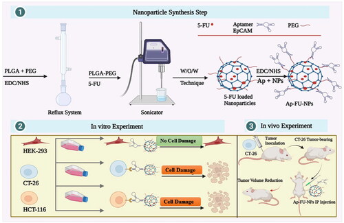
The present study was carried out in the three steps summarised: (1) synthesis and characterisation. At first, PEG was conjugated to PLGA polymers by EDC/NHS chemistry method, then the resulting polymers were used to prepare PLGA-PEG NPs via the W/O/W technique. Finally, EpCAM Aptamers were attached to fabricated NPs. (2) In the second step cytotoxicity of formulated NPs was evaluated in vitro experiments in HEK-293, CT-26 and HCT-116 cell lines. (3) In the third step in vitro results further was investigated in CT-26 tumour-bearing BALB/c mice. ‘Created with BioRender.com’.
Acknowledgements
This study has been adapted from a Ph.D. thesis at Hamadan University of Medical Sciences. The authors are extremely grateful to the efforts of the supervisor of this study Mr. Professor Massoud Saidijam, who passed away while conducting this study.
Ethics approval
All methods and protocols and also, the animal using protocol was reviewed and approved by the Institutional Ethics and Research Advisory Committees of Hamadan University of Medical Sciences (IR.UMSHA.REC.1397.874).
Consent for publication
Not Applicable
Availability of data and materials
The data are available on reasonable request from the corresponding author
Disclosure statement
The authors report no conflicts of interest in this work.
Authors’ contributions
Conceptualisation; Bahram Yavari, Seyyed Shamsadin Athari, Massoud Saidijam; Data curation: Bahram Yavari, Seyyed Shamsadin Athari; Formal analysis: Bahram Yavari; Akram Jalali; Funding acquisition: Rezvan Najafi; Investigation: Bahram Yavari, Seyyed Shamsadin Athari, Yadollah Omidi; Methodology: Bahram Yavari, Yadollah Omidi, Massoud Saidijam, Rezvan Najafi; Project administration: Massoud Saidijam, Rezvan Najafi; Resources: Bahram Yavari, Akram Jalali; Software: Bahram Yavari, Akram Jalali; Supervision: Massoud Saidijam, Rezvan Najafi; Validation: Bahram Yavari, Seyyed Shamsadin Athari; Visualisation: Bahram Yavari, Seyyed Shamsadin Athari; Roles/Writing – original draft: Bahram Yavari, Akram Jalali; Writing - review & editing: Bahram Yavari, Seyyed Shamsadin Athari, Akram Jalali.