Abstract
Based on the research on small gap bridging peripheral nerve injury in SD rats, we propose to investigate the possibility of bridging peripheral nerve injury with small gap using a de-acetyl chitin conduit in primates. The median nerves of 8 rhesus monkeys were cut at 2 cm above the elbow; the right sides were subjected to small gap (2 mm) bridging to repair the nerve with chitin conduit (conduit inner diameter 4 mm; length 10 mm); the left sides were subjected to traditionary epineurium suture. The electrophysiology analysis was conducted after the 3rd month and 6th month, respectively. The adhesions condition of biological conduit was only a little after the 15 3rd month; the conduit can remain cast contour; vessels can be seen on the conduit 16 surface and nerve intumescentia was not obvious. The adhesion and intumescentia condition can display better biocompatibilities than traditional suture methods. The motor nerve conduction velocity was only 1/2 of the control group. Although the motor nerve conduction velocity of the conduit group was a little higher than the epineurium suture group, there was no statistically significant difference (P>0.05) at the 3rd month (). The conduit cast contour disappeared after 6 months. The motor nerve conduction velocity was only 4/5 of the control group. The motor nerve conduction velocity of the conduit group was higher than the epineurium suture group; there were statistically significant differences (P<0.05) at 6 months. The nerve trunk conduction velocity of biological conduit was higher than the epineurium suture group at the 6th month, and there were statistically significant differences (P<0.05) (). The biocompatibility of the biological chitin conduit in primate rhesus monkeys was quite good. The electrophysiological results of biological conduit in primate rhesus monkeys were better than the traditional epineurium suture. The biological conduit can be used in primate rhesus monkeys to substitute for the traditional epineurium suture methods.
Epineurial neurorrhaphy and lamellar sheath suture are clinically traditional repair methods after peripheral nerve injury. In recent years, small gap bridging peripheral nerve injury has gradually become a hot area of research. The microenvironment that biological conduit constructed at the broken ends of fractured nerve profits nerve fiber thoroughly educe selectivity regenerative action. Based on early experiments in SD rats, we are going to progress experiment about biological conduit sleeve bridging median nerve injury with small gap in primate rhesus monkey. We devised this method to substitute the traditional epineurial neurorrhaphy.
MATERIALS AND METHODS
Materials and Groups
1. Materials
Hollow cylindrical conduit (a de-acetyl chitin conduit invented by Beijing University People's Hospital and the Chinese Textile Academy, the State Patent No.: 01136314.2). Size: tube length 10mm, thickness 1mm, inner diameter 4mm.
2. Groups
8 male rhesus monkeys were anesthetized with the Sumianxin II (0.11ml/Kg i.m (Military Veterinary Research Institute of Military Medical Academy); after the anaesthesia, bilateral elbow median nerves were cut at 2 cm above the elbow; the right sides were subjected to small gap (2mm) bridging to repair the nerve with chitin conduit (conduit inner diameter 4mm; length 10 mm); the left sides were subjected to traditional epineurium suture. The electrophysiology analysis was conducted after the 3rd month and 6th month, respectively.
3. Surgical Procedures
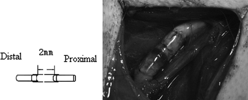
The suture method of the biological conduit group was shown in .The epineurium suture group: after cutting off the median nerve, the proximal and distal ends were relatively rotated 180 degrees and epineurium sutured with a 9-0 Nylon Suture; every one stump seamed two needles.
Figure 1. Model of biological conduit bridging with small gap.
Detection Project
The bilateral elbow median nerves were exposed, then the motor nerve conduction velocity was tested as normal nerve conduction velocity data in the first surgery.
General ObservationAfter anesthesia, we exposed the bilateral median nerve of all the experimental groups, and observed the general form of the stitching, the conduit and adhesions of soft tissue after the 3rd month and 6th months, respectively.
Electrophysiological StudyElectrophysiological assessment was conducted at the 3rd month and 6th month, respectively. At the 3rd month, we placed a central stimulating electrode at the distal end of the stitching along the nerve conduit, and placed a receiver electrode between thenar muscle then measured the distance and latency differences between the electrodes, and determined motor conduction velocity with Oxford EMG. At the 6th month, we tested motor conduction velocity in the way mentioned above, and tested conduction velocity of postrepaired nerve trunk after exposing the median nerve in wrist.
Statistical AnalysisThe data were analyzed and processed using SPSS 11.0 and compared using the T test methods. A probability where p <0.05 was considered significant for all statistical comparison. All values are presented with the mean±SD. The differences of nerve conduction velocity among groups were compared.
RESULTS
1. General Observation
Three months after the surgery, the biological conduit that can maintain its inherent shape was partly absorbed, and the vascular network was clearly visible on the conduit surface. Suture intumescentia were relatively obvious in the epineurium suture group.
2. Distal Limb Motor Function Recovery
At the 3rd month, thumb functions of the epineurium suture group and biological conduit group both rehabilitated in certain degrees; we can see flexion movement of the first, second and third fingers; however, feeding mainly depends on both hands as the action. On the other hand, at 6 months’ time, thumbs of both groups have better functional recovery. The flexion movement of the first, second and third fingers can often be seen; in addition, feeding can be completed with one hand at 6 months time.
3. Motor Nerve Conduction Velocity
Motor nerve conduction velocity of the epineurium suture group was a little lower than the biological conduit group, but there were no statistical significant differences between the data. However, the motor nerve conduction velocity was significantly different between the two groups and control group. The former two were only 1/5 to 1/4 of normal conduction velocity at the 3rd month. Based on the contrast of motor nerve conduction velocity at the 6th month, we found that statistical differences existed between the biological conduit group and epineurium suture group. The restoration of the former group was significantly better than the latter one, but only l/2 to 2/3 of the normal control group. However, based on the contrast of nerve trunk conduction velocity at the 6th month, we found that although the data of the biological conduit group was better than the epineurium suture group, there were not statistically significant differences between them.
DISCUSSION
Epineurium and perineurium sutures are the most common methods used to repair peripheral nerve injury Citation[1], Citation[2]. Nerve fibers axon regeneration and effective domination to the target organ are two most important aspects in peripheral nerve reparation. The selective regeneration of peripheral nerves provides a new practical way to good neural docking. Jiang Baoguo et al. Citation[3], Citation[4] first introduce and authenticate that small gap (2–3mm) conduit can promote reparation of peripheral nerve injury of SD rats.
Table 1.Motor nerve conduction velocity 3 months after surgery
Table 2. Conduction velocity of nerve cord and motor nerve 6 months after surgery
On the basis of animal experiments, Jiang Baoguo and Sun Yushan invented a conduit material, which was made of marine life, non-toxic, easily absorbed and degradated in vivo. In addition, it has good biocompatibility and has patents of The State Intellectual Property Office Citation[5], Citation[6].
In preliminary animal experiments, we found that therapeutic effects of biological conduit bridging with small gap (observation indicators were functional recovery, partial adhesion of the nerve injury, medullated nerve fibers, number of stump distal end, motor nerve conduction velocity) were significantly better than the epineurium suture with stump rotated in the sciatic nerve injury model in the SD rats, and were similar with the results of mutilated nerve suture in situ Citation[7–9]. The ability to regenerate in SD rats, primates, and humans were different. Were the results in primates consistent with the results in rats? We haven't seen such reports at home and abroad yet.
In rhesus monkey experiments, we have chosen the median nerve injury in the elbow model. After mutilation, the right sides were subjected to small gap (2mm) bridging to repair the nerve with chitin conduit (conduit inner diameter 4mm; length 10 mm); the left sides were subjected traditional epineurium suture. Three months after surgery, the adhesions condition and nerve intumescentia was relatively obvious in the epineurium suture group. The general observation results indicated that the biological conduit has good biocompatibility and can effectively reduce the ectomycorrhizal phenomenon of nerve fiber and reduce neuromatous formation opportunities. There were no statistically significant differences in motor nerve conduction velocity between the biological conduit group and epineurium suture group at the 3rd month. However, the motor nerve conduction velocity of both groups was only 1/5 to 1/4 of the normal group. Results above showed nerve conduction velocity and thumb functions recovered to a certain extent at the 3rd month.
Nerve trunk conduction velocity reflects recovery of neural trunk nerve fibers to some extent; although the data of the biological conduit group was better than the epineurium suture group, there was no statistical difference between them. However, differences in nerve conduction velocity between the biological conduit group and epineurium suture group were statistically significant at 6 months; restoration of the former group was better than the latter, but only l/2 to 2/3 of a normal control. As for the distal limb (hand) functional recovery at 6 months, recovery of thumb function was fine and the activities of the first, second and third fingers were basically near normal, but the nails of the residual finger were distorted or ablated in six of eight monkeys.
In the rhesus median nerve injury model, although the results of the biological conduit group were better than the epineurium suture group, the differences between the two were not statistically significant. However, the biological conduit group still has tremendous advantages compared to the suture considering its good biocompatibility, simple surgical operation, light local intumescentia in vivo. The biological conduit sleeve bridging peripheral nerve injury with small gap (2mm) can replace traditional epineurium sutures in rhesus monkey experiments.
Due to the limitations of clinical impaired tissue and the restrictions on microsurgical technique and equipment level, it is difficult to suture nerve sections in situ but in varying degrees of rotation clinically. So, we suture the stump of the epineurium suture group animal with relative rotation.
Acknowledgements
This work was Supported by China National Natural Science Funds for Distinguished Young Scholar (No.30625036), China 973 Projection (No.2003CB515306), and was also funded by China Nature Science Youth Foundation (Number: 30801169) and the China Research Fund for the Doctoral Program of Higher Education (No.20070001780).
Related Research Data
References
- Lundborg G. A 25-year perspective of peripheral nerve surgery: Evolving neuroscientific concepts and clinical significance. J Hand Surg [Am] 2000; 25: 391–414
- Baoguo Jiang, Shuhuan Wang, Chuanhan Feng. 1994. The comparison study of small gap artery bridging and epineurial neurorrhaphy in peripheral nerve injury. Peking University Medical College Journal, 26: 249–250.
- Baoguo Jiang, Yasuji Yoshida 1998. The immunohistochemical study on gap artery sleeve bridging in peripheral nerve. Chinese Microsurgery Journal, 14: 39–42.
- Baoguo Jiang, Bing Wang, Shousheng Hu, , et al. 1999. The comparison of different small gap artery bridging in peripheral nerve. Chinese Orthopaedic Surgery Journal, 6: 115–117.
- Yushan Sun, Baoguo Jiang, Qingsong Zhu. 2002. The experimental functional medical material in animal body. Spinning and Weaving Science Research, 13: 7–22.
- Jian Li, Baoguo Jiang, Dianying Zhang, , et al. 2003. Biological tube small gap bridging repair peripheral nerve injury. Chinese Hand Surgery Journal, 19((2)): 118–120.
- Jiang B., Zhang P., Zhang D., Fu Z., Yin X., Zhang H. Study on small gap sleeve bridging peripheral nerve injury. Artif Cells Blood Substit Immobil Biotechnol. 2006; 34(1)55–57
- Zhang P., He X., Zhao F., Zhang D., Fu Z., Jiang B. Bridging small-gap peripheral nerve defects using biodegradable chitin conduits with cultured schwann and bone marrow stromal cells in rats. J Reconstr Microsurg. 2005; 21(8)565–571
- Peixun, Zhang, Baoguo, Jiang, Fuqiang, Zhao, Zhongguo, Fu, Dianying, Zhang, Chan, Du, Hongbo, Zhan. 2005. Chitin biological tube bridging the peripheral nerve with a small gap. Chinese Surgery Journal, 43((20)): 1345–1347.