Abstract
Erythrocyte shape and biomechanical properties have close relation to its physiological function. In this research the erythrocyte was reconstructed with natural structure protein and lipids based on cellular mechanics and hemorheology concepts. The biomechanical properties of the reconstructed erythrocyte were determined with the micropipette aspiration system. The shapes of reconstructed erythrocyte were obtained with electron scanning microscope. The oxygen carrying-releasing function was analyzed with the HEMOX analyzer from TCS, the experimental results indicated that the reconstructed erythrocytes were similar to the natural erythrocyte: having biconcave disc shape, good deformability and carrying-releasing oxygen function.
INTRODUCTION
The blood substitute has been a hot researching focus around the world. As a blood substitute, its function should be comparable to that of natural hemoglobin, carrying and releasing oxygen around the human bodies. The present known blood substitutes are perfluorocarbon, various kinds of modified hemoglobin by chemical technology and gene engineering. However, the perfluorocarbon molecule is hard to metabolize and imposes harmful effects on human health due to its accumulation in vivo. Gene engineering produced human hemoglobin from tobacco plant root and has been used to produce human hemoglobin, but the high cost and low yield hinder the high production. In addition, liposome has been used to envelope hemoglobin to be used as a blood substitute. However, the procedure has only been carried out in labs at present Citation[1], Citation[2].
Erythrocyte shape and biomechanical properties have close relation to the oxygen carrying-releasing dynamics function. But up to now little research on artificial blood substitutes has realized this viewpoint and mainly focused the studies on how to use blood substitute simulating physiology of normal red blood cells Citation[3], Citation[4]. The main problem of these products lies in the lack of natural configuration, and deformability needed in circulation like those in mature RBCs that would hinder its oxygen carrying-releasing dynamics function.
In this research, we have studied the biorheological properties of reconstructed erythrocytes and their function of carrying-releasing oxygen. The reconstruction of red blood cells in vitro could be of great significance in the biomedical research field and may potentially solve the shortage problems of blood supply.
MATERIALS AND METHODS
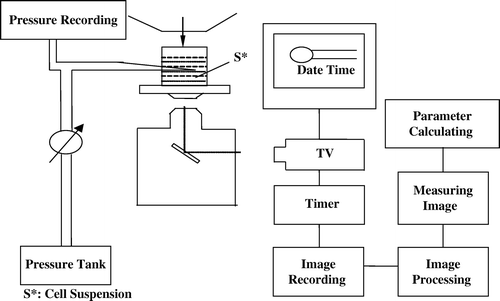
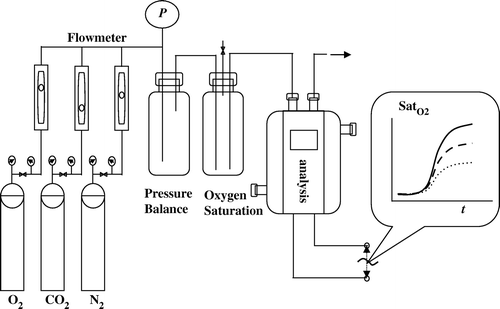
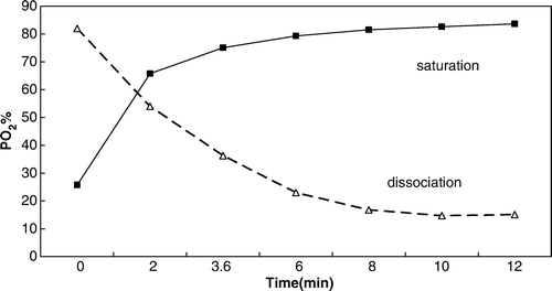
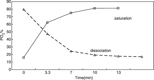
In this research the biomechanical properties of the reconstructed RBC were determined with the micropipette aspiration system, etc Citation[5], Citation[6]. The image of reconstructed RBC was obtained with an electron scanning microscope. The dynamics function of carrying-releasing oxygen was analyzed with the HEMOX analyzer from TCS and the dynamics analytical system established in our lab ( and ).
RESULTS AND DISCUSSION
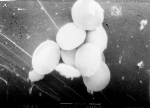
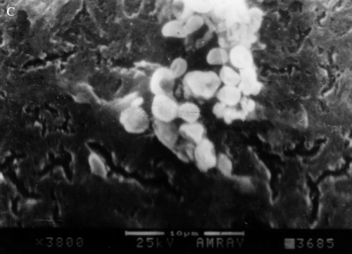
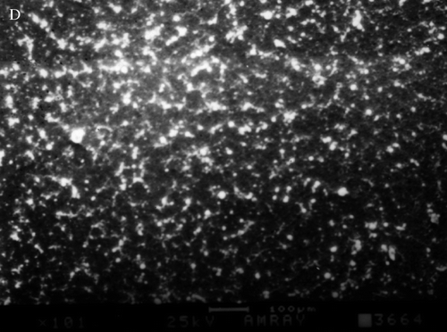
The experimental results indicated that the reconstructed erythrocytes have a biconcave disc form that was similar to the natural erythrocyte (). Compared with normal erythrocytes the reconstructed erythrocytes have similar biomechanical properties (µ: from 4.562 to 7.892×10⁻³ dyn/cm and η from 0.175 to 0.372×10⁻⁴ dyn·s/cm), having good deformability (). The reconstructed erythrocyte demonstrated greater resistance to osmotic lysis. The reconstructed erythrocytes have carrying-releasing oxygen function. The reconstructed erythrocytes blood can carry oxygen (oxygen saturation reaching up to 86%) and can release oxygen by alternatively being replaced with carbon dioxide (oxygen saturation declining lowering down to 15%). Animal experimental results show that in hemorrhagic shock the reconstructed erythrocytes blood has a good resuscitation function. The survival time of Wistar rats was very near to that of rats receiving normal blood ( and , ).
Figure 3B. The electron scanning microscope patterns of reconstructed erythrocyte with different size. 5500×.
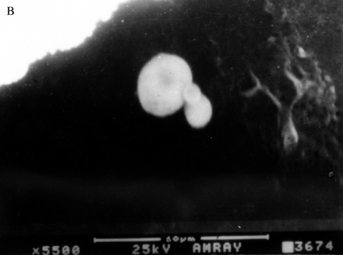
Table 1. Biomechanical properties of reconstituted erythrocyte based on Hemispherical cap model (from Chien's theory)
Table 2. Anti-hemorrhagic shock results of reconstituted erythrocyte blood
CONCLUSIONS
Biorheological properties of reconstructed erythrocytes are closely related to the function of carrying-releasing oxygen. The reconstructed erythrocytes with bioconcave disc shape have oxygen carrying-releasing function and good deformability. The study indicated the physiological function of reconstructed erythrocyte was similar to that of natural erythrocyte.
Acknowledgements
Dr. Xiang Wang was supported by National Natural Science Foundation of China (NSFC 10572159). This research is also partially supported by “111 Project” entitled Biomechanics & Tissue Repair Engineering (No.: B06023) and Chongqing Science & Technology Council (CSTC 2006ba5010).
References
- Chang, T.M 2000. Artificial cell biotechnology for medical applications. Blood Purif:;18((2)):91–6. Chang, T.M. (2002). Artificial cells for artificial organs. Artif. Cells Blood Substit. Immobil. Biotechnol., 30(5 & 6): 469–497.
- Cheung, Anthony T.W., Driessen, Bernd, Jahr, Jonathan S., Duong, Patricia L., Ramanujam, Sahana, Chen, Peter C. Y. and Gunther, Robert A. (2004). Blood substitute resuscitation as a treatment modality for moderate hypovolemia, Artif. Cells Blood Substit. Immobil. Biotechnol, 32((2)):189–207.
- Spahn D.R., Pasch T. Physiological properties of blood substitutes. News Physiol Sci. 2001; 16: 38–41
- Sung K.L.P., Chien S. Viscous and elastic properties of human red cell membrane. American Institute of Chemical Engineers 1977; 18: 54
- Sung K.L.P., Chien S. Viscous and elastic properties of human red cell membrane. The American Institute of Chemical Engineers Symposium Series: Biorheology No. 182 1978; 74: 81
- Chien S., Sung K.L.P., Skalak R., Usami S., Tozeren A. Theoretical and experimental studies on viscoelastic properties of erythrocyte membrane. Biophys. J 1978; 24: 463
