Abstract
Based on the research on small gap bridging peripheral nerve injury in SD rats, we decided to investigate the histological analysis possibility of bridging peripheral nerve injury with small gap using a de-acetyl chitin conduit in primate. Median nerves of 8 rhesus monkeys were cut at 2 cm above the elbow, and the right sides were subjected to small gap (2 mm) bridging to repair the nerve with chitin conduit (conduit inner diameter 4 mm; length 10 mm); the left sides were subjected to traditionary epineurium suture. Histology detections were conducted after 6th month. The conduit was almost absorbed and the conduit cast contour disappeared after 6 months. The histological analysis displayed that the regenerated nerve fibers in conduit grew forward in fasciculation. The re-myelinated nerve axons number per unit area in the conduit group distal segment was higher than that of traditionary epineurium suture group. The biocompatibility of biological chitin conduit in primate rhesus monkeys was quite good. The regenerated nerve fibers in conduit grew forward in fasciculation. The histological analysis results of biological conduit in primate rhesus monkeys were better than the traditionary epineurium suture. The biological conduit can be used in primate rhesus monkeys to substitute the traditionary epineurium suture methods.
In recent years, small gap bridging peripheral nerve injury Citation[1–4] has gradually become a hot area of research. The biological conduit constructed a microenvironment that can thoroughly profit selective regeneration of nerve fibers at the broken segments of impaired nerves. Based on previous experiments in SD rats, we performed experiments about biological conduit sleeve bridging median nerve injury with small gap in primate rhesus monkeys to analyze the growth rule of nerve fibers in conduit.
MATERIALS AND METHODS
Materials
Hollow cylindrical conduit (a de-acetyl chitin conduit invented by Beijing University People's Hospital and the Chinese Textile Academy, the State Patent No.: 01136314.2). Size: tube length 10 mm, thickness 1 mm, inner diameter 4 mm.
Groups
Eight male rhesus monkeys were anesthetized with the Sumianxin II (0.11ml/Kg i.m) (Military Veterinary Research Institute of Military Medical Academy); after anaesthesia, the bilateral elbow median nerves were cut at 2 cm above elbow, and the right sides were subjected to small gap (2 mm) bridging to repair the nerve with chitin conduit (conduit inner diameter 4 mm; length 10 mm); the left sides were subjected to traditionary epineurium suture. Histology detections were conducted after 6 months.
Surgical Procedures
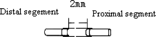
The biological conduit group suture method is shown in ; traditionary epineurium suture group: after impairing the median nerve, the proximal and distal segments were relatively rotated 180 degrees and the epineurium sutured with a 9-0 Nylon Suture thread, each stump seamed two needles.
Figure 1. The ideograph of biogradable conduit small gap bridging methods.
Detection Project
Histological analysis 6 months postoperatively: the results of HE staining and osmium acid staining were observed separately, which could show the nerve fibers’ course and assess the quality of regenerative nerve fibers.
Statistical Analysis
The data were analyzed by SPSS 11.0 and compared by t-test method. A probability where p < 0.05 was considered significant for all statistical comparisons. All values were presented as the mean±SD. The numbers and cross-sectional areas of distant and proximal myelined nerve fibers were compared.
RESULTS
Histology Detection Results
Histology staining results showed that the regenerative nerve fibers had grown through the gap along the biological conduit sleeve at six months. Longitudinal sections showed that regenerative nerve fibers were positively stained by S100 in the conduit sleeve, while in the cross-sections HE staining and osmium acid staining showed that the nerve fiber regenerated forward in fasciculation.
Figure 2. Conduit group S100 staining 10X.
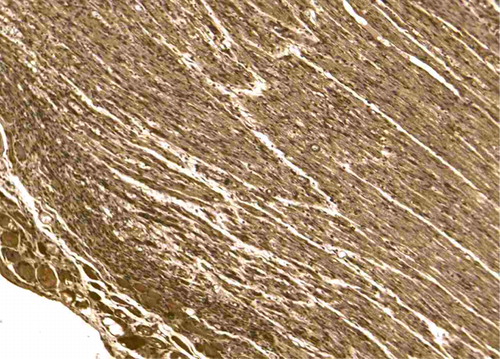
Figure 3. Conduit group HE staining 4X. Nerve fibers’ course in conduit 6 months operatively (longitudinal section).
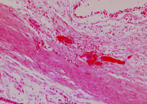
Figure 4. The course of nerve fibers in conduit 6 months operatively (cross section) 20X.
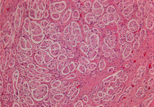
Figure 5. Regenerative nerve fiber in fasciculation in conduit group.
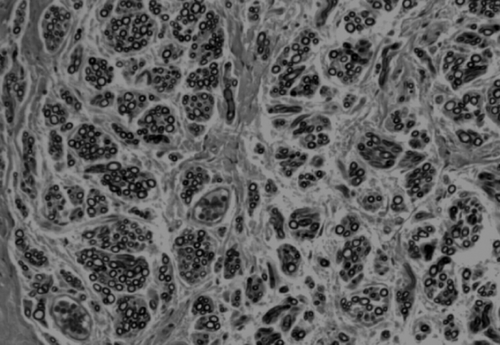
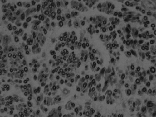
Figure 6. Regenerative nerve fiber in diffused distribution in traditionary epineurium suture group.
Statistical Analysis
Statistical data included:
Ratio of myelinated nerve number of distal and proximal segments in the unit area at six months postoperatively;
Ratio of axonal cross-section area of distal and proximal segments in the unit area at six months postoperatively.
Table 1. Numbers of myelinated nerve fibers in all the experimental groups
Based on the ratio of myelinated nerve number and axonal cross-section area of distal and proximal segments in the unit area at six months postoperatively, the conduit group was better than the traditionary epineurium suture group; the differences between them were statistically significant. Distal myelined nerve fibers were more than proximal myelined nerve fibers in the conduit; the results manifested the phenomenon that a small amount of nerve fibers controlled remote large amount of nerve fibers may exist during the regeneration. The distal myelined nerve fiber area significantly exceeded the proximal myelined nerve fiber area; the area ratio was greater than 1, and the histological results illustrated good results of restoration.
DISCUSSION
Peripheral nerve injury is common clinically; although the microsurgical technique has been noticeably improved, epineurial neurorrhaphy and interfascicular suture have been the main therapy during the past 100 years. If nerve fibers connect with the wrong type of nerve fibers, taking a motor nerve connecting with sensory nerve for example, it will lead to failure of nerve regeneration and therefore affect the neurological function recovery. Restoration after peripheral nerve injury mainly depends on effective and accurate docking (effective connection) and the re-innervation with target organ (re-innervation). So how to accurately and effectively connect different types of nerve stumps becomes the hot research.
Peripheral nerve selective regeneration based on “Y-tube experiments” provided a new, practical way in good docking of nerve fibers during regeneration. The technical limits of epineurial neurorrhaphy and the peripheral nerve selective regeneration phenomenon indicated that we could take full advantage of the different characteristics of peripheral nerve fiber regeneration; therefore biological conduit sleeve bridging nerve injury with small gap could substitute epineurial neurorrhaphy and interfascicular suture, which had been used for nearly a hundred years.
Jiang Baoguo et al. Citation[1–4] developed a degradable conduit sleeve, and they were the first to confirm that conduit sleeve bridging small gap (2-3 mm) could promote the restoration of SD rat sciatic nerve injury; observation index (functional recovery, the local nerve adhesion, myelined nerve fiber number of the distant ends, motor neuron conduction velocity) were significantly better than stump epineurial neurorrhaphy with relative rotation, which were similar with clinic suture after nerve injury. What's more, we made a further investigation on the comparison between sutures with small space and the traditional epineurial neurorrhaphy. We also investigated the most suitable choice of the small space and the biological characteristics of peripheral nerve regeneration through the small gap and the conduit sleeve materials.
The regenerative abilities were very different between SD rats and humans Citation[5]. We have found no reports about biodegradable conduit sleeve healing primate nerve injury at home or abroad. In the experiment of primate rhesus monkeys, we cut the median nerve at the elbow; the left sides were subjected to epineurial neurorrhaphy with relative rotation; the right sides were bridged small gap (2 mm) with conduit sleeve (diameter 4-5 mm). Early electrophysiological data showed that nerve conduction velocity of the conduit sleeve group was better than that of the traditionary epineurium suture group.
HE staining results showed that the regenerative nerve fibers had grown through the gap, and S100 staining, which labeled Schwann cells in the peripheral nervous system, showed that there were bulks of Schwann cells in the conduit sleeve. Based on the results of HE staining and osmium acid staining in transection, we have found that the regenerative nerve fibers in the conduit sleeve grew forward mainly in fasciculation rather than along the wall of the conduit sleeve. These observations were consistent with the assumptions we made before Citation[6–8]. Myelinated nerve fiber numbers and cross-section areas of proximal and distal segments further illustrated that the phenomenon of a small amount of nerve fibers controlling distal large amounts of nerve fibers may exist during the regeneration in conduit sleeve.
Because of the disadvantages of clinical conditions of damaged tissues, operation equipment and microsurgical technology, it's hard to achieve real in situ suture. Therefore, as far as animal models, we performed segments of epineurial neurorrhaphy with rotation of some degrees Citation[9].
In this experiment, we mainly observed the pattern of nerve fibers regeneration in the conduit sleeve, and also elucidated that biological conduit sleeve bridging nerve injury with small gap is better than stumps epineurial neurorrhaphy with relative rotation based on the histology analysis.
Acknowledgements
This research project was funded by Chinese National Natural Science Fund for Outstanding Youth (30625036), Chinese 973 Project Planning (2005CB522604), Chinese National Natural Science Youth Fund (30801169) , Beijing City Science & Technology New Star Classification A-2008-10 and Chinese ministry of education for Doctor Position New Teacher (20070001780).
Declaration of interest: This research project was funded by Chinese National Natural Science Fund for Outstanding Youth (30625036), Chinese 973 Project Planning (2005CB522604), Chinese National Natural Science Youth Fund (30801169), Beijing City Science & Technology New Star Classification A-2008-10 and Chinese ministry of education for Doctor Position New Teacher (20070001780)
References
- Lundborg G. A 25-year perspective of peripheral nerve surgery: Evolving neuroscientific concepts and clinical significance. J Hand Surg [Am] 2000; 25: 391–414
- Baoguo, Jiang, Shuhuan, Wang, Chuanhan, Feng. 1994. The comparison study of small gap artery bridging and epineurial neurorrhaphy in peripheral nerve injury. Peking University Medical College Journal, 26:249–250.
- Baoguo, Jiang, Yasuji, Yoshida. 1998. The immunohistochemical study on gap artery sleeve bridging in peripheral nerve. Chinese Microsurgery Journal, 14:39–42.
- Baoguo, Jiang, Bing, Wang, Shousheng, Hu, , et al 1999. The comparison of different small gap artery bridging in peripheral nerve. Chinese Orthopaedic Surgery Journal, 6:115–117.
- Yushan, Sun, Baoguo, Jiang, Qingsong, Zhu. 2002. The experimental functional medical material in animal body. Spinning and Weaving Science Research, 13: 7–22.
- Jian, Li, Baoguo, Jiang, Dianying, Zhang, , et al 2003. Biological tube small gap bridging repair peripheral nerve injury. Chinese Hand Surgery Journal, 19(2):118–120.
- Jiang B., Zhang P., Zhang D., Fu Z., Yin X., Zhang H. Study on small gap sleeve bridging peripheral nerve injury. Artif Cells Blood Substit Immobil Biotechnol. 2006; 34(1)55–7
- Zhang P., He X., Zhao F., Zhang D., Fu Z., Jiang B. Bridging small-gap peripheral nerve defects using biodegradable chitin conduits with cultured schwann and bone marrow stromal cells in rats. J Reconstr Microsurg. 2005; 21(8)565–571
- Peixun, Zhang, Baoguo, Jiang, Fuqiang, Zhao, Zhongguo, Fu, Dianying, Zhang, Chan, Du, Hongbo, Zhan. 2005. Chitin biological tube bridging the peripheral nerve with a small gap. Chinese Surgery Journal, 43(20):1345–1347.