
Abstract
Due to the various physical mechanisms of interaction between a worker's body and the electromagnetic field at various frequencies, the principles of numerical simulations have been discussed for three areas of worker exposure: to low frequency magnetic field, to low and intermediate frequency electric field and to radiofrequency electromagnetic field. This paper presents the identified difficulties in applying numerical simulations to evaluate physical estimators of direct and indirect effects of exposure to electromagnetic fields at various frequencies. Exposure of workers operating a plastic sealer have been taken as an example scenario of electromagnetic field exposure at the workplace for discussion of those difficulties in applying numerical simulations. The following difficulties in reliable numerical simulations of workers’ exposure to the electromagnetic field have been considered: workers’ body models (posture, dimensions, shape and grounding conditions), working environment models (objects most influencing electromagnetic field distribution) and an analysis of parameters for which exposure limitations are specified in international guidelines and standards.
1. Introduction
Electromagnetic fields (EMFs) over a wide range of frequencies – from static magnetic fields (e.g., near Magnetic Resonance Imaging devices) through low frequency (LF) and intermediate frequency (IF) fields (e.g., near welding or electrosurgery devices) to radiofrequency (RF) fields (e.g., near plastic sealers) – form one of the environmental factors of the workplace.[Citation1,Citation2] EMF exposure causes direct and indirect exposure effects because of physical interaction between EMF and the worker's body and other exposed objects. This interaction involves various frequency-dependent mechanisms and requires different approaches in their evaluation.
The direct effects of exposure to LF and IF fields include an electric field and an electric current induced inside the human body, and a thermal effect resulting from EMF energy absorption in the body is dominant in RF fields.[Citation2–4] The effects of high-level exposure to EMF are well investigated and known as ‘the established exposure effects’.[Citation5–8] The physical estimators of established exposure effects, such as the induced electric field (Ein) or specific energy absorption rate (SAR)Footnote1, can only be evaluated in the virtual human body models by numerical simulations. The ratio of the value of estimators of EMF exposure effects in the body to the level of exposure affecting the body – represented by E-field (electric field strength) and H-field (magnetic field strength), e.g., SAR/E or Ein/H – depends on the frequency, polarisation and type of dominant component (electric or magnetic) of incident EMF, but also on dimensions, shape, posture, biophysical and bioelectrical properties of the human body.
The indirect effects include contact current (Ic) flowing through the human body that comes into contact with an object (usually metal) of different electric potential.[Citation3] Contact current, expressed in milliamperes (mA), can be evaluated by numerical simulations or by measurements. The values and spatial distribution in the body of this estimator depend on EMF frequency; the dimensions of the exposed metal object; the dimensions, shape, posture, biophysical and bioelectrical properties of the human body; and the surface of the contact between the human body and the object.
1.1. The evaluation of the direct physical effect of EMF exposure – induced electric field (Ein) or SAR
Requirements related to the evaluation of the induced electric field value established by international guidelines – International Commission on Non-Ionising Radiation Protection (ICNIRP) and the Institute of Electrical and Electronics Engineers (IEEE) – are significantly different.[Citation6,Citation8,Citation9] According to the IEEE, the induced electric field should be evaluated in the virtual model of the human body in four groups of regions: (a) brain; (b) heart; (c) hands, wrists, feet and ankles; (d) other tissues.[Citation8] For the purpose of assessing exposure, the calculated induced electric field values should be the maximum from values averaged over a straight line segment, 0.5 cm long, oriented in any direction within the identified tissue. According to ICNIRP 2010 requirements, the limitations are provided for the Central Nervous System in the head or in other tissues.[Citation6,Citation9] The exposure assessment is based on the value of the internal electric field, which is the 99th percentile value (1% of the highest calculated in the model values is declined) averaged over 2×2×2 mm3 contiguous tissue in the model.[Citation6] The requirements given in European Directive 2013/35/EU for evaluating the internal electric field had not been specified, but in recital [Citation15] it is stated that ‘the physical quantities, laid down in Directive are based on the recommendations of the ICNIRP and should be considered in accordance with ICNIRP concepts, save where this Directive specifies otherwise’.[Citation9]
The thermal effect of exposure to RF EMF is estimated using the SAR – averaged over the whole body (WBA) or located in the head and trunk or in the limbs. According to the IEEE, Directive 2013/35/EU and ICNIRP requirements, localised SAR should be averaged over any 10 g of tissue.[Citation5,Citation7,Citation9] The only difference is the suggested algorithms of SAR values averaging, which affect the calculation results. According to the directive and ICNIRP requirements, SAR in the continuous mass is averaged, while, further to IEEE requirements, SAR in the volume with the averaging point at the centre is taken into consideration.[Citation5,Citation7,Citation9] In the process of evaluating the maximum localised SAR, where the particular cube of the human body model is located partly in the air (i.e., it belongs to the body surface representation), the volume is invalid for SAR assessment, and SAR is taken from the nearest point with the maximum SAR where the averaging volume contains body tissues only.[Citation7] McIntosh and Anderson reported that the shape of the averaging volume (a cube in IEEE) is of minor importance in localised SAR calculations and the easiest shape should be used.[Citation10]
1.2. Evaluation of the indirect physical effect of EMF exposure – contact current
According to the IEEE, the evaluation of contact current refers to two cases: ‘touch contact’ (1 cm2 surface contact of human body and metal object exposed to EMF) and ‘grasp contact’ (15 cm2 surface contact of human body and exposed metal object).[Citation8] ICNIRP guidelines refer to ‘point contact’ and do not specify any detailed information about the contact surface.[Citation5] The European Directive 2013/35/EU follows the ICNIRP approach.[Citation9]
1.3. Numerical simulations for EMF exposure evaluation
Numerical simulations are usually the only way to use physical estimators such as Ein or SAR inside the human body, and one of possible options for using Ic to assess the influence of an EMF on a worker. To obtain reliable results for a worker's exposure through evaluation by numerical simulations of biophysical results caused by exposure to an EMF, it is necessary to use models adequately representing exposure situations (known as exposure scenarios) and specific parameters of numerical models related to various kinds of interaction mechanisms. The use of a fully realistic model is ideal, but in practice the use of very complex models is usually technically impossible or too expensive – because of software and hardware limitations, acceptable time consumption (costs) of creating the model, computer calculations and analysing the results.
Generally, various numerical methods may be applied in evaluating electromagnetic hazards (EN 50413:2008)[Citation11]:
Finite Element Method (FEM) – preferred for LF and IF fields [Citation12,Citation13]
Finite Difference Method (FDM) – preferred for stationary fields [Citation12,Citation13]
Finite Difference Time Domain (FDTD) – preferred for IF and RF fields and dielectric materials (Yee-cells) [Citation14]
Finite Integration Technique (FIT) – suitable for LF, IF and RF fields and multi material object analysis [Citation15]
Method of Moments (MoM) and similar algorithms of Boundary Element Method (BEM) – preferred for LF and IF fields and well conductible materials [Citation12,Citation13,Citation16]
Scalar Potential Finite Difference method (SPFD) – preferred for LF fields, EMF field is transformed into scalar potential form and solved by FDM techniques [Citation17]
Impedance Method (IM) – preferred for RF fields and strongly non-homogenous domains [Citation18]
Frequency Scaling – can only be applied for magnetic fields up to 5 MHz.[Citation11]
In this paper, difficulties in applying numerical simulations to evaluate occupational EMF hazards have been discussed. Difficulties in evaluating estimators of exposure effects have been analysed with regard to: worker's body models, working environment models and parameters in which exposure limits are specified in international guidelines and standards. Problems of RF EMF exposure are presented on the basis of numerical simulations of SAR performed on various human body numerical models – which may exemplify both the difficulties in applying numerical simulations when assessing EMF exposure, as well as the uncertainty of such an assessment. The discussion of the protocols of compliance tests (i.e., an assessment of whether exposure meets limitations) has not been included in the scope of the study, and therefore the SAR obtained from simulations are presented as relative values. Induced electric field from LF magnetic field exposure and induced electric field and contact current from LF and IF electric fields exposure have also been analysed on the basis of the literature.[Citation16,Citation19–25]
2. Material and method
Difficulties in using numerical calculations were disused on the basis of problems in modelling and assessing workers’ exposure while operating dielectric sealers. Dielectric sealers are used for sticking/gluing thermoplastic materials by applying the thermal effects of RF currents. Operating dielectric heaters consists of placing the components from thermoplastic foil between electrodes and switching the RF power on (usually 27 MHz). In the case of manual operation, the worker is sitting or standing in front of the device, usually exposed to a spatially heterogeneous high level EMF.[Citation1,Citation26] Dielectric heaters are high impedance electromagnetic field sources, meaning that the electric component of the EMF is dominant due to the high electric voltage involved in the sticking process. Any assessment of such exposure may be limited to considerations regarding the electric field. The used virtual model for a dielectric sealer was developed at the Central Institute for Labour Protection – National Research Institute (CIOP-PIB) and validated by experimental data.[Citation1,Citation26]
To analyse the role of the worker's body model, simulations were made with using cylindrical, HUGO and CIOP-MAN models.[Citation20] HUGO – The Professional Anatomical Data Set (developed by Medical Virtual Reality Studio GmbH, Germany) is an adult anatomical male model 185 cm in height and with a weight of 105 kg. It was created on the basis of data from the Visible Human Project (VHP).[Citation27] The human body model named CIOP-MAN was developed by CIOP-PIB.[Citation20] It is based on anthropometric data of the 50th percentile Polish males (175 cm tall), but for the presented calculations it was resized to be 185 cm tall, for better comparison with HUGO. CIOP-MAN was used in standing or sitting postures, but HUGO only in a standing posture because it is not flexible (). Both models were used in three grounding conditions: grounded, partly insulated by a model of shoes (4 cm of gum) and fully insulated.
Figure 1. Virtual model of Electromagnetic field (EMF) exposure scenario created with the human virtual model CIOP-MAN in a sitting posture and a model of a dielectric sealer.[26,29]
![Figure 1. Virtual model of Electromagnetic field (EMF) exposure scenario created with the human virtual model CIOP-MAN in a sitting posture and a model of a dielectric sealer.[26,29]](/cms/asset/101bc358-0e45-4118-b90a-e960637043d7/tose_a_1028233_f0001_b.gif)
The following parameters were analysed: whole body average (WBA) SAR and localised (10 g) SAR in head (H), trunk (T) and legs (L). SAR values were obtained by using the IEEE averaging algorithm over a cube of 10 g tissue mass.
In the presented example, the FIT method was used as one of the most universal methods. Dedicated Computed Simulation Technology STUDIO SUITE software was used (CST STUDIO 2012). The uncertainty of the presented numerical calculations’ results was assumed to be ±40%, while the uncertainty of the E-field measurements used for numerical model experimental validation was ±14%.[Citation28–30]
The differences in results obtained from analysed exposure scenarios were assessed with the use of the Student's t test (StatSoft - STATISTICA v.9.0).
3. Results
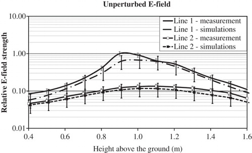
Numerical simulations modelled the workers’ exposure to a heterogeneous E-field of a spatial distribution typical for the workplace near a dielectric sealer. The spatial distribution of the E-field obtained from the dielectric sealer virtual model was validated by a detailed E-field measurement performed in the work environment near a real device (from where the geometrical and dielectric structures were represented in the virtual model) ().[Citation1,Citation26,Citation31] The differences between simulated and measured E-field values are in the range of uncertainties of their determination (±40% and ±14%, respectively) and are statistically not significantFootnote2 (p>.22, Student's t test).
Figure 2. The comparison of simulation (solid lines) and measurement (dash lines) results of spatial distribution of an unperturbed E-field in front of dielectric sealer.
Note: Reference value = highest measured E-field value for line 1; line 1 = at a distance of 30 cm from the powered electrode; line 2 = at a distance of 60 cm.
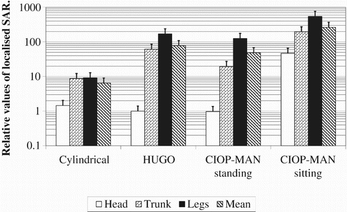
shows the whole body averaged (WBA) SAR values in cylindrical, HUGO and CIOP-MAN standing and sitting models, calculated for various grounding conditions, presented as normalised in relation to the values obtained for the HUGO insulated model, while shows localised (10 g) SAR in head (H), trunk (T) and legs (L) of these grounded models – in relation to the values obtained in the head of the HUGO model.
Figure 3. Relative values of the whole body averaged (WBA) SAR in various human body models (bars) under various grounding conditions (reference value – HUGO insulated model), with the uncertainty of numerical simulations (whiskers).
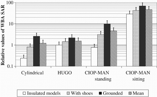
The highest values of WBA SAR were obtained for grounded models, while the lowest values were for insulated models. In the case of the insulated model – WBA SAR normalised values were in the range from 0.25 (in the cylindrical model) up to 29 (in the CIOP-MAN sitting model), (). WBA SAR ranged from 0.8 (cylindrical) up to 42 (CIOP-MAN sitting) or from 2.2 (HUGO) up to 70 (CIOP-MAN sitting) in the case of exposure scenarios involving models ‘with shoes’ or grounded models, respectively ().
In insulated models, the value obtained for the CIOP-MAN standing model was 22% lower than that obtained for the HUGO model.
In grounded models, the highest localised (10 g) SAR normalised values ranged from 9 up to 550 in legs (L), while the lowest values ranged from 0.98 up to 47 in the head (H). Localised (10 g) SAR values in legs (L), presented in , are up to seven times higher than values in the trunk (T) (CIOP-MAN standing) and up to 172 times higher than in the head (H) (HUGO).
Figure 4. Relative values of localised (10 g) SAR in head (H), trunk (T) and legs (L), (bars) under various grounded human body models (reference value – head of the HUGO model); with the uncertainty of numerical simulations (whiskers).
4. Discussion
A numerical model of interaction between a worker's body and an EMF consists of numerical models of: a worker's body, EMF source and the workplace in the vicinity. Differences between results obtained from various models can be significantly higher than the uncertainty of numerical simulations themselves – for the frequency of EMF investigated in the example presented in this paper, the international standards estimate it to be ±40%.[Citation11,Citation28] In the results of such a complex structure, all the elements of the model of the exposure scenario that may influence the obtained results of numerical calculations are discussed below.
4.1. Numerical model of the worker's body
The numerical human body model for modelling situations of a worker's exposure to EMF in the workplace should include: dielectric properties, ergonomics data (posture, shape, dimensions), spatial resolution of the model and its grounding conditions.
4.1.1. Dielectric properties of models
EMFs are absorbed with different effectiveness by different tissues. In the case of RF EMF, exposure relative permittivity (εr), electric conductivity (σ) and tissue density need to be considered.[Citation23] Tissue density can be omitted in the case of the LF electric field, while in the LF magnetic field only electric conductivity is important (in an assessment of EMF exposure results by numerical calculations). The values of dielectric properties of different tissues for various frequencies have been determined in the high frequency range and extrapolated into the low frequency.[Citation32] In the case of using the homogenous model, the value of the dielectric parameter is constant for the whole model – usually corresponding to the muscle properties or the intermediate value between muscle and fat tissues (weighted average value calculated following the relative content of these tissues in the human body).[Citation3,Citation5] In the discussed example, the cylindrical and CIOP-MAN models consist of muscle tissue and the Hugo model consists of 40 various tissues – with dielectric properties based on Gabriel's data.[Citation32]
4.1.2. Models grounding conditions
The analysis of the presented results confirms that the proper representation of the grounding conditions is one of the crucial issues in a numerical simulation-based assessment of a worker's exposure to the electric field of any frequency. The results for grounding models indicate up to over four-fold differences between SAR values for the HUGO and CIOP-MAN models, while in the case of insulated models the difference was just 22%. The SAR value for the grounded HUGO model was over twice as high as for the insulated model, while for CIOP-MAN standing it was over 12 times the difference. Dimbylow reports up to two-fold differences between the induced electric field in grounded and insulated models exposed to LF electric fields.[Citation22] Anatomically-based models have different numbers of voxels in contact with the ground (e.g., the HUGO model looks like it is standing on its toes and only has 150 voxels of 2×2×2 mm3 resolution each in contact with the ground – giving approximately 2.5×2.5 cm square), so the results obtained with the use of such models should be analysed carefully, because of doubts over whether the model of grounding is realistic. When in the workplace, no special insulation measures have been applied, the model of an insulated body is not appropriate. The requirements regarding the grounding conditions of human virtual models have not yet been defined by calculation standards and guidelines.
4.1.3. Models’ shape and posture
The example of calculation results presented in the article shows that SAR values depend on the human body ergonomics data (shape and posture). SAR values for the CIOP-MAN in a sitting posture were up to 36 times as high as for the CIOP-MAN in a standing posture. For an insulated model of a different shape, the calculation results for the HUGO model were similar to the CIOP-MAN in a standing posture, and four times as high for the cylindrical model (). Differences (up to 45%) have been reported also in the case of induced electric field in the brain of two CIOP-MAN models of different dimensions (dimensions of 5th and 95th percentile of adult male) exposed to an LF magnetic field generated by a suspended gun for resistance welding devices.[Citation33] Likewise, Dimbylow has reported up to 20% differences of induced electric field values in the brains of two different anatomically-based models (male of 176 cm height and female of 163 cm) in the case of homogenous LF magnetic field exposure.[Citation22] It should be noted that all those models were in the range of 1.76 m ± 8%, as required by EN 50505:2008.[Citation28]
Results of numerical calculations regarding exposure to RF EMF presented in the article covered the distribution of EMF exposure effects in particular human body parts, e.g., relative distributions in the head, torso and legs, using various human body models. HUGO and CIOP-MAN models allow simulations of SAR distribution to be made in particular body parts (head/trunk/legs). As the cylindrical model is only a rough presentation of a human body, the values of localised SAR can only be calculated in the parts of the cylinder corresponding to the location of the human body part (e.g., the top of cylinder – head, the lower part – legs). Therefore, the use of cylindrical models in such an analysis creates a low quality reconstruction of the exposure scenario, and may produce significantly under- or overestimated results of exposure evaluation. However, simple homogenous (cylindrical, spheroidal or ellipsoidal) models are widely used to validate numerical methods used later with other models (EN 50505:2008), because analytical formulas for SAR estimations are achievable for such models.[Citation28] The same observations refer to LF magnetic and LF and IF electric field.[Citation28] In the same cases, when exposure to EMFs is of a spatially localised nature, e.g., in the vicinity of an EMF source of small dimensions, numerical simulations of exposure to localised RF EMF sources can be limited to the part of the worker's body model under investigation. In recent years, such simulations have been carried out on a wide scale in international studies referring to mobile phone–human head interactions.[Citation23]
The highest values for the CIOP-MAN sitting model indicates that, in the case of a non-realistic posture of the worker's model (a sitting posture is very common when operating dielectric sealers), the exposure evaluation may by significantly underestimated, and to obtain reliable results the calculations should be made with models of postures as close as possible to workers’ postures in a realistic working environment.
Usually the worker is close to the EMF source in various postures. The results univocally show that SAR values in particular body parts are indeed related to human body posture (Figures and ). The use of models allowing posture to be changed also gives advantages in the case of analysing the effects of exposure to LF magnetic or LF and IF electric fields.
The Student's t test shows that differences between SAR values in human body models of various postures are statistically significant (p<.05) and are demonstrated by the mean SAR values on Figures and . Therefore, in evaluation of occupational electromagnetic hazards by numerical simulations, differences involving posture of human body models are the most important ones. The lower level of differences in obtained SAR is related to grounding conditions (p>.45, statistically not significant). The least difference level has been found out for this kind of model (anatomically based – HUGO or homogenous – CIOP-MAN in standing posture), p>.16 (statistically not significant) and indicates that to simplify numerical calculations homogenous models can be recommended.
4.1.4. Spatial resolution models
Another key parameter that should be taken into account is a spatial resolution of the human body model (according to EN 50505:2008, the resolution of a human body model should be in the range of 2–10 mm). Models with the highest resolution (most detailed) give more reliable results. However, a high resolution leads to a longer calculation time (it can be speeded up for LF magnetic field exposure by using the frequency scaling method). Furthermore, the human body model resolution used in calculations should not exceed the dimension of about 1/10 of the wavelength in tissues. For the purpose of calculations at a frequency of 3 GHz, the resolution of the model should not exceed 1 mm, while in the case of the presented example of 27 MHz it should not exceed 110 mm.[Citation10] Because of the complex structure of various tissues in the body, the spatial resolution of models is also important in the case of exposure to EMF at lower frequencies. For example, Hirata reports up to 20% differences between the maximum induced field value in the brain of 1 mm and 2 mm human body resolutions for LF magnetic field exposure.[Citation34] In the case of WBA SAR (Dimbylow) and localised 10 g SAR (Gosselin), there have been reports of up to 10–20% differences between results obtained for 1 mm and 2 mm resolution models.[Citation24,Citation25] Likewise, the various resolutions of developed anatomical models that are currently available for use in numerical simulations can make it impossible to strictly follow the guidelines of averaging localised (10 g) SAR values, e.g., when using the Hugo model with the lowest available resolution of 8×8×8 mm3.[Citation20] The human body models used in the discussed example had resolutions of 4–6 mm.
4.2. Numerical model of the electromagnetic field source and the workplace
A numerical model of EMF source should allow for the reconstruction of the field distribution in the workers’ area of activity. Geometry and the electricity supply of the field source affects the field level and spatial distribution in the vicinity of the source and the worker's exposure level, which determine the value of the internal electric field or SAR inside the worker's body. This is why the shape of particular field source components should be taken into account during numerical modelling. Differences of up to 40% between E-Fields calculated using five numerical models representing the same dielectric heater with various precision of its virtual reconstruction have been found.[Citation31] The parameters of the components of space around the source can also affect the EMF spatial distribution (all electrically conductive elements and objects in the structure of the device emitting EMF, or placed nearby, serve to modify the electric field distribution, while the magnetic field is modified only by ferromagnetic elements and objects) and should be taken into account during numerical modelling. Such an influence of conductive objects on electric field distribution was presented in the Tarao et al.’s investigations.[Citation21]
5. Conclusions
The discussed difficulties are the core of the process of applying numerical simulations in the field of occupational health and safety, e.g., in the area of transposing European Directive 2013/35/EU [Citation9] into systems of national labour law or in compliance analysis regarding EMF exposure at particular workplaces. The outcome from the presented analysis of difficulties in applying numerical simulations for EMF exposure evaluations should be considered when drafting the legislation, as well as the standardisation of detailed exposure assessment protocol for specific types of exposure scenarios in the workplace.
The main area of difficulty in applying numerical simulations to evaluating occupational hazards concerns worker's body models. The presented results shown that an unrealistic worker's posture (up to 36 times higher SAR values in models in a sitting posture in comparison to a standing one), dimension and shape of models (single block model use) and grounding conditions of models (up to 12 times higher SAR values in grounded models than in insulated ones) can significantly under- or overestimate the exposure effects in the body. Furthermore, in the case of anatomically-based models, the problem of possible non-realistic grounding properties of such models has also been found. All these issues should be considered in the process of developing numerical models to achieve models that are suitable for the investigated exposure scenario, and to obtain reliable results of an assessment of a worker's EMF exposure.
Numerical simulations are still a method of limited usefulness when assessing the compliance of individual workplaces with legal requirements regarding exposure to EMFs, because in recent decades the highest attenuation of research was on how to apply such methods to evaluate public exposure, e.g., while mobile phone handsets are used near the body. However, they are gradually becoming more widely applicable in research on electromagnetic health hazards or assessments of the safety of new technologies emitting EMFs in the work environment.
Funding
This paper has been based on the results of a research task carried out within the scope of the second stage of the National Programme “Improvement of safety and working conditions” partly supported in 2011–2013 - within the scope of state services - by the Ministry of Labour and Social Policy. The Central Institute for Labour Protection-National Research Institute (CIOP-PIB) is the Programme's main coordinator
Notes
1. Induced electric field (Ein) – the electric field present in the body (in situ) as a result of exposure to the environmental electromagnetic field, expressed in volt per metre (V/m).Specific energy absorption rate (SAR) – is the rate at which energy is absorbed per unit mass of body tissue, averaged over the whole body or over parts of the body, and is expressed in watts per kilogram (W/kg).[Citation9]
2. Statistically not significant – data with p value of obtained Student's t test statistics higher than significance level of .05.Statistically significant – data with p value of obtained Student's t test statistics lower than significance level of .05.
References
- Hansson Mild K, Alanko T, Gryz K, Hietanen M, Karpowicz J, Decat G, Falsaperla R, Rossi P, Sandström M. Exposure of workers to electromagnetic fields. A review of open questions on exposure assessment techniques. Int J Occup Saf Ergon. 2009;15(1):3–33. DOI:10.1080/10803548.2009.11076785
- Koradecka D, Pośniak M, Jankowka E, Skowroń J, Karpowicz J. Chemical, dust, biological, and electromagnetic radiation hazards. In: Salvendy G, editor. Handbook of human factors and ergonomics. 3rd ed. New York (NY): Wiley; 2006. p. 945–964.
- Reilly PJ. Applied bioelectricity. from electrical stimulation to electropathology. New York (NY): Springer-Verlag; 1998.
- Durney CH. Electromagnetic dosimetry for models of humans and animals: a review of theoretical and numerical techniques. Proc IEEE. 1980;68(1):33–40. doi: 10.1109/PROC.1980.11578
- International Commission on Non-Ionizing Radiation Protection (ICNIRP). Guidelines for limiting exposure to time-varying electric, magnetic, and electromagnetic fields (up to 300 GHz). Health Phys. 1998;74(4):494–522.
- International Commission on Non-Ionizing Radiation Protection (ICNIRP). Guidelines for limiting exposure to time-varying electric and magnetic fields (1 Hz–100 kHz). Health Phys. 2010;99(6):818–836.
- Institute of Electrical and Electronics Engineers (IEEE). Recommended practice for measurements and computations of radio frequency electromagnetic fields with respect to human exposure to such fields, 100 kHz–300 GHz (Standard No. C95.3-2002). New York (NY): IEEE; 2002.
- Institute of Electrical and Electronics Engineers (IEEE). Standard for safety levels with respect to human exposure to radio frequency electromagnetic fields, 3 kHz to 300 GHz (Standard No. C95.1:2005). New York (NY): IEEE; 2006.
- Directive 2013/35/EU of the European Parliament and of the Council of 26 June 2013 on the minimum health and safety requirements regarding the exposure of workers to the risks arising from physical agents (electromagnetic fields) (20th individual Directive within the meaning of Article 16(1) of Directive 89/391/EEC), O.J. nr L-179 of 29 June 2013, Brussels, Belgium
- McIntosh RL, Anderson V. SAR versus VAR, and the size and shape that provide the most appropriate RF exposure metric in the range of 0.5–6 GHz. Bioelectromagnetics. 2011;32(4):312–321. doi: 10.1002/bem.20642
- European Committee for Electrotechnical Standardization (CENELEC). Basic standard on measurement and calculation procedures for human exposure to electric, magnetic and electromagnetic fields (0 Hz–300 GHz). (Standard No. EN 50413:2009). Brussels: CENELEC; 2009.
- Chari MVK, Salon SJ. Numerical methods in electromagnetism. San Diego (CA): Academic Press; 2000.
- Bossavit A. Computational electromagnetism. San Diego (CA): Academic Press; 1998.
- Yee KS. Numerical solution of initial boundary value problem involving Maxwell's equations in isotropic media. IEEE Trans. Antennas Propag. 1966;AP-14(3):302–307.
- Weiland T. A discretization method for the solution of Maxwell's equations for six-component fields. AEU. 1977;31(3):116–120.
- Harrington RF. Field computation by moment methods. New York (NY): MacMillan; 1968.
- Dawson TW, Stuchly MA. High-resolution organ dosimetry for human exposure to low-frequency magnetic fields. IEEE Trans Magn. 1998;34(3):708–718. doi: 10.1109/20.668071
- Orcutt N, Gandhi OP. A 3-D impedance method to calculate power deposition in biological bodies subjected to time-varying magnetic fields. IEEE Trans. Biomed. Eng. 1988;35:577–583. doi: 10.1109/10.4590
- Institute of Electrical and Electronics Engineers (IEEE). Recommended practice for determining the peak spatial average specific absorption rate (SAR) in the human body from wireless communications devices, 30 MHz–6 GHz: general requirements for using the finite difference time domain (FDTD) method for SAR calculations (Standard No. P 62704-1). New York (NY): IEEE; draft.
- Zradziński P. The properties of human body phantoms used in calculations of electromagnetic fields exposure by wireless communication handsets or hand-operated industrial devices. Electromagn Biol Med. 2013;32(2):226–235. doi: 10.3109/15368378.2013.776434
- Tarao H, Korpinen L, Kuisti H, Hayashi N, Elovaara J, Isaka K. Numerical evaluation of currents induced in a worker by ELF non-uniform electric fields in high voltage substations and comparison with experimental results. Bioelectromagnetics. 2012;34(1):61–73. doi: 10.1002/bem.21738
- Dimbylow PJ. Development of the female voxel phantom, NAOMI, and its application to calculations of induced current densities and electric fields from applied low frequency magnetic and electric fields. Phys Med Biol. 2005;50(6):1047–1070. doi: 10.1088/0031-9155/50/6/002
- Gandhi OP, Lazzi G, Furse CM. Electromagnetic absorption in the human head and neck for mobile telephones at 835 and 1900MHz. IEEE Trans. Microw Theory Tech. 1996;44:1884–1897. doi: 10.1109/22.539947
- Dimbylow P, Bolch W, Lee C. SAR calculations from 20 MHz to 6 GHz in the University of Florida newborn voxel phantom and their implications for dosimetry. Phys Med Biol. 2010;55(5):1519–1530. doi: 10.1088/0031-9155/55/5/017
- Gosselin MC, Christ A, Kühn S, Kuster N. Dependence of the occupational exposure to mobile phone base stations on the properties of the antenna and the human body. IEEE Trans Electromagn Compat. 2009;51(2):227–235. doi: 10.1109/TEMC.2009.2013717
- Karpowicz J, Gryz K. Practical aspects of occupational EMF exposure assessment. Environmentalist. 2007;27:525–531. doi: 10.1007/s10669-007-9067-y
- Ackerman MJ. The Visible Human Project. Proc IEEE. 1998 Mar;86(3):504–11. doi: 10.1109/5.662875
- European Committee for Electrotechnical Standardization (CENELEC). Basic standard for the evaluation of human exposure to electromagnetic fields from equipment for resistance welding and allied processes (standard No. EN 50505:2008). Brussels: CENELEC; 2008.
- Zradziński P, Leszko W, Karpowicz J, Gryz K. Ocena narażenia na pola elektromagnetyczne użytkowników przenośnych radiotelefonów, z wykorzystaniem symulacji numerycznych i wymagań dyrektywy 2013/35/UE. [Assessment of the portable radiophone user's exposure to electromagnetic fields, with use of numerical simulations and directive 2013/35/UE requirements]. Med Pr. 2013;65(6):817–827.
- Gryz K, Zradziński P, Karpowicz J, Leszko W. Pomiary i ocena pola elektromagnetycznego przy radiotelefonach przenośnych – w kontekście wymagań dyrektywy europejskiej 2013/35/UE i polskiego prawa pracy. [Measurement and assessment of electromagnetic fields near radiophones in line with provisions for European directive 2013/35/UE and Polish labour law]. Med Pr. 2013;64(5):671–680.
- Gryz K, Karpowicz J, Molenda M, Zradziński P, Więckowski A, Mielniczek E. Analysis of EMF hazards in the vicinity of dielectric heaters – results of measurements and numerical simulations by various methods. Proceedings of the International Workshop on Electromagnetic Fields in the Workplace; 2005 Sept 5–7; Warszawa, Poland: Central Institute for Labour Protection – National Research Institute (CIOP-PIB).
- Gabriel C. Compilation of the dielectric properties of body tissues at RF and microwave frequencies. Brooks Air Force Technical Report AL/OE-TR-1996-0037. Available from: http://niremf.ifac.cnr.it/docs/DIELECTRIC/Report.html
- Zradziński P. The modeling and evaluation of the exposure of the workers operating suspended resistance welders to the simultaneous electromagnetic and biomechanical factors. Acta Bio-Opt Inform Med Inżynieria Biomed [Acta Bio-Opt Inform Med Biomed Eng]. 2012;18(1):50–54 [in Polish].
- Hirata A, Takano Y, Fujiwara O, Dovan T, Kavet R. An electric field induced in retina and brain at treshold magnetic flux density causing magnetophosphenes. Phys Med Biol. 2011;56:4091–4101. doi: 10.1088/0031-9155/56/13/022
