Abstract
Objective: Physiologic alterations during rapid sequence intubation (RSI) have been studied in several emergency airway management settings, but few data exist to describe physiologic alterations during prehospital RSI performed by ground-based paramedics. To address this evidence gap and provide guidance for future quality improvement initiatives in our EMS system, we collected electronic monitoring data to evaluate peri-intubation vital signs changes occurring during prehospital RSI. Methods: Electronic patient monitor data files from cases in which paramedic RSI was attempted were prospectively collected over a 15-month study period to supplement the standard EMS patient care documentation. Cases were analyzed to identify peri-intubation changes in oxygen saturation, heart rate, and blood pressure. Results: Data from 134 RSI cases were available for analysis. Paramedic-assigned prehospital diagnostic impression categories included neurologic (42%), respiratory (26%), toxicologic (22%), trauma (9%), and cardiac (1%). The overall intubation success rate (95%) and first-attempt success rate (82%) did not differ across diagnostic impression categories. Peri-intubation desaturation (SpO2 decrease to below 90%) occurred in 43% of cases, and 70% of desaturation episodes occurred on first-attempt success. The incidence of desaturation varied among patient categories, with a respiratory diagnostic impression associated with more frequent, more severe, and more prolonged desaturations, as well as a higher incidence of accompanying cardiovascular instability. Bradycardia (HR decrease to below 60 bpm) occurred in 13% of cases, and 60% of bradycardia episodes occurred on first-attempt success. Hypotension (systolic blood pressure decrease to below 90 mmHg) occurred in 7% of cases, and 63% of hypotension episodes occurred on first-attempt success. Peri-intubation cardiac arrest occurred in 2 cases, one of which was on first-attempt success. Only 11% of desaturations and no instances of bradycardia were reflected in the standard EMS patient care documentation. Conclusions: In this study, the majority of peri-intubation physiologic alterations occurred on first-attempt success, highlighting that first-attempt success is an incomplete and potentially deceptive measure of intubation quality. Supplementing the standard patient care documentation with electronic monitoring data can identify unrecognized physiologic instability during prehospital RSI and provide valuable guidance for quality improvement interventions.
Introduction
Rapid sequence intubation (RSI) of severely ill or injured patients is a critical intervention performed in various emergency care settings, including the out-of-hospital environment. Although RSI facilitates definitive airway control to protect against aspiration and establish adequate oxygenation and ventilation, the procedure also creates a risk for significant peri-intubation physiologic alterations including oxygen desaturation, cardiac dysrhythmias, and significant hemodynamic changes Citation(1, 2). Such physiologic derangements have been associated with increased morbidity and mortality in patients with some types of acute pathology Citation(3–8), and can increase the risk of peri-intubation cardiac arrest in unstable patients or patients with comorbidities Citation(1, 9, 10). Consequently, prehospital RSI remains a subject of controversy and is presently outside the scope of practice in many North American EMS agencies Citation(11).
Numerous papers have described the observed incidence of physiologic alterations during emergency RSI Citation(9, 10, 12–40). However, due to significant differences in clinical setting, provider level, and case mix, a minimal amount of the existing literature is directly applicable to EMS systems with paramedic-level providers performing RSI in both medical and trauma patients. Most studies describing physiologic alterations during emergency intubation have been conducted in an emergency department or in-hospital setting Citation(9, 10, 12, 15, 17, 21–24, 27, 29–40). The existing prehospital literature largely reflects experience outside the United States where intubation was usually performed by physicians Citation(18, 19, 25, 26, 28, 31). The few studies reporting on paramedic-performed RSI either describe air-medical experience or focus exclusively on traumatic brain injury (TBI) patients Citation(13, 14, 16, 20). Thus while the potential physiologic sequelae of emergency RSI are well established, the existing literature provides minimal insight into the frequency and magnitude of various physiologic alterations that may be expected during prehospital RSI performed by ground-based paramedics. To address this evidence gap and provide guidance for future RSI quality improvement initiatives in our EMS community, we prospectively collected electronic monitoring data to evaluate peri-intubation physiology and vital signs alterations occurring during prehospital paramedic-performed RSI.
Methods
Setting
Spokane County spans an area of 1,781 square miles and has a population of approximately 500,000 citizens distributed across urban, suburban, and rural areas. Emergency Medical Services are provided by 19 Fire Department first-responder agencies operating non-transport vehicles, and three ambulance transport services providing additional 9-1-1 response and all ground transport (approximately 47,000 transports annually) within the county. The present study involved the largest Fire Department, serving the city of Spokane (population 210,000), and the largest ambulance response/transport agency, serving the entire county. These two EMS agencies perform the substantial majority of prehospital RSI procedures in Spokane County. Vehicles for both participating agencies are staffed by teams of one or more EMTs and at least one paramedic.
An RSI protocol has been in place in Spokane County EMS since 1996, permitting paramedics to perform RSI for the indications of 1) airway protection for a patient with decreased level of consciousness (GCS ≤ 8), facial trauma, airway burns, excessive secretions or other airway compromise, or 2) respiratory failure with inability to oxygenate or ventilate adequately by less invasive means. Online medical direction is available but not required prior to attempting intubation. Of roughly 550 prehospital intubations performed annually, approximately a third are RSIs.
As a part of agency Ongoing Training and Evaluation Programs, all paramedics receive advanced airway management education and procedural training annually, comprising computer-based, classroom, and psychomotor instruction. Approximately 180 paramedics are employed across the two participating agencies, and the average paramedic performs three to four prehospital intubations annually. New paramedics in the county are required to perform at least 12 successful prehospital intubations in their first 3-year cycle of certification, and must make up any shortfall in the operating room. Thereafter, paramedics are required to perform at least 6 successful intubations every 3-year cycle. Prior to each recertification, a paramedic must also demonstrate procedural proficiency in a manikin-based simulation to the satisfaction of the Medical Program Director.
RSI is primarily performed using etomidate and succinylcholine for sedation and paralysis, respectively. Midazolam and rocuronium are available if either primary agent is contraindicated or unavailable. Lidocaine is used by protocol in addition to these agents in cases of suspected acute brain injury. Pre-oxygenation modalities include non-rebreather mask and bag-valve mask ventilation; during our study period neither apneic oxygenation nor bag-valve mask PEEP valves were in use. Intubation is predominantly accomplished via direct laryngoscopy; the participating fire department also has video laryngoscopes available in their airway kit. Supraglottic airway devices are available for use as primary or rescue airways.
Data Collection
The present study was a retrospective analysis of data prospectively collected between December 2013 and March 2015 as part of the ongoing quality improvement program in Spokane County EMS. The study was IRB-approved with waiver of the requirement for written informed consent. Beginning in December 2013, we began collecting monitor/defibrillator (LIFEPAK 15, Physio-Control, Inc., Redmond, WA) electronic data files to supplement the standard patient care report documentation for cases in which advanced airway interventions were performed. The ending date for the present analysis was determined by the timing of an upcoming airway management training session and the desire to incorporate pertinent insights from these data into that educational activity. We included all cases of RSI attempted by the participating agencies for which an electronic monitor file had been downloaded. Excluded were cardiac arrest patients receiving RSI as a component of post-arrest management. RSI was defined as administration of a paralytic agent prior to attempted tracheal intubation.
EMS patient care reports (Multi EMS Data System [MEDS] electronic patient care reporting [ePCR] system, American Medical Response, Greenwood Village, CO) were retrospectively reviewed to abstract pertinent patient data along with EMS response and treatment details. Data abstraction was performed by a single author trained on use of the MEDS ePCR system and with substantial experience using this system as both an EMS data analyst and EMS provider. Data abstracted from each patient care report included patient age, gender, estimated weight, chief complaint, the prehospital diagnostic impression assigned by the treating paramedic, times and details of all medications and procedures, transport interval, any noted airway complications, and all manually documented vital signs. For the purpose of uniform documentation, an intubation attempt is defined in our system as advancing the laryngoscope blade past the teeth with the intent of visualizing the glottis. Electronic monitoring data files were automatically synchronized to an atomic clock during the download process, and transferred into commercial data review software (CODE-STAT 10 Data Review, Physio-Control, Inc., Redmond, WA). Raw waveform and vital signs data (ECG, pulse oximetry, capnography, and noninvasive oscillometric blood pressure) were exported into specialized software (MATLAB Release 2015a, MathWorks, Natick, MA) for further analysis.
Measurements and Data Analysis
We assessed each case for peri-intubation changes in oxygen saturation (SpO2), heart rate (HR), and systolic blood pressure (SBP). SpO2 values were obtained via peripheral (finger) pulse oximetry and were trended by the monitor at 30-second intervals, with additional intermittent values recorded in conjunction with device alarms and actions such as event marking, printing, and automated blood pressure measurement. Peri-intubation oxygen saturation measurements were made as follows. Since capnography is a standard and required component of advanced airway placement confirmation in our system, we identified the time of successful airway placement by the appearance of persistent physiologic CO2 waveform data and end-tidal CO2 measurements in the electronic monitor file Citation(13, 20, 31). We then noted any oxygen desaturation during the period from 5 minutes before until 2 minutes after successful airway placement. We defined desaturation as a decrease in SpO2 to below 90%, or a decrease of greater than 10% if the initial value was below 90% Citation(18, 19, 25). We further defined severe desaturation as a decrease in SpO2 to below 80%, or a decrease of greater than 20% if the initial value was below 90%. The initial pre-intubation SpO2 was measured as the maximum SpO2 value observed in the 5 minutes preceding the onset of a peri-intubation desaturation if present, or preceding airway placement success if no desaturation was present. For cases in which the time of induction documented in the patient care report preceded airway placement success or the onset of desaturation by more than 5 minutes, we also assessed for maximum SpO2 in a 4 minute interval straddling the documented time of induction. The SpO2 nadir was measured as the lowest value observed between the initial pre-intubation value and the time of successful airway placement. To account for potential latency in the peripherally-measured SpO2, we extended the SpO2 observation interval for 2 minutes after airway placement success Citation(20). The duration of desaturation was measured as the interval between the first SpO2 reading below 90% (or the first decrease below pre-intubation SpO2 if initial SpO2 was below 90%) and recovery to an SpO2 of at least 90% (or recovery to the pre-intubation level if initial SpO2 was below 90%).
Continuous HR data were derived via post-processing of the continuous ECG signal. Pre-intubation baseline HR was measured at the time point of the pre-intubation SpO2 maximum. We defined peri-intubation bradycardia and profound bradycardia as decreases in HR to below 60 bpm and 40 bpm, respectively. To assess blood pressure responses to RSI, we included both manually-performed measurements and automated oscillometric measurements performed by the monitor. Hypotensive and hypertensive responses were defined as decreases and increases, respectively, in SBP of greater than 20%, measured between the last pre-intubation measurement and the first post-intubation measurement Citation(26, 31). Absolute hypotension was defined as a SBP below 90 mmHg. Pre-intubation shock index was calculated as the ratio of HR to SBP, as measured at the time of the last pre-intubation SBP measurement. Peri-intubation cardiac arrest was defined as patient deterioration requiring initiation of cardiopulmonary resuscitation during the RSI attempt or within 10 minutes following successful airway placement Citation(10, 30).
Statistical Analysis
Continuous data were summarized as mean (SD) or median (25th, 75th percentile). Comparisons were performed with the Mann-Whitney U test for continuous data and chi-square for proportions (Minitab 16, Minitab, Inc., State College, PA). All tests were two-tailed, with p < 0.05 considered statistically significant.
Results
Patient and Airway Management Process Characteristics
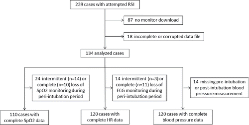
From the 15-month study period, a total of 134 cases were available for analysis (). Patient and airway management process characteristics are described in . Based on the primary impression documented in the EMS patient care report, the paramedic-assigned prehospital diagnostic category was neurologic in 42% of cases, respiratory in 26%, toxicologic in 22%, trauma in 9%, and cardiac in 1%. Nine of the 35 respiratory patients failed CPAP prior to RSI. Successful tracheal intubation was achieved in 95% (n = 127) of cases. Success was achieved on the first laryngoscopic attempt in 110 cases (82%), by the second attempt in 125 cases (93%), and on the third attempt in the remaining 2 cases. All 7 intubation failures were rescued via placement of a supraglottic airway device after 2 (n = 4), 3 (n = 2), or 4 (n = 1) failed intubation attempts. There were no significant differences across prehospital diagnostic categories in first-attempt (p = 0.74) or overall (p = 0.66) intubation success.
Table 1. Patient and airway management process characteristics, grouped by paramedic-assigned prehospital diagnostic impression categoryTable Footnote*
Peri-intubation Oxygen Saturation
Twenty-four cases exhibited loss of SpO2 monitoring during the peri-intubation interval, preventing determination of pre-intubation SpO2 and identification of any desaturation events. Among the remaining 110 cases, the median (25th, 75th percentile) pre-intubation SpO2 maximum was 98% (92%, 100%).
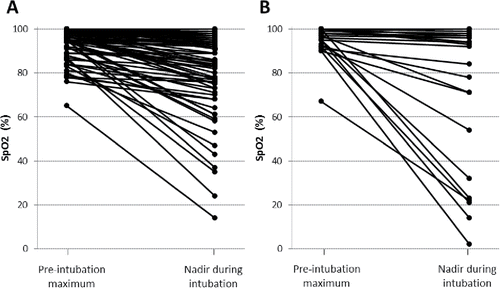
Peri-intubation desaturation occurred in 43% (n = 47) of cases (). The majority (68%, n = 32) of these events were severe desaturations, which occurred more frequently in cases with a prehospital respiratory diagnostic impression compared to other diagnostic impression categories (p = 0.03). There was no difference in the incidence of desaturation between RSIs performed on scene versus during transport (43% vs. 41%, respectively; p = 0.81).
Figure 2. Percentage of cases (with 95% confidence interval) in each prehospital diagnostic impression category with any desaturation (light grey) and severe desaturation (dark grey). Median (25th, 75th percentile) pre-intubation SpO2 maximum is indicated below each category. There was loss of SpO2 data in the 1 cardiac case; thus, results are only depicted for the remaining 4 categories.
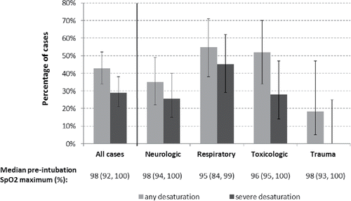
The occurrence of desaturation events was significantly associated with the maximum pre-intubation SpO2 (any desaturation: 95% [90%, 97%] vs. no desaturation: 99% [96%, 100%]; p < 0.001), and there was a progressive decrease in rates of any desaturation and severe desaturation as pre-intubation SpO2 increased above 90% (). Among the 47 cases with any desaturation, the median nadir SpO2 was 71% (36%, 79%) and the median desaturation duration was 120 (81, 212) seconds. The median nadir SpO2 was markedly lower and the median desaturation duration was markedly longer for respiratory patients compared to patients in other prehospital diagnostic impression categories (). The median desaturation duration was significantly longer for desaturation episodes occurring in cases requiring multiple intubation attempts versus cases with first-attempt success (165 [120, 270] seconds vs. 90 [60, 150] seconds; p < 0.001). Among cases with any desaturation, desaturation duration was at least 2 minutes in 46% of cases with first-attempt success and in 100% of cases requiring multiple attempts.
Figure 3. Probability (with 95% confidence interval) of any desaturation (light grey) and severe desaturation (dark grey) associated with different ranges of maximum pre-intubation SpO2. Median (25th, 75th percentile) change in SpO2 from pre-intubation maximum to nadir during intubation is indicated below each SpO2 range.
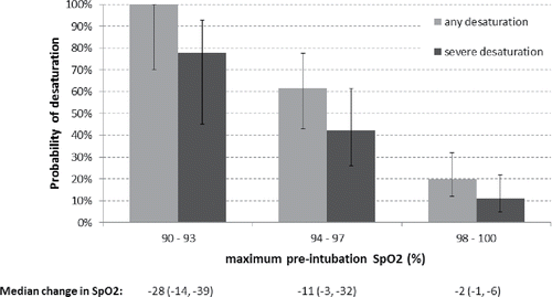
Figure 4. SpO2 nadir and desaturation duration for all desaturation events, grouped by prehospital diagnostic impression category.
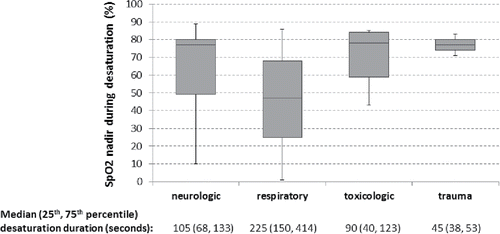
Although the frequency of desaturation was significantly higher in cases requiring multiple laryngoscopic attempts versus a single attempt (70% vs. 37%; p = 0.01), 70% of all desaturations occurred on first-attempt intubation success (). Only 11% of desaturations were reflected in the EMS patient care report.
Peri-intubation Heart Rate
Fourteen cases exhibited loss of ECG monitoring during the peri-intubation interval, preventing determination of pre-intubation HR and identification of peri-intubation HR changes. Among the remaining 120 cases, the baseline pre-intubation HR was 96 (84, 116) bpm. Changes in HR during the peri-intubation period were variable, with HR increasing in 54% and decreasing in 28% of cases; 18% of cases exhibited both increases and decreases from baseline at different times during the peri-intubation period. Decreases in HR were significantly more common (62% vs. 36% of cases, p < 0.01) and larger in magnitude (−28% [−9%, –56%] versus −14% [−6%, −20%] change from baseline; p < 0.01) among cases with versus without desaturation, respectively.
Overall, 15 patients (13%) developed bradycardia during the peri-intubation period, with eight (7%) progressing to profound bradycardia. The frequency of bradycardia was not statistically significantly different for cases requiring multiple laryngoscopic attempts versus a single attempt (25% vs. 9%; p = 0.08). Sixty percent of bradycardia events occurred on first-attempt intubation success, and 53% occurred in patients with a prehospital respiratory diagnostic impression. Of the 14 bradycardia cases with available SpO2 data, 12 (86%) had concomitant desaturations, with a median SpO2 nadir of 30% (12%, 44%) and median desaturation duration of 324 (227, 447) seconds. Similarly, 50% of the profound bradycardia events occurred on first-attempt success, 88% occurred in respiratory patients, and all occurred in conjunction with severe desaturation. Pre-intubation shock index did not differ between cases with versus without peri-intubation bradycardia (0.7 [0.6, 0.8] vs. (0.7 [0.6, 0.9], respectively; p = 0.52). No instances of peri-intubation bradycardia were reflected in the EMS patient care report.
Peri-intubation Blood Pressure
Paired pre-intubation and post-intubation blood pressure measurements were available for analysis from 120 cases. Median pre-intubation SBP was 133 (115, 153) mmHg, measured a median of 4 (2, 8) minutes prior to the documented time of induction, and median post-intubation SBP was 143 (117, 175) mmHg, measured a median of 4 (2, 6) minutes after intubation.
A hypotensive response to RSI was observed in 17 (14%) cases, with a median decrease in SBP of 48 (35, 63) mmHg from a baseline of 155 (130, 175) mmHg. Absolute hypotension developed following RSI in 8 cases (7%). The frequency of hypotension was not statistically significantly different for cases requiring multiple laryngoscopic attempts versus a single attempt (13% vs. 5%, p = 0.18). Five of the eight episodes of hypotension occurred on first-attempt success, five occurred in respiratory patients, and three occurred in conjunction with episodes of severe desaturation and profound bradycardia. Pre-intubation shock index did not differ significantly between cases with versus without post-intubation hypotension (0.8 [0.8, 0.9] vs. (0.7 [0.6, 0.9], respectively; p = 0.22).
Overall, two of 134 patients (1.5%) developed cardiac arrest requiring CPR following RSI. Both cardiac arrests occurred in respiratory patients, following successful intubation requiring one and three laryngoscopic attempts.
A hypertensive response to RSI was observed in 40 (33%) cases, with SBP increasing from a baseline of 118 (97, 145) mmHg to 169 (139, 196) mmHg. A hypertensive response was observed more frequently when the first available post-intubation SBP was acquired ≤3 minutes after intubation versus > 3 minutes (46% vs. 28%; p = 0.05). There was no difference in the likelihood of a hypertensive response between cases with versus without first-attempt success (33% vs. 35%, respectively, p = 0.87). Among the 16 cases with lidocaine pretreatment, paired blood pressure measurements were available in 12, and a hypertensive response was observed in two (17%). Paramedics customarily documented each intermittently acquired blood pressure measurement, and thus 75% of the instances of absolute hypotension, 88% of the instances of a hypotensive response, and 85% of the instances of a hypertensive response were evident in the EMS patient care report.
Discussion
We conducted an observational study of vital signs alterations occurring during prehospital RSI performed by ground-based paramedics in a metropolitan county EMS system. Our primary goal was to leverage continuous physiologic monitoring data to attempt to identify quality improvement opportunities pertinent to our EMS system that are not evident from standard documentation of the patient encounter. Several significant observations emerged from analysis of this cohort of 134 RSI procedures. Oxygen desaturation during intubation was common, more than half of the desaturation events were severe, and most desaturations occurred on first-attempt intubation success. The incidence of desaturation varied among prehospital diagnostic impression categories, with a respiratory diagnostic impression associated with more frequent, more severe, and more prolonged desaturations, as well as a higher incidence of accompanying bradycardia, hypotension, and peri-intubation cardiac arrest. Within the constraints of intermittent oscillometric blood pressure measurements, peri-intubation hypotension was observed infrequently, while a significant peri-intubation hypertensive response occurred in one-third of cases. Finally, peri-intubation derangements of continuously-monitored physiologic parameters (SpO2 and HR) were rarely captured in the standard patient care report.
Details of peri-intubation physiologic alterations have been reported in many studies, spanning most of the clinical settings in which emergency RSI is performed Citation(9, 10, 12–40). However, there is substantial heterogeneity in both the characteristics and findings of these studies. Important differences in factors such as case mix, drug regimens, provider level, procedural exposure volume among individual providers, and clinical environment complicate extrapolation of particular study findings to disparate circumstances. Additionally, variability in data sources has been identified as a factor that likely contributes to the wide variability in reported incidence of peri-RSI physiologic changes Citation(13, 24, 34). Among studies of prehospital paramedic-performed RSI, most have focused on procedural success rates and have not reported on physiologic derangements during the RSI attempt Citation(41–43). The few exceptions have been limited to cohorts of TBI or air medical transported patients, which likely do not reflect the full spectrum of baseline characteristics and pathology among patients meeting RSI indications in a system such as ours Citation(13, 14, 16, 20). These considerations motivated us to prospectively study peri-RSI physiology in our own EMS community, both to address this gap in the published evidence and to provide more pertinent guidance for our quality improvement program.
In our study, oxygen desaturation during RSI occurred in 43% of cases, with 29% of cases exhibiting a drop in SpO2 to below 80%. A number of studies in other patient cohorts or clinical settings have similarly reported frequent occurrences of desaturation during emergency RSI, particularly when electronic vital signs data capture was employed. For example, in a cohort of 54 TBI patients receiving prehospital RSI, Dunford et al. Citation(13) observed desaturation in 57% of cases, and in a cohort of 110 air medical transported patients, Davis et al. Citation(20) reported a 45% incidence of desaturation to an SpO2 ≤ 93%. Among 166 patients receiving RSI in an emergency department setting, Bodily et al. Citation(32) found a 36% incidence of desaturation, and in a cohort of 117 TBI patients receiving RSI in a trauma resuscitation unit, Yeatts et al. Citation(29) reported a 36% incidence of desaturation to an SpO2 < 80%.
In patients with severe TBI, episodes of hypoxemia are a potent cause of secondary brain injury and have been associated with significantly worse outcomes Citation(3–6), particularly when there is coexisting hypotension Citation(8). Avoiding desaturation during intubation is thus a critical goal in these patients. A similar hazard likely exists for patients with other types of acute brain injury Citation(7). On the other hand, in patients without acute brain injury, the impact on patient outcomes of a transient or shallow desaturation without other concomitant deterioration has not been well elucidated. Exposure to brief episodes of hypoxemia is understood to be not harmful in healthy individuals, provided there is no coexisting systemic hypotension or vital organ ischemia Citation(44). However, for individuals who are already physiologically compromised and undergoing emergency RSI, the development of desaturation may presage imminent hemodynamic deterioration Citation(1, 2, 9). Thus even when peri-intubation desaturations do not occur in the presence of acute brain injury and are not severe enough to produce hemodynamic sequelae, they serve to indicate “near miss” events of significance for patient safety and quality improvement initiatives Citation(45).
Various studies have described a significant association between multiple intubation attempts and an increased rate of procedural complications, including peri-intubation physiologic derangements Citation(15, 27, 32, 46). Consequently, in the ongoing discourse around emergency airway management, a strong emphasis exists on achieving first-attempt intubation success in order to minimize such complications. What may be obscured by this focus on the risks associated with multiple intubation attempts is the large absolute number of physiologic derangements occurring on first-attempt success. In our study, 70% of all desaturations, 60% of bradycardia episodes, 63% of hypotension episodes, and one of the two cardiac arrests occurred on first-attempt success. In the study by Bodily et al., while desaturation was three times more likely to occur in cases with multiple intubation attempts than with first-attempt success, 59% of all desaturations actually occurred on first-attempt success Citation(32). In another emergency department study, Kim et al. reported that 36 of 41 (88%) peri-intubation cardiac arrests occurred on first-attempt success Citation(30). While these observations are a mathematical consequence of the fact that first-attempt success is achieved in a large proportion of cases, they also reinforce the fact that first-attempt success – the traditional measure of intubation quality – is not a reliable indicator of patient safety. Thus while there may be a greater risk of physiologic derangements with multiple intubation attempts, there may be a greater absolute number of such derangement events occurring with first-attempt success, and for maximum quality improvement impact it would be important to understand and address specifically these “near miss” events occurring on first-attempt success.
The combination of standard written documentation and electronic monitoring data that was the basis of our analysis does not allow definitive determination of the reason for the high rate of physiologic derangements on first-attempt success. However, one contributing factor would appear to be a prolonged duration of these successful intubation attempts. In our study, nearly half of desaturation episodes occurring on first-attempt success lasted at least 2 minutes. Similarly, the presence of prolonged desaturations on first pass success – indicative of prolonged attempts – can be inferred from the data reported in several prior studies Citation(13, 32, 33). Prolonged first-attempt intubations precipitating prolonged desaturation might be a consequence of lack of awareness of the passage of time during an intubation attempt, or lack of awareness of the occurrence of desaturation. It is known that the stress of complex procedures and critical incidents can compromise an individual's situational awareness Citation(47–49). In an emergency department study, Cemalovic et al. found that physicians performing RSI significantly underestimated the duration of their intubation attempts, and also under-recognized the occurrence of desaturation, even when declining saturation values were announced out loud by an assistant during the procedure Citation(33).
Alternatively, prolonged desaturations on first-attempt success could be an unintended consequence of the focus on first-attempt success itself and the common use of first-attempt success as a primary measure of intubation quality. Particularly in a setting of diminished situational awareness, this emphasis on first-attempt success might inadvertently encourage unwarranted prolongation of first intubation attempts, paradoxically contributing to the physiologic derangements that such a focus on procedural efficiency is intending to prevent. From our data we cannot discern whether a primacy of first-attempt success over other procedural goals may have affected intra-attempt decision-making. However, anecdotally we have found this to occur, and others have noted the potential for inappropriate extrapolation from the notion of first-attempt success as a vital prospective goal – which warrants optimal planning and preparation for an RSI procedure – to a continued prioritization of first-attempt success even after an intubation attempt is well underway Citation(50). Given the significant number of “near miss” events on first-attempt success that are evident in our study and others, it may be valuable in the discourse surrounding emergency RSI to more explicitly emphasize that the goal of targeting first-attempt success in preparation for an intubation attempt is superseded by a goal of prompt and appropriate response to developing physiologic derangements once the intubation attempt has commenced.
Abrupt and often transient changes in blood pressure can occur as a consequence of anesthesia induction, laryngoscopy, tracheal intubation, and initiation of positive pressure ventilation Citation(1, 51–56). While intermittent oscillometric blood pressure measurement is not an ideal technique for characterizing the precise amplitude and temporal profile of such changes, it has been the basis of most prior explorations of peri-intubation hemodynamics during emergency RSI Citation(12, 14, 17, 22, 23, 26, 31). In our study, a significant increase in blood pressure in response to RSI occurred in one-third of cases. Prior investigations of prehospital RSI have also observed a high incidence of this hypertensive response. In a cohort of trauma patients, using a similar anesthesia regimen to ours, Perkins et al. observed a significant hypertensive response in 70% of cases, with a similar magnitude of SBP increase as we observed (mean 41 [95% CI: 31–51] mmHg, vs. median 41 [31, 55] mmHg in our study) Citation(26). We observed a hypertensive response more frequently among cases in which the first post-intubation blood pressure was measured promptly following intubation, suggesting that the true incidence was higher than the 33% that we were able to detect. Similarly, the intermittent nature and variable timing of the blood pressure measurements likely resulted in an underestimate of the true magnitude of acute blood pressure elevation in these cases. While such large spikes in blood pressure may be well tolerated by many individuals, they may be less safe in certain patients such as those with acute cardiovascular or intracranial pathology Citation(1, 57, 58).
On the other hand, hypotension in response to RSI poses a hazard for all patients. Hypotension can exacerbate the impact of hypoxemia in vulnerable patients Citation(8), increase in-hospital length of stay and mortality Citation(22), and directly precipitate cardiac arrest Citation(1, 2). Of the 8 cases we observed with hypotension developing promptly after intubation, 4 recovered to normotensive status by the next blood pressure measurement, two remained hypotensive through arrival at the emergency department, and two continued to deteriorate into cardiac arrest. Peri-intubation arrest rates of up to 3–4% have been reported in studies of emergency RSI, underscoring that peri-intubation physiologic derangement in any patient represents a potentially critical safety threat Citation(10, 21, 28).
Among the cases in our cohort with any combination of desaturation, bradycardia, or hypotension, desaturation was the sole finding or a component of the physiologic derangement in 85%. These desaturations occurred predominantly on first-attempt intubation success, appeared to be rarely recognized by the providers, and thus represented the most significant target for quality improvement. Various strategies have been identified in the literature to optimize pre-oxygenation, maximize safe apnea time, and reduce the incidence of peri-RSI desaturation. In response to our findings, a number of such strategies were incorporated into a new Spokane County EMS “Optimal Sequence Intubation” (OSI) protocol Citation(59), including: head-elevated patient positioning Citation(60), non-rebreather mask oxygen delivered at the maximum available flowmeter setting Citation(61), use of CPAP or bag-valve-mask with a PEEP valve if SpO2 > 93% cannot be achieved via non-rebreather mask Citation(62), and apneic oxygenation via nasal cannula Citation(63). In addition, the OSI protocol implemented a “pit crew” approach, similar to that already in use in our system for cardiac arrest management, that includes tasking BLS providers with ensuring patient sensor attachment throughout the procedure, maintaining vigilance on vital signs, accurately recording key process steps with time-stamped events on the monitor, and providing a countdown to the onset of paralysis to avoid initiating laryngoscopy too early.
Limitations
Our study was conducted in a single county EMS system with specific provider and RSI protocol characteristics, and our findings may not be generalizable to EMS systems or emergency airway management settings with significantly different characteristics. Multiple components of emergency RSI can contribute to peri-intubation physiologic derangement, and our data are insufficient to determine which specific factors were most responsible for the vital signs alterations we observed. While there were no associations between RSI medication doses and any observed derangements, the standard EMS documentation did not allow us to confidently determine other factors such as the duration of pre-oxygenation or duration of individual laryngoscopic attempts.
We were unable to obtain monitor data for a sizable number of RSIs performed during our study period, and therefore cannot exclude the possibility of selection bias impacting our findings. However, beyond the cases with technical issues on data download, which we believe occurred randomly, there were several additional considerations that gave us confidence that our analyzed cohort provided a representative baseline of RSI performance in our system useful for guiding quality improvement efforts. A number of cases were downloaded by supervisory personnel with a strong interest in the success of the study, rather than by the intubating paramedics, and capture of such cases was influenced by the random availability of those personnel to perform the download before the data were overwritten in the device memory. Download of the monitor data was a new process introduced at the beginning of the data collection period, and while the rate of successful downloads increased through the study, there was no difference in the rates of any desaturation or severe desaturation between the first half and second half of the data collection period (p = 0.98 and p = 0.77, respectively). Finally, there were no differences in the proportion of cases with an RSI indication of respiratory failure (p = 0.45) or in the rate of first-attempt success (p = 0.32) between cases with versus without monitor downloads.
The sampling interval of the SpO2 data we analyzed was not ideal for precise characterization of the contour of SpO2 changes during peri-intubation desaturation. It is possible that some brief desaturations occurred which we could not detect. It is also likely that the nadir SpO2 values we measured sometimes underestimated the true nadir. However, any desaturations that went undetected in our study were by definition very transient and therefore also likely to be relatively shallow. The median desaturation duration of 120 seconds that we measured is in a similar range as the median durations of 160 Citation(13), 176 Citation(20), and 80 Citation(32) seconds reported in studies using shorter SpO2 sampling intervals. Extrapolating from the data reported by Bodily et al., we would have detected more than 90% of the desaturations they identified with their SpO2 sampling interval of 5 seconds Citation(32).
Similarly, it is likely that the intermittent non-invasive blood pressure measurements sometimes underestimated the magnitude of peri-intubation blood pressure changes. During our study, EMS providers typically measured blood pressure in preparation for anesthesia induction and then again only after confirmation of endotracheal tube placement. The true proportion of cases with hypertensive or hypotensive responses during the RSI sequence may be higher than we observed.
Finally, we did not have hospital care or disposition data for our patient cohort, and thus our study is unable to address the question of whether the physiologic alterations we observed during the prehospital peri-RSI period impact downstream patient outcomes.
Conclusions
In this study of paramedic-performed RSI, peri-intubation oxygen desaturation was common, with the most severe and prolonged desaturations, and the highest incidence of accompanying cardiovascular instability, occurring disproportionately in patients with a respiratory diagnostic impression. Few of the peri-intubation physiologic alterations identified from electronic monitoring data were evident in the standard patient care documentation, while the majority of such events occurred on first-attempt intubation success, reinforcing the notion that first-attempt success is an incomplete and potentially deceptive measure of intubation quality. Supplementing the standard patient care documentation with electronic monitoring data can identify numerous unrecognized “near miss” patient safety events and provide valuable guidance for quality improvement interventions.
References
- Mort TC. Complications of emergency tracheal intubation: hemodynamic alterations–part I. J Intensive Care Med. 2007;22(3):157–65.
- Mort TC. Complications of emergency tracheal intubation: immediate airway-related consequences: part II. J Intensive Care Med. 2007;22(4):208–15.
- Chesnut RM, Marshall LF, Klauber MR, Blunt BA, Baldwin N, Eisenberg HM, Jane JA, Marmarou A, Foulkes MA. The role of secondary brain injury in determining outcome from severe head injury. J Trauma. 1993;34(2):216–22.
- Stocchetti N, Furlan A, Volta F. Hypoxemia and arterial hypotension at the accident scene in head injury. J Trauma. 1996;40(5):764–7.
- Davis DP, Dunford JV, Poste JC, Ochs M, Holbrook T, Fortlage D, Size MJ, Kennedy F, Hoyt DB. The impact of hypoxia and hyperventilation on outcome after paramedic rapid sequence intubation of severely head-injured patients. J Trauma. 2004;57(1):1–10.
- Chi JH, Knudson MM, Vassar MJ, McCarthy MC, Shapiro MB, Mallet S, et al. Prehospital hypoxia affects outcome in patients with traumatic brain injury: a prospective multicenter study. J Trauma. 2006;61(5):1134–41.
- Rowat AM, Dennis MS, Wardlaw JM. Hypoxaemia in acute stroke is frequent and worsens outcome. Cerebrovasc Dis. 2006;21(3):166–72.
- Spaite DW, Hu C, Bobrow BJ, Chikali V, Barnhart B, Gaither JB, et al. The effect of combined out-of-hospital hypotension and hypoxia on mortality in major traumatic nrain injury. Ann Emerg Med. 2017;69:62–72.
- Mort TC. The incidence and risk factors for cardiac arrest during emergency tracheal intubation: a justification for incorporating the ASA Guidelines in the remote location. J Clin Anesth. 2004;16(7):508–16.
- Heffner AC, Swords DS, Neale MN, Jones AE. Incidence and factors associated with cardiac arrest complicating emergency airway management. Resuscitation. 2013;84(11):1500–4.
- Riyapan S, Lubin J. The variability of statewide prehospital drug-facilitated intubation protocols in the United States. Am J Emerg Med. 2016;34(12):2459–60.
- Sivilotti ML, Ducharme J. Randomized, double-blind study on sedatives and hemodynamics during rapid-sequence intubation in the emergency department: the SHRED Study. Ann Emerg Med. 1998;31(3):313–24.
- Dunford J V, Davis DP, Ochs M, Doney M, Hoyt DB. Incidence of transient hypoxia and pulse rate reactivity during paramedic rapid sequence intubation. Ann Emerg Med. 2003;42(6):721–8.
- Deitch S, Davis DP, Schatteman J, Chan TC, Vilke GM. The use of etomidate for prehospital rapid-sequence intubation. Prehosp Emerg Care. 2003;7(3):380–3.
- Mort TC. Emergency tracheal intubation: complications associated with repeated laryngoscopic attempts. Anesth Analg. 2004;99(2):607–13.
- Tiamfook-Morgan TO, Harrison TH, Thomas SH. What happens to SpO2 during air medical crew intubations? Prehosp Emerg Care. 2006;10(3):363–8.
- Lin C-C, Chen KF, Shih C-P, Seak C-J, Hsu K-H. The prognostic factors of hypotension after rapid sequence intubation. Am J Emerg Med. 2008;26(8):845–51.
- Newton A, Ratchford A, Khan I. Incidence of adverse events during prehospital rapid sequence intubation: a review of one year on the London Helicopter Emergency Medical Service. J Trauma. 2008;64(2):487–92.
- Nakstad AR, Heimdal H-J, Strand T, Sandberg M. Incidence of desaturation during prehospital rapid sequence intubation in a physician-based helicopter emergency service. Am J Emerg Med. 2011;29(6):639–44.
- Davis DP, Aguilar S, Sonnleitner C, Cohen M, Jennings M. Latency and loss of pulse oximetry signal with the use of digital probes during prehospital rapid-sequence intubation. Prehosp Emerg Care. 2011;15(1):18–22.
- Jabre P, Avenel A, Combes X, Kulstad E, Mazariegos I, Bertrand L, Lapostolle F, Adnet F. Morbidity related to emergency endotracheal intubation — A substudy of the KETAmine SEDation trial. Resuscitation. 2011;82(5):517–22.
- Heffner AC, Swords D, Kline JA, Jones AE. The frequency and significance of postintubation hypotension during emergency airway management. J Crit Care. 2012;27(4):417. e9–13.
- Green RS, Edwards J, Sabri E, Fergusson D. Evaluation of the incidence, risk factors, and impact on patient outcomes of postintubation hemodynamic instability. Can J Emerg Med. 2012;14(2):74–82.
- Kerrey BT, Rinderknecht AS, Geis GL, Nigrovic LE, Mittiga MR. Rapid sequence intubation for pediatric emergency patients: higher frequency of failed attempts and adverse effects found by video review. Ann Emerg Med. 2012;60(3):251–9.
- Helm M, Kremers G, Lampl L, Hossfeld B. Incidence of transient hypoxia during prehospital rapid sequence intubation by anaesthesiologists. Acta Anaesthesiol Scand. 2013;57(2):199–205.
- Perkins ZB, Gunning M, Crilly J, Lockey D, O'Brien B. The haemodynamic response to prehospital RSI in injured patients. Injury. 2013;44(5):618–23.
- Sakles JC, Chiu S, Mosier J, Walker C, Stolz U. The importance of first pass success when performing orotracheal intubation in the emergency department. Acad Emerg Med. 2013;20(1):71–8.
- Bloomer R, Burns BJ, Ware S. Improving documentation in prehospital rapid sequence intubation: investigating the use of a dedicated airway registry form. Emerg Med J. 2013;30(4):324–6.
- Yeatts DJ, Dutton RP, Hu PF, Chang YW, Brown CH, Chen H, Grissom TE, Kufera JA, Scalea TM. Effect of video laryngoscopy on trauma patient survival: a randomized controlled trial. J Trauma Acute Care Surg. 2013;75(2):212–9.
- Kim WY, Kwak MK, Ko BS, Yoon JC, Sohn CH, Lim KS, Andersen LW, Donnino MW. Factors associated with the occurrence of cardiac arrest after emergency tracheal intubation in the emergency department. PLoS One. 2014;9(11):e112779.
- Lyon RM, Perkins ZB, Chatterjee D, Lockey DJ, Russell MQ. Significant modification of traditional rapid sequence induction improves safety and effectiveness of prehospital trauma anaesthesia. Crit Care. 2015;19(1):134.
- Bodily JB, Webb HR, Weiss SJ, Braude DA. Incidence and duration of continuously measured oxygen desaturation during emergency department intubation. Ann Emerg Med. 2016;67(3):389–95.
- Cemalovic N, Scoccimarro A, Arslan A, Fraser R, Kanter M, Caputo N. Human factors in the emergency department: Is physician perception of time to intubation and desaturation rate accurate? Emerg Med Australas. 2016;28(3):295–99.
- Rinderknecht AS, Dyas JR, Kerrey BT, Geis GL, Ho MH, Mittiga MR. Studying the safety and performance of rapid sequence intubation: data collection method matters. Acad Emerg Med. 2017;24(4):411–21.
- Reid C. The who, where, and what of rapid sequence intubation: prospective observational study of emergency RSI outside the operating theatre. Emerg Med J. 2004;21(3):296–301.
- Jaber S, Amraoui J, Lefrant J-Y, Arich C, Cohendy R, Landreau L, Calvet Y, Capdevila X, Mahamat A, Eledjam J. Clinical practice and risk factors for immediate complications of endotracheal intubation in the intensive care unit: a prospective, multiple-center study. Crit Care Med. 2006;34(9):2355–61.
- Griesdale DEG, Bosma TL, Kurth T, Isac G, Chittock DR. Complications of endotracheal intubation in the critically ill. Intensive Care Med. 2008;34(10):1835–42.
- Bowles TM, Freshwater-Turner DA, Janssen DJ, Peden CJ. Out-of-theatre tracheal intubation: prospective multicentre study of clinical practice and adverse events. Br J Anaesth. 2011;107(5):687–92.
- Mayo PH, Hegde A, Eisen LA, Kory P, Doelken P. A program to improve the quality of emergency endotracheal intubation. J Intensive Care Med. 2011;26(1):50–6.
- Simpson GD, Ross MJ, McKeown DW, Ray DC. Tracheal intubation in the critically ill: a multi-centre national study of practice and complications. Br J Anaesth. 2012;108(5):792–9.
- Hubble MW, Brown L, Wilfong DA, Hertelendy A, Benner RW, Richards ME. A Meta-Analysis of Prehospital Airway Control Techniques Part I: orotracheal and Nasotracheal Intubation Success Rates. Prehosp Emerg Care. 2010;14(3):377–401.
- Lossius HM, Røislien J, Lockey DJ. Patient safety in prehospital emergency tracheal intubation: a comprehensive meta-analysis of the intubation success rates of EMS providers. Crit Care. 2012;16(1):R24.
- Crewdson K, Lockey DJ, Røislien J, Lossius HM, Rehn M. The success of prehospital tracheal intubation by different prehospital providers: a systematic literature review and meta-analysis. Crit Care. 2017;21:31.
- Bickler PE, Feiner JR, Lipnick MS, Batchelder P, MacLeod DB, Severinghaus JW. Effects of acute, profound hypoxia on healthy humans. Anesth Analg. 2017;124(1):146–53.
- National Quality Forum. 2009. National quality forum patient safety terms and definitions. [accessed 2017 Mar 02] Available at: https://www.qualityforum.org/Topics/Safety_Definitions.aspx.
- Bernhard M, Becker TK, Gries A, Knapp J, Wenzel V. The first shot is often the best shot: first-pass intubation success in emergency airway management. Anesth Analg. 2015;121(5):1389–93.
- Endsley MR. Toward a theory of situation awareness in dynamic systems. Hum Factors. 1995;37(1):32–64.
- Gaba DM, Howard SK, Small SD. Situation awareness in anesthesiology. Hum Factors. 1995;37(1):20–31.
- Schulz CM, Krautheim V, Hackemann A, Kreuzer M, Kochs EF, Wagner KJ. Situation awareness errors in anesthesia and critical care in 200 cases of a critical incident reporting system. BMC Anesthesiol. 2016;16:4.
- Limkakeng A, Broder JS, Theiling BJ. Chicken or egg? Risks of misattribution of cause-effect relationships in studies of association. Acad Emerg Med. 2013;20(9):965.
- King BD, Harris LC, Greifenstein FE, Elder JD, Dripps RD. Reflex circulatory responses to direct laryngoscopy and tracheal intubation performed during general anesthesia. Anesthesiology. 1951;12(5):556–66.
- Prys-Roberts C, Greene L, Meloche R, Foex P. Studies of anaesthesia in relation to hypertension II: haemodynamic consequences of induction and endotracheal intubation. Br J Anaesth. 1971;43(6):531–47.
- Hassan HG, El-Sharkawy TY, Renck H, Mansour G, Fouda A. Hemodynamic and catecholamine responses to laryngoscopy with vs. without endotracheal intubation. Acta Anaesthesiol Scand. 1991;35(5):442–7.
- Hickey S, Cameron AE, Asbury AJ, Murray GD. Timing of peak pressor response following endotracheal intubation. Acta Anaesthesiol Scand. 1992;36:21–4.
- Weiss-Bloom LJ, Reich DL. Haemodynamic responses to tracheal intubation following etomidate and fentanyl for anaesthetic induction. Can J Anaesth. 1992;39(8):780–5.
- Manthous CA. Avoiding circulatory complications during endotracheal intubation and initiation of positive pressure ventilation. J Emerg Med. 2010;38(5):622–31.
- Kovac AL. Controlling the hemodynamic response to laryngoscopy and endotracheal intubation. J Clin Anesth. 1996;8(1):63–79.
- Stein DM, Hu PF, Brenner M, Sheth KN, Liu KH, Xiong W, Aarabi B, Scalea TM. Brief episodes of intracranial hypertension and cerebral hypoperfusion are associated with poor functional outcome after severe traumatic brain injury. J Trauma. 2011;71(2):364–74.
- Spokane County EMS & Trauma Care Council. 2016. Spokane County EMS Protocols. [accessed 2017 Jul 23] Available at: http://www.emsoffice.com/protocols/ems-protocols.pdf.
- Ramkumar V, Umesh G, Philip FA. Preoxygenation with 20-degree head-up tilt provides longer duration of non-hypoxic apnea than conventional preoxygenation in non-obese healthy adults. J Anesth. 2011;25(2):189–94.
- Driver BE, Prekker ME, Kornas RL, Cales EK, Reardon RF. Flush rate oxygen for emergency airway preoxygenation. Ann Emerg Med. 2017;69(1):1–6.
- Weingart SD, Levitan RM. Preoxygenation and prevention of desaturation during emergency airway management. Ann Emerg Med. 2012;59(3):165–175.
- Wimalasena Y, Burns B, Reid C, Ware S, Habig K. Apneic oxygenation was associated with decreased desaturation rates during rapid sequence intubation by an Australian Helicopter Emergency Medicine Service. Ann Emerg Med. 2015;65(4):371–6.

