
ABSTRACT
Background
None of the various treatment modalities for failed back surgery syndrome (FBSS) is satisfactory.
Aim of the study
Comparison of subjective and laboratory outcomes of the 3-in-1 procedure to percutaneous screw and rod spinal fixation (PSRF) for FBSS patients determined at the end of Phase II of the study (3-year follow-up after surgery).
Patients & Methods
This is phase II (follow-up at 3 years) of patients undergoing 3-in-1 procedure (Group B) or PSRF (Group A) as regard pain and disability scores and cytokines levels.
Results
At phase of the study, pain and disability scores were still significantly lower in all patients with significantly lower scores in group B than in group A. In comparison to the results obtained at the end of Phase I, the 3-in-1 procedure provided better outcome than PSRF only and number of patients graded the outcome as excellent-to-good was significantly higher than in patients of group B than group A. At the end of both phases, serum inflammatory cytokines’ levels were significantly decreased and anti-inflammatory cytokines’ levels were significantly increased in all patients compared to preoperative levels with significant difference in favor of patients of group B. the percentages of change in pain and disability scores at end of Phase II were significantly correlated with the percentage of change of serum anti-inflammatory cytokines’ levels in both groups.
Conclusion
After 36 m after the 3-in-1 procedure pain scores were significantly decreased with improved disability scores and inflammatory milieu than in patients having FBSS only.
1. Introduction
The incidence of failed back-surgery syndrome (FBSS) ranges between 10% and 40% with or without fusion. [Citation1,Citation2] Normally, back is dynamic structure, while the failed back is no longer dynamic [Citation3]; such discrepancy results in recurrence of symptoms that may be aggravated than before the first surgery [Citation4] and patients may become unresponsive to medical conservative treatment and their quality of life (QOL) could be easily compromised [Citation5].
Various medications, reoperations and interventions had been suggested as treatment options for FBSS, unfortunately, evidence of its clinical outcome has not been clearly determined. [Citation6] Thus, treatment of each FBSS patient must be selective to provide long-term pain control with reduction of costs and avoiding fewer effective modalities. [Citation7]
Potential advantages of minimally invasive techniques for spinal surgery include reduction of intraoperative blood loss, requirements for postoperative (PO) analgesia and length of hospital stay, so as to allow early resumption of daily activities and return to work. [Citation8,Citation9] Screws act as a rigid and stable anchor points that are bridged and connected with a rod as part of a construct for gripping spinal segment to get spinal fusion. [Citation10]
Lumbar spine facet joint injection where the superior articular process connects to the base of the transverse process is the ideal site for medial branch infiltrations. [Citation11] Injection mixture of corticosteroid and local anesthetic allows pain reduction [Citation12] and proper selection of patient who may get benefit on thermal radiofrequency neurotomy (TRFN) [Citation13] Efficacy facet joint injections depends on the anti-inflammatory and antiedematous effect of steroids in addition to its immunosuppressive action and to the inhibition of neural transmission within the nociceptive C fibers through reduction of lamina II GABAergic synaptic transmission resulting in a selective decrease in the processing of nociceptive inputs. [Citation14]
Thermal techniques including TRFN induce mechanical decompression with thermally induced modifications of intradiscal cytokines involved in disc disease. [Citation15] TRFN also destroy nociceptors in the periphery of the annulus. [Citation13] Moreover, at temperature more than 70°C, fusion of collagen fibers in the annulus occurs with shrinkage at the periphery of the disc. [Citation16] Combined TRFN and steroid nerve block produced substantial improvement in terms of long-term pain relief and QOL. [Citation17]
The 3-in-1 procedure was previously applied by the authors (Phase I) and entailed percutaneous TRNF of the median branch of the facet nerve, percutaneous screw and rod insertion and finally interlaminar epidural injection of triamcinolone. The 3-in-1 procedure showed superior short outcome than percutaneous screw and rod spinal fixation (PSRF). [Citation18] The purpose of this study was to compare subjective and laboratory outcomes of the 3-in-1 procedure (applied during Phase I of the study) to the PSRF alone for FBSS patients at the end of 30-m follow-up.
2. Patients & methods
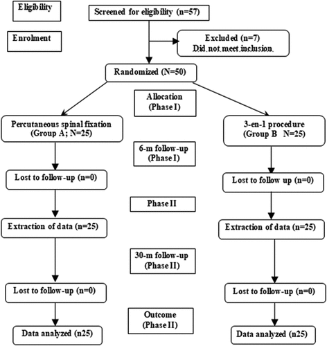
is a flow diagram that shows the progress of our study phases. This prospective comparative follow-up study was carried out in Anesthesia Departments, Faculty of Medicine, Tanta & South Valley Universities in conjunction with Medical Biochemistry Department, Faculty of Medicine, Benha University and Rejuvenation Center, Benha, Egypt, between 2017 and 2020.
Figure 1. Consort flow diagram of the different study phases
The study protocol was approved by the Local Ethical Committee to continue follow-up evaluation of patients previously had the 3-in-1 procedure (Group B) or PSRF (Group A) as interventional management of FBSS patients. [Citation18] Patients accepted to attend the follow-up visits, to give blood sample for estimation of assigned laboratory investigations and signed fully informed written consents were included in the study. Preoperative and 6-m PO data of the enrolled patients were extracted out of their hospital files to be compared to data obtained at re-evaluation sessions till end of 30-m PO (Phase II).
3. Clinical outcome measures
3.1. Primary outcome
Pain severity was assessed using an 11-point numeric rating scale (NRS) where 0 indicates no pain and 10 indicates worst pain imaginable. [Citation19,Citation20] Back and leg pain during day and night was assessed and total NRS pain score was calculated.
4. Secondary outcomes
Disability secondary to pain was assessed using the Oswestry Low Back Pain Disability Questionnaire [Citation21,Citation22] that covers 10 items for evaluation of pain intensity, personal care, lifting, walking, sitting, standing, sleeping, sex life, social life and travelling. Each item was scored from 0 to 5 according to increased disability and a total Oswestry Disability index (ODI) scores was calculated. Disability was graded as minimal (ODI = 0–20), moderate (ODI = 20–40), severe (ODI = 40–60), crippled (ODI score = 60–80) and ODI score of 80–100 indicates that the patient is either bed-bound or exaggerating his or her symptoms [Citation22,Citation23]. Pain and disability scores were compared to previous scores and percentages of change in relation to preoperative and 6-m PO scores were evaluated. [Citation24,Citation25]
Pain medication requirements were recorded using a 0- 4-point scale with 0 indicates no medication used; 1 indicates occasional use of pain medications; 2 indicate regular use of non-opioid medications, 3 indicates occasional use of opioid medications and 4 indicates regular use of opioid medications.
Patients’ evaluation of outcome was graded according to Odom’s criteria into Excellent: relief of all preoperative symptoms and all abnormal findings were improved; Good: minimal persistence of preoperative symptoms and all abnormal findings were improved or unchanged; Fair: definite relief of some preoperative symptoms, while other symptoms were unchanged or slightly improved; Poor: all preoperative symptoms and signs were unchanged or exacerbated. [Citation26]
5. Blinding
The anesthetist who gathered data from patients at 36 m post-operatively was not aware of the study protocol.
6. Sampling & investigations
Venous blood samples (5 ml) were collected from the antecubital vein under complete aseptic conditions and were kept in a plan container, allowed to clot and then serum was separated by centrifugation at 3000 rpm for 10 minutes, collected in sterile Eppendorf tube and stored at −80°C till be assayed. Blood samples were collected and numbered by an assistant who was blinded about type of surgery.
7. Laboratory tests
Serum levels of estimated markers were measured using enzyme-linked immunosorbent assay (ELISA) kits according to the manufacturer’s instructions and were read using a 96 well microplate ELISA reader (Dynatech. MR 7000).
Human TNF-α was measured with the enzyme linked immunoassay (ELISA) kit (catalogue no. ab179886, abcam Inc., Cambridge, USA) by quantitative sandwich enzyme immunoassay technique. [Citation27]
Human IL-1β with the enzyme linked immunoassay (ELISA) kit (catalogue no. ab46052, abcam Inc., Cambridge, USA) by quantitative sandwich enzyme immunoassay technique. [Citation28]
Human IL-6 with the enzyme linked immunoassay (ELISA) kit (catalogue no. ab46042, abcam Inc., Cambridge, USA) by quantitative sandwich enzyme immunoassay technique. [Citation29]
Human IL-10 was measured with the enzyme linked immunoassay (ELISA) kit (catalogue no. ab215089, abcam Inc., San Francisco, USA) by quantitative sandwich enzyme immunoassay technique. [Citation30]
8. Statistical analysis
Obtained data were presented as mean±SD, ranges, numbers and percentages. Results were analyzed using paired t-test, One-way ANOVA Test and Chi-square test. Possible relationships were investigated using Pearson linear regression analysis. Statistical analysis was conducted using the IBM SPSS (Version 23, 2015) for Windows statistical package. P value <0.05 was considered statistically significant.
9. Results
showed no statistically significant difference between studied groups as regard demographic data. At the end of Phase II (30-m of follow-up), total NRS pain scores were still significantly lower, in patients of both groups, in comparison to their preoperative scores with significantly lower total pain scores of patients of group B in comparison to patients of group A. The median percentage of decrease at the end of Phase II, in relation to preoperative pain scores, was significantly higher in patients of group B in comparison to that of patients of group A. On contrary, the median percentage of change in NRS scores at the end of Phase II in comparison to that recorded at the end of Phase I (6th month’ scores) were non-significantly better in group B in comparison to group A. Unfortunately, 25 patients showed worsened pain scores at the end of Phase II with non-significantly higher frequency of patients had worsened pain in group A in comparison to group B; 60% vs. 44% ().
Table 1. Demographic and clinical data of studied patients
Table 2. Outcome data of patients of both groups determined at 36-m PO
At the end of Phase II, ODI scores showed non-significant difference in comparison to scores determined at the end of Phase I in both groups. However, ODI scores that were determined at the end of Phase II were significantly lower in patients of group B on comparison to scores of patients of group A. Moreover, the percentage of change was significantly better in patients of group B in comparison to patients of group A. Lastly, the frequency of patients had improved disability was higher in patients of group B, while the frequency of patients had worsened disability was higher among patients of group A ().
At end of Phase II, patients’ requirements for analgesia were significantly reduced in group B, but non-significantly increased in group A in comparison to their respective requirement scoring at the end of Phase I. Moreover, analgesia requirement scoring for patients of group B was significantly lower than that of patients of group A. Furthermore, the frequency of patients required analgesia was decreased in group B, while the frequency of patients required analgesia in group A was increased with significant difference in favor of groups B ().
According to Odom’s criteria, the frequency of patients of group B who found the outcome excellent was significantly higher than in group A; 52% vs. 24%, while the frequency of patients found the outcome fair-to-good was significantly lower in group B in comparison to group A; 8% vs. 32% ()
Serum levels of TNF-α and IL-1β were significantly decreased, while serum levels of IL-6 were non-significantly decreased and serum IL-10 were non-significantly increased in samples of patients of group A that obtained at the end of Phase I and Phase II in comparison to their preoperative levels. On the other hand, in samples of patients of group B that were obtained at the end of Phase I and Phase II, serum levels of TNF-α, IL-1β and IL-6 were significantly decreased, while serum levels of IL-10 were significantly increased in comparison to their preoperative levels. Moreover, the percentage of change of estimated levels of studied cytokines in relation to preoperative levels showed significant differences between both groups in favor of group B. Estimated levels of TNF-α and IL-1β in 36-m samples of patients of both groups were non-significantly higher in comparison to 6-m sample levels. In patients of group A, serum levels of IL-6 were non-significantly increased, while serum levels of IL-10 were non-significantly decreased in comparison to 6-m levels, while in patients of group B, the difference was significant ().
Table 3. Serum levels of studied cytokines estimated at the end of the study phases
The percentages of change in NRS and ODI scores at the end of Phase I and Phase II in relation to preoperative scores were significantly correlated with the percentage of decrease in serum levels of TNF-α and IL-6 and the percentage of increase of serum IL-10 levels in both groups ().
Table 4. Pearson’s correlation between % of change of NRS and ODI scores and percentage of change in studied cytokines’ serum levels at the end of Phase II in patients of both groups in relation to preoperative cytokine’s levels
10. Discussion
The results obtained at the end of Phase II of the study (36-m after surgery) indicated the efficacy of spinal fixation using percutaneous screw and rod spinal fixation, alone or as a part of the 3-in-1 procedure for management of FBSS patients as evidenced by the maintained significant reduction of pain and disability scores till 36-m PO in comparison to preoperative scores. These findings supported the previous studies that documented the long-term improvement of ODI and pain scores after instrumented fusion of FBSS patients [Citation31–35].
The 3-in-1 procedure maintained significantly lower ODI and pain scores in comparison to preoperative scores and to the 36-m scores of patients had PSRF only. Moreover, in comparison to the 6-m scores the 3-in-1 procedure provided better outcome than PSRF only. This maintained efficacy could be attributed to the complimentary effect of TRFN and local steroid injection in addition to the effect of PSRF. These results support that previously documented by Rahimzadeh et al. [Citation36] who reported the effectiveness of adding hyaluronidase to the epidural injectate for management of chronic back pain (CBP) in patients with FBSS. Also, Rapčan et al. [Citation37] and Ceylan et al. [Citation38] reported significant improvement of leg and back pain after 12-months of epiduroscopic hyaluronidase and steroid injection with mechanical lysis. The reported efficacy of steroid and hyaluronidase injection was attributed by Helm Ii & Racz [Citation39] to the ability of hyaluronidase to facilitate the spread of medications in extracellular matrix by breaking down polysaccharides in the interstitial space.
Concerning the effectiveness of RF, the obtained results are in accordance with Arsanious et al. [Citation40] who found combined pulsed RF followed by TRFN of medial branch of lumbar dorsal ramus improves outcome. Also, Do et al. [Citation41] reported superior pain relief by intra-articular RF than by steroid injection for CBP. Moreover, Chen et al. [Citation42] reported significantly greater improvement in ODI and pain scores and QOL that led to improved function of patients had chronic lumber and sacroiliac joint pain and treated by RFN than controls.
In line with the maintained effect of RF, Lee et al. [Citation43] and Çetin & Yektaş [Citation44] documented that conventional RFN of medial branch in patients with lumbar facet joint pain effectively decreases pain scores and allowed better QOL and daily activities till 12 [Citation43] and 24 months [Citation44], respectively. Thereafter, Ibrahim et al. [Citation45] found RFN of sensory nerve branches along S1-3 lateral foramina and L4-S1 medial branches is minimally invasive procedure that significantly relieved lumbar back pain for 24 months. Recently, Speldewinde [Citation46] reported sustained success rate of 69% in reduction of sacroiliac ligament/joint complex pain with improvement in physical and psychological function for 12 months after TRFN. Further, in support of safety of RF application after insertion of the pedicle and screws, Elwood et al. [Citation47] reported no complications due to hardware temperature and Eckmann et al. [Citation48] detected no motor weakness after thermal neurotomy.
Forty patients (80%) found the outcome was excellent-to-good with significantly higher frequency with the 3-in-1 procedure (92%) in comparison to PSRF alone (68%). Similarly, Woiciechowsky & Richter [Citation49] reported that RFN for CBP resulted in acceptable-to-excellent results in 68% of patients with pain reduction rate of 47% for an average duration of 7.8-m and Arıcı & Kılıç [Citation50] documented that TRF for facet arthropathy is a safe and effective procedure and about 69% found outcome is excellent-to-good with a median duration of pain relief of 10.2 months.
Preoperative serum levels of inflammatory cytokines; IL-1β, IL-6 and TNF-α were significantly higher, while serum levels of anti-inflammatory cytokine, IL-10, were significantly lower in comparison to volunteers’ levels. At 6- and 36-m PO, serum levels of inflammatory cytokines were significantly decreased and serum IL-10 levels were significantly increased in all patients in comparison to preoperative levels with significant difference in favor of patients had the 3-in-1 procedure. These findings indicating either a possible role of disturbed cytokines’ milieu in pathogenesis of the primary disc disease, or in failure of the previous spinal surgery or it may be secondary to failed surgery and this disturbance could be corrected by proper treatment of disc disease. In support of this assumption, percentages of change in NRS and ODI scores at 36-m were significantly correlated with percentage of decrease in serum levels of TNF-α and IL-6 and increase of serum IL-10 levels in both groups.
These findings and attribution are in accordance with that experimentally reported by Gorth et al. [Citation51] who documented the role of inflammatory cytokines in pathogenesis of intervertebral disc disease and Zhong et al. [Citation52] who found spinal Src family of protein tyrosine kinases contribute to radicular pain by activation of p38 MAPK pathway and increasing spinal expression of inflammatory cytokines in rats with autologous nucleus pulposus. Recently, Zhang et al. [Citation53] (2020) detected high expression rates of TNF-α and IL1-β in degenerate goat discs. Clinically, the obtained results are coincident with Bäckryd et al. [Citation54] who detected significantly higher serum IL-6 levels in patients with FBSS with peripheral neuropathic pain and Kamieniak et al. [Citation55] who reported significantly higher and lower serum levels of TNF-α and IL-10, respectively in patients had FBSS. Moreover, Stover et al. [Citation56] suggested the need for targeting of IL-6, TNF-α, and IL-1β for pain modulation in degenerative intervertebral discs and multiple clinical trials evaluated the efficacy of TNF-α and IL-6 inhibitors in treating CBP. [Citation57]
11. Conclusion
36 min after surgery, the 3-in-1 procedure approved its efficacy for reduction of pain and disability scores of FBSS patients and improved the associated disturbance in cytokines’ milieu with lessened inflammatory reaction. Thus, it is recommended as a safe and effective management strategy for FBSS patients especially those intolerants to or had contraindications for medical treatment. However, follow-up for longer duration or wider scale studies are mandatory to establish the obtained results.
Declaration
The Authors declare that there is no conflict of interest.
Disclosure statement
No potential conflict of interest was reported by the author(s).
References
- Sorrell RG, Muhlenfeld J, Moffett J, et al. Evaluation of pulsed electromagnetic field therapy for the treatment of chronic postoperative pain following lumbar surgery: a pilot, double-blind, randomized, sham-controlled clinical trial. J Pain Res. 2018;11:1209–1222.
- Sebaaly A, Lahoud MJ, Rizkallah M, et al. Etiology, evaluation, and treatment of failed back surgery syndrome. Asian Spine J. 2018;12(3):574–585.
- Carney AL. Failed back surgery syndrome: foreword. Neurochirurgie. 2015;61(Suppl 1):S1–4.
- Nikitin AS. Failed back surgery syndrome. Zhurnal Nevrologii I Psikhiatrii Im. S.S. Korsakova. 2016;116(5):112–118.
- Graziano F, Gerardi RM, Lo Bue E, et al. Surgical back risk syndrome and spinal cord stimulation: better safe than sorry. World Neurosurg. 2020Jan;133:e658–e665.
- Cho JH. Treatment outcomes for patients with failed back surgery. Pain Physician. 2017;1(21;1):E29–E43.
- Amirdelfan K, Webster L, Poree L, et al. Treatment options for failed back surgery syndrome patients with refractory chronic pain: an evidence based approach. (Phila Pa 1976) Spine (Phila Pa 1976). 2017;42Suppl 14:S41–S52.
- Mobbs RJ, Sivabalan P, Li J. Technique, challenges and indications for percutaneous pedicle screw fixation. J Clin Neurosci. 2011;18(6):741–749.
- Mobbs RJ, Phan K. History of retractor technologies for percutaneous pedicle screw fixation systems. Orthop Surg. 2016;8(1):3–10.
- Solitro GF, Amirouche F. Innovative approach in the development of computer assisted algorithm for spine pedicle screw placement. Med Eng Phys. 2016;38(4):354–365.
- Choi YK. Lumbar foraminal neuropathy: an update on non-surgical management. The Korean Journal of Pain. 2019;32(3):147–159.
- Filippiadis DK, Kelekis A. A review of percutaneous techniques for low back pain and neuralgia: current trends in epidural infiltrations, intervertebral disk and facet joint therapies. Br J Radiol. 2016;89(1057):20150357.
- Schneider BJ, Doan L, Maes MK, et al. Systematic review of the effectiveness of lumbar medial branch thermal radiofrequency neurotomy, stratified for diagnostic methods and procedural technique. Pain Med. 2020 Feb 10. pii: pnz349. 10.1093/pm/pnz349. Epub ahead of print.
- Zell V, PÉ J, Hanesch U, et al. Corticosterone analgesia is mediated by the spinal production of neuroactive metabolites that enhance GABAergic inhibitory transmission on dorsal horn rat neurons. Eur J Neurosci. 2015;41(3):390–397.
- Kallewaard JW, Terheggen MAMB, Groen GJ, et al. 15. Discogenic low back pain. Pain Pract. 2010;10(6):560–579.
- Buy X, Gangi A. Percutaneous treatment of intervertebral disc herniation. Semin Intervent Radiol. 2010;27(2):148–159.
- Roy C, Chatterjee N, Ganguly S, et al. Efficacy of combined treatment with medial branch radiofrequency neurotomy and steroid block in lumbar facet joint arthropathy. J Vasc Interv Radiol. 2012;23(12):1659–1664.
- Mohamed AA, Salama KA, Salem EA, et al. Management for failed back surgery syndrome: three-in-one procedure versus percutaneous spinal fixation alone. Southern African Journal of Anaesthesia and Analgesia. 2017;23:2:26–31.
- Williamson A, Hoggart B. Pain: a review of three commonly used pain rating scales. J Clin Nurs. 2005;14(7):798–804.
- Fairbank JC, Couper J, Davies JB, et al. The Oswestry low back pain disability questionnaire. Physiotherapy. 1980;66(8):271–273.
- Fairbank JC, Pynsent PB. The Oswestry disability index. Spine (Phila Pa 1976). 2000;25(22):2940–2953.
- Davidson M, Keating J. A comparison of five low back disability questionnaires: reliability and responsiveness. Phys Ther. 2002;82(1):8–24.
- Manchikanti L. Comparative Effectiveness of a One-Year Follow-Up of Thoracic Medial Branch Blocks in Management of Chronic Thoracic Pain: a Randomized, Double-Blind Active Controlled Trial. Pain Physician. 2010;6;13(6;12):535–548.
- Odom GL. Neurosurgical care of the severely injured patient. J Am Med Assoc. 1958;168(16):2087–2090.
- Manchikanti L, Cash KA, McManus CD, et al. Fluoroscopic caudal epidural injections with or without steroids in managing pain of lumbar spinal stenosis: one-year results of randomized, double-blind, active-controlled trial. J Spinal Disord Tech. 2012;25(4):226–234.
- Avellanal M, Diaz-Reganon G, Orts A, et al. One-year results of an algorithmic approach to managing failed back surgery syndrome. Pain Res Manag. 2014;19(6):313–316.
- Coughlan MT, Oliva K, Georgiou HM, et al. Glucose-induced release of tumour necrosis factor-alpha from human placental and adipose tissues in gestational diabetes mellitus. Diabet Med. 2001;18(11):921–927.
- Németh K, Patthy M, Fauszt I, et al. Effect of peptide aldehydes with IL-1β converting enzyme inhibitory properties on IL-1α and IL-1β production in vitro. Int J Immunopharmacol. 1995;17(12):985–993.
- Helle M, Boeije L, de Groot E, et al. Sensitive ELISA for interleukin-6. Detection of IL-6 in biological fluids: synovial fluids and sera. J Immunol Methods. 1991;138(1):47–56.
- Lin X, Deng J, Lu L. IL-10 detection in murine B cells: pros and cons of the different techniques. Methods Mol Biol. 2014;1190:55–69.
- Turunen V, Nyyssönen T, Miettinen H, et al. Lumbar instrumented posterolateral fusion in spondylolisthetic and failed back patients: a long-term follow-up study spanning 11–13 years. Eur Spine J. 2012;21(11):2140–2148.
- Tian W, Xu Y, Liu B, et al. Lumbar spine superior-level facet joint violations: percutaneous versus open pedicle screw insertion using intraoperative 3-dimensional computer-assisted navigation. Chin Med J (Engl). 2014;127(22):3852–3856.
- Koshimune K, Ito Y, Sugimoto Y, et al. Minimally invasive spinopelvic fixation for unstable bilateral sacral fractures. Clin Spine Surg. 2016;29(3):124–127.
- Han S-H, Hyun S-J, Jahng T-A, et al. Posterior osteosynthesis of a spontaneous bilateral pedicle fracture of the lumbar spine. SPINE141207 J Neurosurg Spine. 2016;243:398–401.
- Cuéllar JM, Field JS, Bae HW. Distraction Laminoplasty With Interlaminar Lumbar Instrumented Fusion (ILIF) for lumbar stenosis with or without grade 1 spondylolisthesis: technique and 2-year outcomes. (Phila Pa 1976) Spine (Phila Pa 1976). 2016;41Suppl 8:S97–S105.
- Rahimzadeh P, Sharma V, Imani F. Hamid Reza Faiz HR, Ghodraty MR, Nikzad-Jamnani AR, Nader DN. Adjuvant hyaluronidase to epidural steroid improves the quality of analgesia in failed back surgery syndrome: a prospective randomized clinical trial. Pain Physician. 2014;17(1):E75–82.
- Rapčan R, Kočan L, Mláka J, et al. A Randomized, Multicenter, Double-Blind, Parallel Pilot Study Assessing the Effect of Mechanical Adhesiolysis vs Adhesiolysis with Corticosteroid and Hyaluronidase Administration into the Epidural Space During Epiduroscopy. Pain Med. 2018;19(7):1436–1444.
- Ceylan A, Aşık İ, Özgencil GE, et al. Evaluation of the efficacy of epiduroscopic adhesiolysis in failed back surgery syndrome. Turk J Med Sci. 2019;49(1):249–257.
- Helm IS. Hyaluronidase in Neuroplasty: a Review. Pain Physician. 2019;6(22;6):555–560.
- Arsanious D. Pulsed Dose Radiofrequency Before Ablation of Medial Branch of the Lumbar Dorsal Ramus for Zygapophyseal Joint Pain Reduces Postprocedural Pain. Pain Physician. 2016;7;19(7;9):477–484.
- Do KH, Ahn SH, Cho YW, et al. Comparison of intra-articular lumbar facet joint pulsed radiofrequency and intra-articular lumbar facet joint corticosteroid injection for management of lumbar facet joint pain: a randomized controlled trial. Medicine (Baltimore). 2017;96(13):e6524.
- Chen C-H, Weng P-W, Wu L-C, et al. Radiofrequency neurotomy in chronic lumbar and sacroiliac joint pain: a meta-analysis. Medicine (Baltimore). 2019;98(26):e16230.
- Lee C-H, Chung CK, Kim CH. The efficacy of conventional radiofrequency denervation in patients with chronic low back pain originating from the facet joints: a meta-analysis of randomized controlled trials. Spine J. 2017;17(11):1770–1780.
- Çetin A, Yektaş A. Evaluation of the Short- and Long-Term Effectiveness of Pulsed Radiofrequency and Conventional Radiofrequency Performed for Medial Branch Block in Patients with Lumbar Facet Joint Pain. Pain Res Manag. 2018;2018:7492753.
- Ibrahim R, Telfeian AE, Gohlke K, et al. Endoscopic Radiofrequency Treatment of the Sacroiliac Joint Complex for Low Back Pain: a Prospective Study with a 2-Year Follow-Up. Pain Physician. 2019;22(2):E111–E118.
- Speldewinde GC. Successful Thermal Neurotomy of the Painful Sacroiliac Ligament/Joint Complex—A Comparison of Two Techniques. Pain Med. 2020 Mar 1;21(3):561–569.
- Ellwood S. A Retrospective Review of Spinal Radiofrequency Neurotomy Procedures in Patients with Metallic Posterior Spinal Instrumentation – is it Safe? Pain Physician. 2018;1(21;1):E477–E482.
- Eckmann MS, Lai BK, Uribe MA. 3rd, Patel S, Benfield JA: thermal Radiofrequency Ablation of the Articular Branch of the Lateral Pectoral Nerve: a Case Report and Novel Technique. A&A Practice. 2019;13(11):415–419.
- Woiciechowsky C, Richter LM. Endoscopic 4-MHz radiofrequency treatment of facet joint syndrome is more than just denervation: one incision for three facets. J Neurol Surg A Cent Eur Neurosurg. 2020. DOI:10.1055/s-0039-1698397. Jan 14. Epub ahead of print.
- Arıcı T, Kılıç E. Distal approach for percutaneous radiofrequency thermocoagulation of lumbar medial branches in patients with lumbar facet arthropathy: a retrospective analysis. Agri. 2020 Jan;32(1):31–37.
- Gorth DJ, Ottone OK, Shapiro IM, et al. Differential Effect of Long-Term Systemic Exposure of TNFα on Health of the Annulus Fibrosus and Nucleus Pulposus of the Intervertebral Disc. J Bone Miner Res. 2019 Dec 4. Epub ahead of print. 10.1002/jbmr.3931
- Zhong Y, Huang Y, Hu Y, et al.: SFKs/p38 pathway is involved in radicular pain by promoting spinal expression of pro-inflammatory cytokines in a rat model of lumbar disc herniation Herniation LD. Spine (Phila Pa 1976) 2019; 44(19):E1112–E1121.
- Zhang C, Gullbrand SE, Schaer TP, et al. Inflammatory cytokine and catabolic enzyme expression in a goat model of intervertebral disc degeneration. J Orthop Res. 2020 Feb 24. Epub ahead of print. 10.1002/jor.24639
- Bäckryd E, Ghafouri B, Larsson B, et al. Plasma pro-inflammatory markers in chronic neuropathic pain: a multivariate, comparative, cross-sectional pilot study. Scand J Pain. 2016;10(1):1–5.
- Kamieniak P, Bielewicz J, Grochowski C, et al. The Elevated Serum Level of IFN- γ in Patients with Failed Back Surgery Syndrome Remains Unchanged after Spinal Cord Stimulation. Dis Markers. 2019;2019:2606808.
- Stover JD, Farhang N, Berrett KC, et al. CRISPR Epigenome Editing of AKAP150 in DRG Neurons Abolishes Degenerative IVD-Induced Neuronal Activation. Mol Ther. 2017;25(9):2014–2027.
- Dénes K, Arányi Z, Csillik A, et al. Szérumbiomarkerek akut lumbalis-lumbosacralis fájdalomban. Orv Hetil. 2020 Mar;161(13):483–490.