Abstract
A new species, Otoplana oxyspina, sp. nov., collected in the “Otoplanen‐Zone” of the Ligurian Sea, is described. Although the specimens show the typical morphologic peculiarities of the subfamily Otoplaninae (“Turbellaria”, Proseriata), they nevertheless differ clearly from the species already described. The body length reaches 3.3–4 mm and the anterior end is characterized by the presence of a swelling between the two couples of thick retractile tactile bristles. The rhabdites are grouped into longitudinal rows along the body with the exception of the anterior end. The vitellaries, present on both sides from the anterior end to the pharynx opening, consist of two longitudinal rows. Internally, two longitudinal rows of testes reach the paired germaries located in front of the pharynx. The male copulatory organ consists of a pointed median aculeus (56 µm long) and 20 spines of different shape and length (36–65 µm). This new species differs from O. intermedia and O. truncaspina in its body dimensions, the shorter path of the vitellaries, the reduction of testes, and especially in the characteristics of the male sclerotic apparatus, but it appears more similar to O. bosporana collected on the Black Sea.
Introduction
The family Otoplanidae (Plathelminthes, Rhabditophora, Proseriata) is represented by a group of typically marine flatworms inhabiting the sandy‐breaker zone of sea coasts known as the “Otoplanen‐Zone” of Remane (Citation1933).
Four subfamilies, differentiated by peculiar features used for their taxonomic classification, are recognized:
Archotoplaninae, characterized by a completely ciliate body and a long cylindrical pharynx, situated horizontally along the ventral zone. | |||||
Bulbotoplaninae, characterized by a partially ciliate body, a ciliate sole or “Kriechsohle” and a ventrally located bulbous pharynx. | |||||
Otoplaninae, characterized by a partially ciliate body, a ciliate creeping sole and a long cylindrical pharynx situated horizontally along the ventral zone. Some genera (Otoplana and Kata) possess male genital pores for discharging of the surplus of spermatozoa. | |||||
Parotoplaninae, characterized by a partially ciliate body, a ciliate creeping sole and a transversally located short collar‐shaped pharynx. | |||||
Otoplanidae species have been collected in different zones worldwide:
Seawater:
| |||||
Brackish and fresh water:
| |||||
The new species Otoplana oxyspina belongs to the subfamily Otoplaninae essentially on account of the orientation of the cylindrical pharynx lying longitudinally along the body axis.
Materials and methods
The specimens were collected in May 2003 at Caletta Beach (Castiglioncello, Leghorn; 43°23′46″ N, 10°25′38″E), where the sandy‐breaker zone is characterized by coarse sand mixed with gravel. The meteorological conditions were optimal with a temperature of 25–26°C, a calm sea and a light breeze. All samples collected were transported to the laboratory in plastic containers.
Each organism was first anaesthetized with a solution of one‐third of MgCl2 21% and two‐thirds tap water. Subsequently, at least 15 specimens were studied in vivo by slight squeezing under the coverslip, in order to draw the habitus with the aid of the Camera Lucida. Finally, by compressing the coverslip more forcefully, the sclerotic apparatus spines were examined.
For histological procedures, five specimens were fixed in Stieve solution. The sections were stained with Heidenhain's hematoxylin, using eosin as counterstain.
A graphical elaboration was been used to support the microscopic study.
Taxonomic accounts
Phylum PLATHELMINTHES Schneider, Citation1873
Class RHABDITOPHORA Ehlers, Citation1985
Superdorder SERIATA Bresslau, 1928–Citation33
Order PROSERIATA Meixner, Citation1938
Suborder LITOPHORA Steinböck, Citation1925
Family OTOPLANIDAE Hallez, Citation1892
Subfamily OTOPLANINAE Hallez, Citation1910
Genus Otoplana Du Plessis, Citation1889
Otoplana oxyspina, sp. nov.
Material examined
Holotype: one sagittally sectioned specimen is deposited in the Electron Microscopy Laboratory Collection of the Dipartimento di Biologia, Unità di Etologia (Università di Pisa).
Description
The length varies from 3.3 to 4 mm. The maximum width, measured at germarium level, is about 1 mm. The body appears fusiform, dorsally convex, ventrally flat, colourless and transparent.
The anterior end is characterized by two couples of robust tactile bristles (“Tastborsten”) (tb) retractable into the respective wide pockets: the anterior one is constituted by relatively conspicuous tactile hairs (th), while the posterior one is more extended (Figures ).
Figures 1–2. Habitus(1) and superficial dorsal view (2) of Otoplana oxyspina sp. nov.: th = tactile hairs, tb = “Tastborsten” or tactile bristles, ci = cephalic intestine, sta = statocyst, b = brain, te = testes, vi = vitellaries, i = intestine, ge = germaries, ph = pharynx, sd = spermiductus or deferent ductus, esv = external seminal vesicle, s = sclerotic apparatus, vg = vesicula granulorum, vs = vesicula seminalis, r = rhabdites and ap = adhesive papillae.
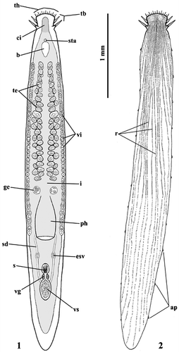
The rhabdoids, a variety of secretions in epidermal cells or in subepidermal glands, are present as true rhabdites (r), grouped into longitudinal lines along the body, with the exception of the anterior end, where they are randomly scattered (Figure ).
The brain (b) is ovoidal, 35–45 µm long (Figure ).
The testes (te), located anteriorly to the pharynx, consist of two rather close series of follicles along the longitudinal axis. They are numerous (30–35 per side), not in a single line, and of medium size (Figure ).
Two rows of small vitellaries (vi) are present laterally to the testes. They start from the same level as the testis follicles and reach the pharynx opening maintaining a regular distribution (Figure ).
Two germaries (ge), at some distance from each other, are present in front of the pharynx, posteriorly to the last testis follicles. They are globoid, larger than the testes and contain numerous egg cells (Figure ).
The pharynx (ph), situated in the second half of the body, shows the so‐called bell‐shaped organization, or “Glöckchen”, typical of the genus (Figure ).
The sacciform intestine (i) is a caecum at both ends, anteriorly possessing a tract, called cephalic intestine (ci) (Figure ).
In the postpharyngeal zone, the localizations of vesicula seminalis (vs), vesicula granulorum (vg) and external seminal vesicles (esv) are similar to those reported for the species already described in literature (Ax Citation1956; Figure ).
The caudal end appears sharpened and provided with numerous adhesive papillae (ap) (Figure ).
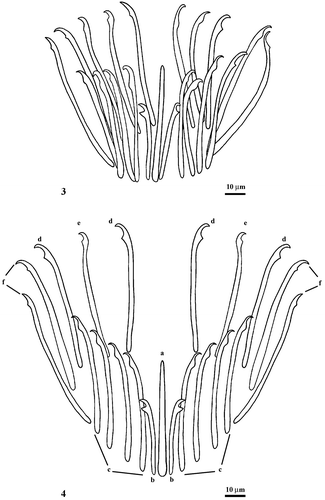
The male copulatory organ is characterized by a sclerotic apparatus (s) with 21 spines of different shape and length (Figures ):
a “Medianstachel” (a) or median aculeus (56 µm long), pointed at the distal end, is situated in the center of the sclerotic complex; | |||||
20 spines weakly curved with pointed tip bent outward:
| |||||
Figures 3–4. Otoplana oxyspina sp. nov.: spines of the male sclerotic apparatus (3) and their spatial distribution (4).
Conclusive remarks
Eighteen species have been historically classified in the genus Otoplana, but most belong to different genera or are incompletely described (Table ).
Table I. Otoplana species
With the description of the new species, O. oxyspina, the valid species of the taxon Otoplana now known are four altogether: O. intermedia Du Plessis, Citation1889 (Ax Citation1956) collected in the Ligurian and Tyrrhenian Seas, O. bosporana Ax, Citation1959 collected in the Bosphorus (Black Sea) and O. truncaspina Lanfranchi, Citation1969 sampled at Monte Rosso al Mare (Ligurian Sea).
The study of our species habitus evidenced a body length shorter than in O. intermedia (8 mm) and O. truncaspina (4.5–5 mm). On the contrary, O. bosporana has a body length comprised between 2.5 mm and 3 mm, and is thus shorter than our specimens. The body presents the maximum width at the germarium level located more anteriorly than in the other species.
The organization of the cephalic zone and the distribution of the rhabdites show great similarity with the other species; however, as with regard to the length, the thickness of the robust tactile bristles and the irregular distribution of the rhabdites on the most anterior region, evidence a marked affinity with O. truncaspina. The rhabdites, structures generally employed for defence or prey capture, are membrane‐bounded lamellate rhabdoids and show the typical distribution observed in O. truncaspina.
The yolk follicles path, in a single longitudinal row from the anterior end to the pharynx opening on both body sides, is a peculiarity of the present species, as is their number. On the contrary, their dimensions are rather similar to those observed in O. intermedia.
The position of the testes is shared with all the species of the genus, while their extension is less. The dimensions and irregular distribution of the testes in our species correspond to those of O. intermedia and O. truncaspina, but their number is lower. On the contrary, O. bosporana shows two regular series of aligned large follicles.
The paired germaries, situated in front of the pharynx, show localization and dimensions similar to those observed in O. truncaspina and O. intermedia. No information is available about O. bosporana.
The pharynx has the typical characteristics observed in all the genera of the subfamily, except for its more central position in the organism.
The sacciform intestine is similar to that of all the species of the genus.
The locations of the vesicula seminalis, vesicula granulorum and penis papilla appear more anterior than in the other species already described.
The spines of the male copulatory organ display a different organization compared to that of all the other species. They are constituted by a pointed “Medianstachel”, 56 µm long, encircled by 20 bristles, with variable shape and length (36–65 µm). The median aculeus (a) is different from that of O. truncaspina, which is truncated and on average 50 µm long. In O. intermedia, the “Medianstachel” is lacking, while in O. bosporana it is similar and longer (63 µm), but with a less wide proximal end. Worthy of note is the presence of two smaller bristles (b) 36 µm long, near the median aculeus, only found in O. truncaspina. These have a length of 28–36 µm and smaller cuneiform denticle. The bristles (c) present a sub‐terminal rounded prominence and a distal end larger than those observed in the spines (33–59 µm long) of O. bosporana. All the spines (d, e and f) have a hooked distal end similar to those of O. truncaspina, but the denticle or sub‐terminal cuneiform prominence is more developed than that of O. truncaspina and on the concave side. The total number of spines observed (21) in O. oxyspina is lower than that of O. bosporana (30–33), O. intermedia (24) and O. truncaspina (23).
On the basis of the data presented, we conclude that our species differs from O. intermedia, O. bosporana and O. truncaspina in its body dimensions, the lesser extent of the vitellaries, the scarcity of the testes and, above all, the characteristics of the sclerotic apparatus.
References
- An der Lan , H. 1964 . Zur Turbellarien‐Fauna der Donau. . Archiv Hydrobiologie , 27 (supplement) : 3 – 27 .
- Ax , P. 1951 . Die Turbellarien des Eulitorals der Kieler Bucht. . Zoologische Jahrbucher Abteilung für Systematik , 80 : 277 – 378 .
- Ax , P. 1956 . Monographie der Otoplanidae (Turbellaria): Morphologie und Systematik. . Akademie der Wissenschaften und der literatur Abhandlungen der Mathematisch‐Naturwissenschaftlichen klasse , 13 : 159 – 278 .
- Ax , P. 1959 . Zur Systematik, Ökologie und Tiergeographie der Turbellarienfauna in den Ponto‐kaspichen Brackwassermeeren. . Zoologische Jahrbücher Abteilung für Systematik, Ökologie und Geographie der Tiere , 87 : 71 – 89 .
- Ax , P. and Armonies , W. 1990 . Brackish water Plathelminthes from Alaska as evidence for the existence of boreal brackish water community with circumpolar distribution. . Microfauna Marina , 6 : 7 – 109 .
- Ax , P. and Ax , R. 1967 . Turbellaria Proseriata von der Pazifikküste der USA (Washington). . Zoologische Morphologie Tière , 61 : 215 – 254 .
- Ax , P. and Ax , R. 1974 . Interstitielle fauna von Galapagos V. Otoplanidae (Turbellaria, Proseriata). . Mikrofauna des Meeresbodens , 27 : 573 – 598 .
- Ax , P. and Sopott‐Ehlers , B. 1987 . Otoplanidae (Plathelminthes, Proseriata) von Bermuda. . Microfauna Marina , 3 : 261 – 281 .
- Ax , P. , Sopott‐Ehlers , B. and Weidemann , E. 1978 . Zur morphologie sublitoraler Otoplanidae (Turbellaria, Proseriata) von Helgoland und Neapel. . Zoomorphologie , 90 : 113 – 133 .
- Bresslau , E. 1928–33 . “ Turbellaria. ” . In Handbuch der Zoologie, Walter de Gruyter, Berlin, vol. II (I) Edited by: Kükenthal , W and Krumbach , T . 52 – 304 .
- Calandruccio , S. 1897 . Anatomia e sistematica di due specie nuove di Turbellaria. . Atti dell'Accademia Gioenia di Scienze Naturali di Catania Ser. 4 , 10 (16) : 1 – 18 .
- Du Plessis , G. 1889 . Note sur l'Otoplana intermedia. . Zoologische Anzeiger , 12 : 339 – 342 .
- Giard , M. A. 1904 . Sur une faunule caractéristique des sables à Diatomées d'Ambleteuse (Pas‐de‐Calalis) III. Les Gastrotriches aberrants. . Comptes Rendus de Sciences, Société de Biologie , 56 : 1063 – 1065 .
- Gieysztor , M. 1938 . Über einige Turbellarien aus dem Süβwasserpsammon. . Archiv Hydrobiologie und Ichtyologie , 11 : 364 – 382 .
- Graff von , L. 1913 . Turbellaria II. Rhabdocoelida. . Tierreich , 35 : 449 – 452 .
- Hallez , P. 1892 . Classification des Ticlades. . Bulletin de la Société Zoologique de France , 17 : 106 – 109 .
- Hallez , P. 1910 . Un nouveau type d'Alloiocoele (Bothriomolus constrictus n. g. n. sp.). . Archive de Zoologique Expérimental et Général , 3 : 611 – 664 .
- Karling , T. G. 1964 . Marine Turbellaria from the Pacific coast of North America (III) Otoplanidae. . Arkiv för Zoologi , 16 : 527 – 541 .
- Karling , T. G. 1973 . Anatomy and taxonomy of a new Otoplanid (Turbellaria, Proseriata) from south Georgia. . Mikrofauna des Meeresbodens , 16 : 362 – 269 .
- Lanfranchi , A. 1969 . Nuovi otoplanidi (Turbellaria, Proseriata) delle coste della Liguria e della Toscana. . Bollettino di Zoologia , 36 : 167 – 188 .
- Lanfranchi , A. 1978 . Morphology and taxonomy of two new otoplanids (Turbellaria, Proseriata) from the Ligurian Sea. . Zoologica Scripta , 7 : 249 – 254 .
- Luther , A. 1960 . Die Turbellarien Ostfennoskandiens. I. Acoela, Catenulida, Macrostomida, Lecithoepiteliata, Prolecithophora und Proseriata. . Fauna Fennica , 7 : 1 – 155 .
- Marcus , E. 1949 . Turbellaria Brasileiros (7). . Boletins de Faculdade de Filosofia, Ciências e Letras, Universidade de Sâo Paulo, Zoologia , 14 : 7 – 156 .
- Marcus , E. 1950 . Turbellaria Brasileiros (8). . Boletins de Faculdade de Filosofia, Ciências e Letras, Universidade de Sâo Paulo, Zoologia , 15 : 5 – 192 .
- Marcus , E. 1952 . Turbellaria Brasileiros (10). . Boletins de Faculdade de Filosofia, Ciências e Letras, Universidade de Sâo Paulo, Zoologia , 17 : 5 – 188 .
- Martens , P. M. and Schockaert , E. R. 1981 . Sand dwelling Turbellaria from the Netherlands Delta area. . Hydrobiologia , 84 : 113 – 127 .
- Meixner , J. 1938 . “ Turbellaria (Strudelwürmer) I. ” . In Die Tierwelt der Nord‐ und Ostsee, Teil IVb , Edited by: Grimpe , G and Wagler , E . 1 – 146 . Leipzig : Akademische Verlagsgesellschaft .
- Noreña , C. , Damborenea , C. and Brusa , F. 2005 . New freshwater interstitial Otoplanidae (Platyhelminthes: Proseriata) from the Paraná and Uruguay rivers, South America. . Journal of Natural History , 39 : 1457 – 1468 .
- Remane , A. 1933 . Verteilung und Organisation der benthonischen Mikrofauna der Kieler Bucht. . Wissenschaftlich Meeresuntersuchungen (N.F.), Abteilung Kiel , 21 : 161 – 221 .
- Riemann , F. 1965 . Turbellaria Proseriata mariner Herkunft aus Sanden der Flubsohle im liminischen Bereich der Elbe. . Zoologische Anzeiger , 174 : 299 – 312 .
- Schneider , A. 1873 . Untersuchungen über Plathelminthen. Separatabdruck aus d. 14. . Jahresbericht Oberhessen Gesellschaft für Natur‐ und Heilkunde Giessen , 14 : 1 – 78, Tab 3–7 .
- Sopott‐Ehlers , B. 1972 . Systematik und Ökologie von Proseriaten (Turbellaria) der deutschen Nordseekuste. . Mikrofauna des Meeresbodens , 13 : 221 – 229 .
- Sopott‐Ehlers , B. 1976 . Interstitielle Macrostomida und Proseriata (Turbellaria) von der französichen Atlantikkuste und den kanarischen Inseln. . Mikrofauna des Meeresbodens , 60 : 558 – 568 .
- Sopott‐Ehlers , B. 1985 . Zwei neue Proseriata (Platyhelminthes) von der französischen Atlantikküste. . Microfauna Marina , 2 : 380 – 396 .
- Sopott‐Ehlers , B. and Ehlers , U. 1980 . Zur Systematik und geographischen Verbreitung interstieller Turbellarien der kanarischen Inseln. . Mikrofauna des Meeresbodens , 80 : 585
- Steinböck , O. 1925 . Zur Systematik der Turbellaria metamerata, zugleich ein Beitrag zur Morphologie des Tricladen‐Nervensystems. . Zoologische Anzeiger , 64 : 165 – 192 .
- Steinböck , O. 1931 . Marine Turbellaria. . Zoology of the Faroe , 8 : 1 – 26 .
- Steinböck , O. 1932 . Die Turbellarien des arktischen Gebietes. . Fauna arctica , 6 : S295 – S342 .
- Tajika , K. I. 1983a . Zwei neue interstitielle Turbellarien der Gattung Archotoplana (Proseriata, Otoplanidae) aus Hokkaido, Japan. . Journal of the Faculty of Sciences Hokkaido University Series VI Zoology , 23 : 179 – 194 .
- Tajika , K. I. 1983b . Zwei neue Otoplaniden (Turbellaria, Proseriata) aus Hokkaido, Japan. . Annotationes zoologicae Japonenses , 56 : 100 – 110 .
- Tajika , K. I. 1983c . Zur Kenntnis Gattung Notocayoplana Steinböck, 1935 (Turbellaria, Proseriata, Otoplanidae). . Bulletin of the National Science Museum Series A (Zoology) , 9 : 97 – 104 .
- Tajika , K. I. 1984 . Eine neue Art der Gattung Itaspiella Ax, 1956 (Turbellaria, Proseriata, Otoplanidae) aus Hokkaido, Japan. . Bulletin of the Liberal Arts and Science Course Nimon University School of Medicine , 12 : 25 – 33 .