 ?Mathematical formulae have been encoded as MathML and are displayed in this HTML version using MathJax in order to improve their display. Uncheck the box to turn MathJax off. This feature requires Javascript. Click on a formula to zoom.
Abstract
The performance of a Collison nebulizer delivering viable H1N1 influenza aerosols was assessed in terms of particle size distribution (PSD) and survivability of the virus upon generation. An H1N1 influenza virus preparation in egg allantoic fluid was diluted in sterile deionized water to a concentration of 3.4 × 10⁶ TCID50/mL. The virus suspension was aerosolized at air flow rates of 2, 6 and 12 L/min using a 1-jet, 3-jet and 6-jet Collison nebulizer, respectively. A scanning mobility particle sizer measured the PSD of the viral aerosol after steady-state delivery times of 1, 15, 30, 45 and 60 min. After 60 min of continuous aerosolization, the viral titre was unchanged and the count median diameter (CMD) of the aerosol PSD was ∼38 nm for the 2 L/min flow rate, ∼35 nm for the 6 L/min flow rate and ∼33 nm for the 12 L/min flow rate. The CMDs were much smaller than the influenza virus (80–120 nm), indicating the aerosol distribution comprised mainly nonviable materials. The PSD produced by the Collison nebulizer exhibited a 20% increase in peak particle concentration after 60 min of continuous operation at 12 L/min. This progressive increase in particle counts may be attributed to a combination of evaporation and shear and impact stresses imparted on components by the Collison nebulizer. The possible slight loss in H1N1 influenza viability over the course of 60 min of continuous aerosolization at 12 L/min is consistent with previous bioaerosol studies using a Collison nebulizer.
Introduction
Bioaerosols are airborne biological agents, including viruses, bacteria, fungi, atmospheric environmental pollutants and pulmonary drug particles suspended in air or another gas.[Citation1] Their generation has numerous applications in pharmaceutical inhalers, in air filtration and sanitization, and in research on contamination, infection, toxicology and immunology. Recent concerns for improving inhaled drug delivery methodologies and indoor air quality, protecting hospital patients and staff from spreading infection, and installing counter-bioterrorism measures have fuelled interest in bioaerosol research. The capability of lab-generated bioaerosols to simulate organisms in a clinical environment is of utmost importance for the validity and applicability of research results. Therefore, it is desirable to use bioaerosol generation techniques that enable control over such significant aerosol parameters as suspension concentration, particle size and count, output stability of the aerosol and survivability of microorganisms.
The Collison nebulizer has dominated aerosol generation research since its invention in 1932 by W.E. Collison.[Citation2–10] The aerosolization process that occurs in a Collison nebulizer is well characterized.[Citation11] In principle, the Collison nebulizer is a pneumatic device for aerosol generation that uses a compressed air jet to atomize particle suspensions or solutions into droplets,[Citation12] of which the smallest are entrained in the air stream exiting the reservoir and the remainder are recycled. May [Citation2] has given a detailed description of the operation and performance of the Collison nebulizer and reported that most of these droplets are further broken down by impaction on the internal walls of the glass reservoir.
The ‘quality’ of the bioaerosol produced by a Collison nebulizer has recently come under scrutiny. Ulevicius et al. [Citation13] hypothesized that the liquid dispersed by the device's high-velocity air streams and solid wall impingement is subject to severe shear and impact forces. Furthermore, in his classical paper, May [Citation2] pointed out that within a Collison nebulizer containing 20 mL of liquid, most of the fluid is recirculated approximately every 6 s in the glass reservoir. Thus, 10 episodes of shear forces are experienced each minute by components in the liquid suspension from this repeated ‘recycling’. These large shear and impact stresses are suspected of causing cumulative metabolic injury to bacteria and viruses during extended periods of aerosolization from a Collison nebulizer.[Citation14–24] Whereas delivery from six-jet Collison nebulizers was shown to be consistent under a fixed set of conditions,[Citation25] delivery rates from Collison nebulizers are sensitive to operating conditions and can yield a range of droplet sizes, which vary over time, potentially rendering aerosol characterization and inter-experimental comparison difficult.[Citation2,Citation22,Citation23,Citation26] Moreover, foaming of propagation media during aerosolization from a Collison nebulizer may require additional sample preparation, such as dialysis or centrifugation. Hogan et al. [Citation27] proposed that the large electrical charge carried by bioaerosols emerging from a Collison nebulizer could also be associated with compromised structural integrity of the aerosolized particles originating from nebulization-induced mechanical stresses.
Tuttle et al. [Citation28] evaluated a nose-only inhalation exposure system for studies of aerosolized viable H5N1 viruses in ferrets. Tuttle et al.'s system comprised a bioaerosol nebulizing generator (BANG) manufactured by BGI, Waltham, MA, USA. In the BANG nebulizer, a high-velocity air stream creates a Venturi effect that siphons liquid through a tube from the nebulizer reservoir. As the liquid exits the tube at the top of the nebulizer, the air stream interacts with the liquid and shears it, creating an aerosol from which the larger particles settle and are recycled. This particular device was selected for Tuttle et al.'s [Citation28] experiments based on the claim that it minimizes potential damage to and clumping of the viral agent, and efficiently produces a uniform particle size distribution (PSD) with a lower rate of fluid use and smaller volume of virus suspension than similar bioaerosol generators. However, those claims were not substantiated by their data and operation of the BANG nebulizer appears to closely resemble that of the Collison.
Reports on the particle size and viability measurements of aerosolized viruses remain scarce in the literature, most likely due to their ultrafine size (25–400 nm), elaborate preparation and safe handling protocols. Nevertheless, airborne viruses (e.g., poxviruses, influenza, etc.) are of particular concern because of their ability to cause rapid infection via respiratory exposure. Hogan et al. [Citation27] investigated aerosolization and collection methodologies of MS2 and T3 bacteriophages. The authors argued that airborne virus PSDs were rarely available in the literature because samplers commonly used to collect virus particles were designed for the collection of micrometre-sized particles.[Citation29,Citation30] Hence, Hogan et al. [Citation27] developed a method to determine the size distribution function of viable virus-containing particles utilizing differential mobility selection. Most published viral aerosol research focused on biological particle penetration through respiratory filters challenged with MS2 bacteriophage.[Citation31–33] However, extrapolation of results obtained with MS2 to other viruses (e.g., H1N1) could be misleading due to the differences in size and other characteristics.[Citation34]
The ability to generate a viable, narrowly dispersed, pathogenic aerosol is essential in evaluating the efficiency of collection and control methods for various airborne microorganisms. In addition, it is important to diminish the formation of residue particles resulting from the fragmentation of the microorganisms by any stresses associated with the aerosolization process, because these residues can be enumerated only by the most size-sensitive particle size spectrometers.[Citation35–37] Furthermore, the implementation of H1N1 virus as a pathogen in the present investigation is of primary interest due to public health concerns about another potential H1N1 influenza pandemic.[Citation38] The ability to perform well-controlled studies of aerosolized viable H1N1 may contribute to an improved understanding of factors responsible for the acquisition of viral respiratory infections by humans and the virulence and lethality relevant to route of transmission.
In this investigation, a Collison nebulizer was employed as the aerosol-generating device due to its widespread use in the research laboratories and industry.[Citation2–10,Citation39,Citation40] The objectives of this study were (1) to investigate the possibility of generating an aerosol comprising a narrow PSD of highly viable H1N1 influenza particles, using the Collison nebulizer; (2) to assess the performance of the Collison nebulizer by measuring PSDs and the viability of the aerosolized H1N1 virus over time.
Materials and methods
H1N1 influenza
H1N1 influenza A/PR/8/34 VR-1469 (ATCC VR-95) virus was propagated in embryonic chicken eggs and titred using a tissue culture median infectious dose assay (TCID50), according to standard World Health Organization protocols.[Citation40] A virus stock was prepared demonstrating an infectivity level of approximately 109 TCID50/mL.
Aerosol system
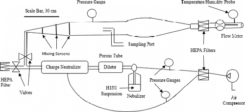
A laboratory-scale aerosol tunnel (LSAT, ) was used to conduct the aerosol experiments for this study.[Citation41] The LSAT consisted of a test chamber with a cylindrical duct having a diverging conical entrance containing three uniformly spaced mixing screens, and a converging conical outlet. The duct section expanded near its outlet and the wide section was flanked by identical sections; the upstream section was plumbed with an isokinetic sampling port. Compressed air was supplied to the LSAT via a compressor that was fitted with a pressure regulator. The compressed air passed through a HEPA (high-efficiency particulate arrestance) filter before being split to feed the nebulizer and the porous tube dilution unit. The air stream from the nebulizer then entered a 85Kr charge neutralizer (TSI, Inc., Shoreview, MN, USA) and merged with the air stream from the porous tube diluters. For this study, no dilution airflow was delivered into the porous tube diluters. Two overflow valves located downstream of the porous tube diluter and upstream of the aerosol tunnel diverted the airflow out of the LSAT through a HEPA filter. Two air pressure gauges (Dwyer, Michigan City, IN, USA) separately monitored the pressure of the air entering the nebulizer and in the aerosol tunnel.
The compressed air lines to the nebulizer and dilution unit were fitted with valves for airflow control. The exiting airflow was HEPA filtered and a digital flow metre (4000 series, TSI, Inc., Shoreview, MN, USA) measured the flow rate of air exiting the test chamber. An integrated hygrometer–thermocouple probe (VWR, San Diego, CA, USA) recorded the exhaust air temperature and humidity.
Aerosol studies
Frozen (−80 °C) H1N1 virus was thawed and diluted in sterile deionized water to a concentration of 3.4 × 10⁶ TCID50/mL. A 50 mL portion of the diluted virus suspension was pipetted into a Collison nebulizer (Model MRE CN24/25, BGI, Waltham, MA, USA). Three jet configurations of the Collison nebulizer were evaluated: (1) single-jet at 2 L/min, (2) 3-jet at 6 L/min, and (3) 6-jet at 12 L/min. The LSAT was configured to divert the aerosol through a HEPA filter while operation of the Collison nebulizer stabilized. Filtered compressed air (20 psi = 138 kPa) was applied to the nebulizer and the system was operated for 10 min initially to bring it to steady state. The LSAT overflow valves were then reconfigured to direct the aerosol to the aerosol tunnel for 10 min. For the 12 L/min flow rate only, aerosol samples were collected from the isokinetic sampling ports into all-glass impingers (AGI-30, Ace Glass Inc., Vineland, NJ, USA) for 5 min each at 12.5 L/min at time points of 1, 15, 30, 45 and 60 min after reaching steady state. Each impinger contained 20 mL of serum-free Eagle's minimum essential medium (sf-EMEM, HyClone™, Thermo Fisher Scientific Inc., Waltham, MA, USA) for viable sampling. Following each impinger sample, a scanning mobility particle sizer (SMPS 3034, TSI, Inc., Shoreview, MN, USA) was used to determine the PSD in the aerosol tunnel. The SMPS measured particles in the size range of 10–487 nm and the concentration range of 102–107 particles/cm3 in up to 54 channels at an inlet flow rate of 0.6 L/min. Impinger samples were serially diluted 1/10 in sf-EMEM, plated according to standard protocol,[Citation42] and incubated for 96 h at 35 °C and 5% CO2. Viability was determined by microscopic observation and crystal violet–glutaraldehyde staining. All experiments were performed in triplicate in a class II biological safety cabinet.
Data analysis
The concentration of viable virus (log10 TCID50/mL) collected in the impingers was determined using the Spearman–Kärber formula.[Citation43] EquationEquation (1)(1)
(1) was used to determine the total amount of virus recovered from each sample (20 mL impinger volume),
(1)
(1) where Cs is the total virus concentration of the sample; C is the viable H1N1 counts expressed in units of log10 TCID50 per milliliter; V is the impinger sample volume, here 20 mL. Equation (Equation2
(2)
(2) ) computed the concentration of viable virus per volume of air,
(2)
(2) where CL is the virus concentration per volume of air expressed in units of TCID50/Lair; Qs is the sampling flow rate (12.5 L/min); and t is the sample collection time (5 min).
Standard statistical analysis methods including calculations of sample mean, standard deviation and analysis of variance (ANOVA) were performed using Microsoft Excel software.
Results and discussion
Particle size distribution (PSD)
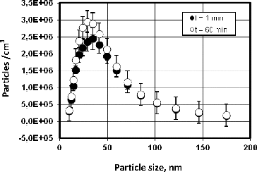
depicts the PSD of the 12 L/min aerosol at 1 and 60 min after aerosol stabilization, for which 10 min was allowed. Therefore, the nebulization times in correspond to approximately 11 and 70 min, respectively. This difference is important because earlier research showed that substantial damage to microorganisms can occur during the first five minutes of nebulization.[Citation10] shows that the peak particle concentration of the PSD occurs at ∼33 nm for the 12 L/min air flow rate after a 60 min aerosolization period. As a single influenza virus measures 80–120 nm in size, any smaller particles are interpreted as fragments. It is clear that the PSD of these samples is dominated by the size of residues from suspended virus fragments, other dissolved and suspended materials and soluble materials, obscuring the virus. indicates that the peak particle concentration produced by the Collison nebulizer increased by about 20% after 60 min of continuous aerosolization. Two factors appear to contribute to this observation: (1) the Collison nebulizer loses volume over time as water evaporates from the liquid, thereby increasing the concentration of the solutes in the suspension; (2) the suspension in the reservoir is constantly recycled through the nozzle(s), subjecting the viruses and fragments to shear and impact stresses inside the Collison nebulizer, thereby producing smaller fragments and increasing the total particle concentration.
Figure 2. SMPS data for H1N1 influenza aerosol generated with the Collison nebulizer at an air flow rate of 12 L/min.
Mainelis et al. [Citation23] showed that the fragment concentration emitted from the Collison nebulizer increased 3–3.5 times after 90 min of continuous bacterial aerosol generation using the Gram-negative rod-shaped bacterial species, Pseudomonas fluorescens. Comparing Mainelis et al.'s [Citation23] work to this work, it is evident that the greater increase in the ratio of fragments to original microbial agent size in Mainelis et al.'s [Citation23] experiments may be attributed to greater momentum of the larger size (∼1 µm) bacterium (compared to H1N1 virus) and the longer aerosolization period (90 vs. 60 min). The larger size would fragment due to an increase in impact momentum and extended aerosolization time increases the number of impacts. The H1N1 virus appears to retain greater viability than the P. fluorescens bacterium in the Collision nebulizer, which is attributed to the same factors that produce a lower level of fragmentation of the virus. Both Gram-negative bacteria and lipid viruses are known to lack environmental stability in general, but it is not clear how their environmental stability relates to shear degradation found in the Collison nebulizer.[Citation44]
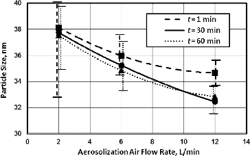
shows the count median diameter (CMD) of the particle as a function of aerosolization air flow rates for the single-jet (2 L/min), 3-jet (6 L/min) and 6-jet (12 L/min) Collison nebulizers for nebulization times of 1, 30 and 60 min. Due to the large standard deviation at the 2 L/min flow rate data points, a one-way ANOVA using all 3 flow rates between the 2 and 12 L/min flow rates was performed and confirmed that the results in are statistically significant. It is observed that the particle size decreased as the air flow increased and/or nebulization time increased. These results are consistent with virus fragmentation stemming from shear and impact stresses, as discussed for the data of .
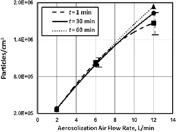
Figure 3. Variation in virus particle size with aerosolization air flow rate and time for the Collison nebulizer. Note: The data markers represent averages of three replicate tests. The trend lines were drawn by fitting the data to second-order polynomials, with R2 = 0.99.
plots the aerosol particle concentration versus airflow rate for the three aerosolization time points. It is clear that the concentration of aerosol particles increases with time, which may be expected because, as noted above, the concentration of particles in suspension inside the Collison nebulizer increases with time due to fragmentation and to progressive evaporation as the liquid continues to recycle through the nozzle. This trend is more pronounced at larger aerosolization flow rates because a higher air flow rate proportionally increases the aerosol output and decreases the time between repeated passages through a nozzle in proportion to the number of jets fed from the reservoir. This result is consistent with the work of Mainelis et al. [Citation23], which used polystyrene latex spherical particles of different sizes to evaluate the particle output from a Collison nebulizer at variable air flow rates.
Virus viability
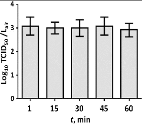
exhibits H1N1 virus viability data for the 12 L/min flow rate collected at intervals of 1, 15, 30, 45 and 60 minutes after reaching steady-state conditions. shows that viability of the virus does not decrease significantly (p = 0.96) with nebulization time. Chilling of the reservoir implies some evaporative concentration, but the concurrent shift of the mode in the PSD to smaller particles suggests that this loss is relatively minor.
Whereas Mainelis et al. [Citation23] reported 50% loss of viability by P. fluorescens over a period of 90 min of delivery from a Collison nebulizer, Kim et al. [Citation45] reported no loss of viability by a suspension of an enveloped coronavirus (80–160 nm) in a Collison during nebulization periods of 10–30 min. Kim et al. [Citation45] suggested that viruses are so small that they have little inertia and do not experience much physical stress due to acceleration, deceleration and/or impaction during nebulization; however, their data at 30 min show a 15% decrease in titre, consistent with the suggestion above that a small amount of loss may actually occur. Similarly, Hermann et al. [Citation46] reported no change (p = 0.89) in titre of enveloped porcine reproductive and respiratory syndrome virus (40–80 nm) during 55 min of nebulization in a 24-jet Collison unit. Given the differences in dimensions and mechanical sensitivity of the diverse microbes studied, the present results with an actual pathogen are consistent with the observations of Kim et al. [Citation45] and Hermann et al. [Citation46] and support the interpretation that viruses are mechanically more robust than bacteria to atomization.
Conclusions
This study assessed the performance of a Collison nebulizer generating a viable H1N1 influenza aerosol. The CMD of the aerosolized virus particles was approximately 33 nm (at an air flow rate of 12 L/min), a value much smaller than the diameter of a single H1N1 virus (80–120 nm). The reduced CMD size was attributed to residues from the allantoic fluid growth medium, rather than the fragmentation of viruses because viability did not decrease significantly during 60 min of nebulization, suggesting a minimal loss of virus particles. The concentration of 34 nm sized particles increased by 20% during a 60 min period of continuous aerosolization at an air flow rate of 12 L/min, and the CMD likewise decreased. Occurrence of these trends is attributed to a combination of evaporation and progressive fragmentation of medium components, possibly including a small amount of the virus, with residence time in the nebulization process. The titre of H1N1 virus in cultures of fluid in the reservoir remained constant over a 60 min period, suggesting a minimal loss of virus particles. The results of the research described in this paper are consistent with the reported literature on Collison nebulizer experiments using enveloped viruses and support the hypothesis that the greater size of P. fluorescens is a factor in its extensive fragmentation reported under similar aerosolization conditions. In terms of virology, there are many other viruses that could be studied in the range of 80–120 nm and in the same conditions of aerosol, concentration or rate of fragmentation as this work.
Disclosure statement
No potential conflict of interest was reported by the authors.
Additional information
Funding
References
- Hinds WC. Aerosol technology. New York, NY: Wiley; 1999.
- May KR. The Collison nebulizer: description, performance, and application. J Aerosol Sci. 1973;4:235–243.
- Jensen PA, Todd WF, Davis GN, et al. Evaluation of eight bioaerosol samplers challenged with aerosols of free bacteria. Am Ind Hyg Assoc J. 1992;53:660–667.
- Chen SK, Vesley D, Brosseau LM, et al. Evaluation of single-use masks and respirators for protection of health care workers against microbacterial aerosols. Am J Infect Control. 1994;22:65–74.
- Forney TL, Bell EC, Bowdle DA. Evaluation of Erwinia herbicola as a surrogate biological warfare agent (BW) aerosol. Columbus, OH: Battelle Memorial Institute; 1997.
- Heidelberg JF, Shahamat M, Levin M, et al. Effect of aerosolization on culturability and viability of gram-negative bacteria. Appl Environ Microbiol. 1997;63:3585–3588.
- Mainelis G, Willeke K, Baron P, et al. Electrical charges on airborne microorganisms. J Aerosol Sci. 2001;32:1087–1110.
- Mainelis G, Gorny RL, Reponen T, et al. Effect of electrical charges and fields on injury and viability of airborne bacteria. Biotech Bioeng J. 2002;79:229–241.
- Agranovski IE, Agranovski V, Reponen T, et al. Development and evaluation of a new personal sampler for culturable airborne microorganisms. Atmos Environ. 2002;36:889–898.
- Stone RC, Johnson DL. A note on the effect of nebulization time and pressure on the culturability of Bacillus subtilis and Pseudomonas fluorescens. Aerosol Sci Technol. 2002;36:536–539.
- BGI Inc. Collison nebulizer – instructions [Internet]. Waltham. MA; 2002. Available from: http://bgi.mesalabs.com/wp-content/uploads/sites/35/2014/10/Collison.pdf
- John W. The characteristics of environmental and laboratory-generated aerosols. In: Willeke K, Baron PA, editors. Aerosol measurement: principles, techniques and applications. New York, NY: Van Nostrand Reinhold; 1993. p. 54–76.
- Ulevicius V, Willeke K, Grinshpun SA, et al. Aerosolization of particles from a bubbling liquid: characteristics and generator development. Aerosol Sci Technol. 1997;26:175–190.
- Cox CS. The aerosol survival and cause of death of Escherichia coli K12. J Gen Microbiol. 1968;54:169–175.
- Cox CS. The cause of loss of viability of airborne Escherichia coli K12. J Gen Microbiol. 1969;7:77–80.
- Israeli E. Effect of aerosolization and lyophilization on macromolecular synthesis in E. coli. In Sixth International Symposium on Aerobiology; 1972 September 3–7; Technical University at Enschede, The Netherlands. New York, NY: Wiley; 1973.
- Israeli E, Gitelman J, Lighhart B. Death mechanisms in microbial bioaerosols with special reference to freeze-dried analog. In: Lighthart B, Mohr AJ, editors. Atmospheric microbial aerosols, theory and applications. New York, NY: Chapman and Hall; 1994. p. 166–191.
- Marthi B, Fieland VP, Walter M, et al. Survival of bacteria during aerosolization. Appl Environ Microbiol. 1990;56:3463–3467.
- Griffiths WD, Decosemo GAL. The assessment of bioaerosols: a critical review. J Aerosol Sci. 1994;25:1425–1458.
- Griffiths WD, Stewart IW, Reading AR, et al. Effect of aerosolization, growth phase and residence time in spray and collection fluids on the culturability of cells and spores. J Aerosol Sci. 1996;27:803–820.
- Stewart SL, Grinshpun SA, Willeke K, et al. Effect of impact stress on microbial recovery on an agar surface. Appl Environ Microbiol 1995;61:1232–1239.
- Reponen T, Willeke K, Ulevicius V, et al. Techniques for dispersion of microorganisms into air. Aerosol Sci Technol. 1997;27:405–421.
- Mainelis G, Berry D, An HR, et al. Design and performance of a single-pass bubbling bioaerosol generator. Atmos Environ. 2005;39:3521–3533.
- Rule AM, Schwab KJ, Kesavan J, et al. Assessment of bioaerosol generation and sampling efficiency based on Pantoea agglomerans. Aerosol Sci Technol. 2009;43:620–628.
- First MW, Macher J, Gussman R, et al. Nebulizer characteristics for certification tests of biosafety cabinets with bacteria and simulants. J Am Biol Safety Assoc. 1998;3:26–29.
- Zarrin F, Kaufman SL, Socha JR. Droplet size measurements of various nebulizers using differential electrical mobility particle sizer. J Aerosol Sci. 1991;22(S1):S343–S346.
- Hogan CJ, Kettleson EM, Lee MH, et al. Sampling methodologies and dosage assessment techniques for submicrometer and ultrafine virus particles. J Appl Microbiol. 2005;99:1422–1434.
- Tuttle RS, Sosna WA, Daiels DE, et al. Design, assembly, and validation of a nose-only inhalation exposure system for studies of aerosolized viable influenza H5N1 virus in ferrets. Virol J. 2010;7:135.
- Grinshpun SA, Willeke K, Ulevicius V, et al. Effect of impaction, bounce, and re-aerosolization on the collection efficiency of impingers. Aerosol Sci Technol. 1997;26:326–342.
- Willeke K, Lin XJ, Grinshpun SA. Improved aerosol collection by combined impaction and centrifugal motion. Aerosol Sci Technol. 1998;28:439–456.
- Balazy A, Toivola M, Adhikari A, et al. Do N95 respirators provide 95% protection level against airborne viruses, and how adequate are surgical masks? Am J Infect Control. 2006;34:51–57.
- Eninger RM, Adhikari A, Reponen T, et al. Differentiating between physical and viable penetrations when challenging respirator filters with bioaerosols. Clean – Soil, Air, Water. 2008;36:615–621.
- Eninger R, Honda T, Adhikari A, et al. Filter performance of N99 and N95 facepiece respirators against viruses and ultrafine particles. Ann Occup Hyg. 2008;52:385–396.
- Turgeon N, Toulouse MJ, Matel B, et al. Comparison of five bacteriophages as models for viral aerosol studies. Appl Environ Microbiol. 2014;80:4242–4250.
- Verreault D, Moineau S, Duchaine C. Methods for sampling of airborne viruses. Microbiol Mol Biol Rev. 2008;72:413–444.
- Qian Y, Willeke K, Ulevicius V, et al. Dynamic size spectrometry of airborne microorganisms: laboratory evaluation and calibration. Atmos Environ. 1995;29:1123–1129.
- Tang JW. The effect of environmental parameters on the survival of airborne agents. J R Soc Interface. 2009;6:S737–S746.
- Dawood FS, Jain S, Finelli L, et al. Emergence of a novel swine-origin Influenza A (H1N1) virus in humans. New Eng J Med. 2009;360:2605–2615.
- Kim SY, Kim M, Lee S, et al. Survival of microorganisms on antimicrobial filters and the removal efficiency of bioaerosols in an environmental chamber. Microbiol Biotech J. 2012;22:1288–1295.
- WHO manual on animal influenza diagnosis and surveillance [Internet]. Geneva, Switzerland: World Health Organization; 2002. Available from: http://www.who.int/vaccine_research/diseases/influenza/WHOmanual_on_animal-diagnosis_and_surveillance_2002_5.pdf.
- Heimbuch BK, Wallace WH, Kinney K, et al. A pandemic influenza preparedness study: use of energetic methods to decontaminate filtering facepiece respirators contaminated with H1N1 aerosols and droplets. Am J Infect Control. 2011;39:e1–e9.
- Allaire A, Luong MX, Smith KP. Basics of cell culture. In: Stein GS, Borowski M, Luong MX, Shi MJ, Smith KP, Vazquez P, editors. Human stem cell technology and biology: a research guide and laboratory manual. New York, NY: Wiley; 2011. p. 19–32.
- Finney DJ. Statistical methods in biological assays. 2nd ed. New York, NY: Hafner; 1964.
- Kramer A, Schwebke I, Kampfl G. How long do nosocomial pathogens persist on intimate surfaces? BMC Infect Dis. 2006;6:130.
- Kim S, Ramakrishnan M, Raynor P, et al. Effects of humidity and other factors on the generation and sampling of a coronavirus aerosol. Aerobiologia. 2007;23:239–248.
- Hermann JR, Hoff SJ, Yoon, KJ, et al. Optimization of a sampling system for recovery and detection of airborne porcine reproductive and respiratory syndrome virus and swine influenza virus. Appl Environ Microbiol. 2006;72:4811–4818.

