
ABSTRACT
The aim of this retrospective study was to review, analyse and characterize the root canal morphology of maxillary molars, using cone beam computed tomography (CBCT), in a group of the Turkish Cypriot population. The sample for this cross-sectional study consisted of retrospective evaluation of CBCT scans of 290 adult patients (age range 16–80). The number of roots and their morphology, the number of canals per tooth and the root canal configurations were also classified according to the method of Vertucci. Pearson's chi-square test was performed for canal configurations, sides and gender (p < 0.05). Among the 373 first molars, there was no single-rooted specimen, 2 (0.53%) teeth had 2 roots, 365 (97.8%) teeth had 3 roots and 6 ones (1.6%) had 4 roots. Among the 438 second molars, 14 (3.1%) were single-rooted, 26 (5.9%) teeth had 2 roots, 392 (89.4%) teeth had 3 roots and 6 teeth (1.3%) had 4 roots. No sex difference was found in the frequency of additional canals both in the maxillary first and second molars. Occurrence of additional canals did not differ with age. These results provide detailed knowledge of the root canal anatomy of the maxillary molar teeth in this particular population, which is of clinical importance for dental professionals when performing endodontic treatment.
Introduction
The most important stages of the root canal therapy are thorough shaping and cleaning of all pulp spaces and its complete obturation with an inert filling material. Therefore, clinicians should be aware of common root canal configurations and possible anatomic variation.[Citation1] A thorough understanding of the complexity of the root canal system is essential for understanding the principles and problems of shaping and cleaning, for determining the apical limits and dimensions of canal preparations.[Citation2] The inability to detect debris and obturate all extant canals is a major cause of endodontic failure.[Citation3]
A number of techniques, such as sectioning, radiography, dye penetration and clearing, post-treatment clinical examination, to cone beam computed tomography (CBCT), have been applied in studies on the root and canal morphologies of different populations.[Citation2] Although tooth clearing methods and modification of these techniques have been generally considered the gold standard for analysing root canal anatomy, many potential endodontic applications, including analysis of the canal morphology have been reviewed with the introduction of CBCT in the field of endodontics in 1990.[Citation1,Citation4]
Baratto Filho et al. [Citation5] used three methods (ex vivo, clinical and CBCT) to assess the internal morphology of maxillary first molars and concluded that CBCT was effective for initial identification of such morphology. CBCT can also provide personal data, such as sex, age and tooth position, which may have important implications in the preoperative evaluation of canal morphology for non-surgical root canal therapy.[Citation3,Citation6,Citation7]
Many attempts have been made to assess the anatomic characteristics of maxillary molars because of their complex root anatomy and canal morphology.[Citation3,Citation8] Although based on the published results, it is generally accepted that most maxillary molars have three roots, these teeth also exhibit some anatomic variation.[Citation3,Citation8–12] Kottoor et al. [Citation13] reported a case in which the maxillary first molar exhibited three roots and seven canals: three in the mesiobuccal root (MBR), two in the distobuccal root (DBR) and two in the palatal root (PR).
To the best of our knowledge, there are no reports on the root canal morphology of maxillary molars in the Turkish Cypriot population. Thus, the aim of this retrospective study was to analyse and characterize the root canal morphology of the maxillary molars in this population together with the prevalence of additional canals, using CBCT.
Subjects and methods
The cohort for this cross-sectional study consisted of 290 adult patients (age range 16–88) seeking routine dental care at the Near East University Dental Hospital. All subjects agreed to participate in this study and gave their written informed consent. The study was approved by the ethics committee of the School of Medicine at Near East University. Digitized CBCT images of maxillary molars were collected from patients who had undergone CBCT scanning for diagnostic purposes in the period January 2012–January 2015. Maxillary molars with immature apices, apical periodontitis, root canal fillings, post and crown restorations were excluded. Cases where the anatomy was compromised by physiological or pathological processes and unclear root canal morphology were also excluded from the study.
CBCT scans (Newton 3G: Quantitative Radiology s.r.l., Verona, Italy) used a 9-inch field of view to include maxillary anatomy. All CBCT exposures were performed by an experienced licensed radiologist by applying the minimum exposure necessary for adequate image quality. The as low as reasonably achievable protocol was strictly followed. Axial, coronal and cross section images were used for evaluation of root canal anatomy. All constructions and measurements were performed on a 21.3-inch flat-panel colour-active matrix thin-film-transistor medical display (NEC MultiSync MD215MG, Munich, Germany) with a resolution of 2048 × 2560 pxat 75 Hz and 0.17-mm dot pitch operated at 11.9 bits.
All CBCT images were evaluated retrospectively by two endodontists and an oral and maxillofacial radiologist with at least 10 years' experience using the CBCT device and software (NNT 4.6, QR Verona, Italy). An inter-examiner calibration based on the anatomic diagnosis of CBCT images had been previously performed to assess data reliability.
CBCT images were evaluated and the following were observed: (1) the number of roots and canals; (2) the canal configuration in each root using Vertucci's classification [Citation2]; (3) the frequency of additional roots and the frequency of C-shaped canals in the maxillary first and second molars ().
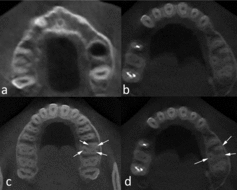
Figure 1. (a) Axial CBCT image showing one root, (b) three canals of the first molar, (c) four canals (arrows) and (d) C-shaped canal of the second molar (arrows).
The observers evaluated the images twice with a one-week interval between the assessments. The intra- and inter-examiner reliability was assessed. Wilcoxon matched-pairs signed rank test was used for intra-observer assessment, while the inter-observer reliability was made by the intra-class correlation coefficient (ICC) and the coefficient of variation (CV). Values for the ICC range were 0–1. ICC values higher than 0.75 were considered to show good reliability and low CV demonstrates the precision error as an indicator for reproducibility.[Citation14] The relationship between the patients' sex or the sides and the incidence of additional canals was determined using the chi-square test. Differences were considered significant when p < 0.05.
Results and discussion
The aim of this retrospective study was to analyse and characterize the root canal morphology together with the prevalence of additional canals in the Turkish Cypriot population, using CBCT. Even though various techniques have been used in root morphology studies, it has been mentioned that the most detailed information can be obtained by demineralization and staining techniques.[Citation2,Citation6,Citation15,Citation16] In recent years, CBCT is widely used in implantology, maxillofacial reconstruction and in endodontic diagnosis before surgical endodontics as well as for assessment of canal preparation, obturation and removal of root fillings. It has been reported that CBCT is as accurate in identifying root canal morphology as the modified canal staining and clearing technique.[Citation11,Citation17]
CBCT findings
In this study, of the 373 first molars, none was single rooted. Two (0.53%) teeth had 2 roots, 365 (97.8%) teeth had 3 roots and 6 teeth (1.6%) had 4 roots. In total (i.e. as a sum in female and male patients), the number of canals per tooth in the maxillary first molars was 2 in 0.5% of the studied teeth, 3in 48.2%, 4 in 50.6% and 5 in 0.5%. Of the 438 second molars, there were 14 (3.1%) single-rooted ones. Twenty-six (5.9%) teeth had 2 roots, 392 (89.4%) teeth had 3 roots and 6 teeth (1.3%) had 4 roots. The number of canals per tooth in the maxillary second molars in total (i.e. as a sum in female and male patients) was 1 in 1.3% of the studied teeth, 2 in 7.7%, 3 in 67.8% and 4 in 22.8%. The frequency distribution of the number of root canals did not appear to differ significantly between females and males (p > 0.05) ().
Table 1. Classification of first and second molars according to the number of roots and the number of canals per tooth.
Among the first molars, in the examined female patients, there were 104 (52.7%) MBRs with a Vertucci Type I root canal anatomy that had 3 and 4 separate roots, whereas there were 81 ones (46.5%) in males (). For both male and female participants, 78 MBRs of teeth had a Type II canal anatomy. Fourteen (7.1%) had a Type IV canal anatomy in females while 12 (6.8%) in males. Only one (0.5%) of the distobuccal roots had Type II canal anatomy in the group of male patients. All the palatal roots (371; 100%) and extra roots (6; 100%) had Type I root canal anatomy. In the group with 2 roots, 100% had a Vertucci Type I anatomy for the buccal and palatal roots. In the group with four roots, all roots had Type I canal anatomy except for one second MBR, which had Type II canal anatomy. Out of 373 first molars in total, 186 ones (49.8%) had an additional mesiobuccal canal.
Table 2. Vertucci classification of first and second molars that had three and four separate roots, depending on their root canal anatomy.
In the second molar group (), 174 (79%) of the MBRs in females and 129 (72.4%) in males were observed to have a Vertucci Type I root canal anatomy. Among the female patients, 42 teeth (19%) had type II canal anatomy, and among the males, this number was 41 (23%). Type IV canal anatomy was found in 4 teeth (1.8%) in the female patients and in 7 ones (3.9%) in the male patients. Only one case of a MBR (0.5%) was found with Type VIII canal anatomy. Of all the second molars, 95 ones (21.6%) had an additional mesiobuccal canal (). Only a single canal (0.22%) was found to be C-shaped in all maxillary second molars. No difference between the two sexes was found in the frequency of additional canals both in the maxillary first and second molars (p > 0.05).
Comparative analysis
Most CBCT studies use the classification system proposed by Vertucci,[Citation2] although additional classification systems have been used as well ().[Citation6,Citation8,Citation18,Citation19] The result obtained in this study that 2.2% of the maxillary first molars do not have three separate roots is consistent with previous findings in Indian, Chinese, Brazilian and Irish populations.[Citation3,Citation8,Citation9,Citation11,Citation20] However, in earlier studies in Thai, Kuwaiti and Burmese populations, there were found three separate roots in all maxillary first molars.[Citation12,Citation18,Citation21] These differences in root canal anatomy highlight the influence of ethnic background on maxillary molar root morphology.[Citation3]
Table 3. Review of some reports on the maxillary root canal anatomy in different populations*.
Our results were similar to those in several other previous investigations [Citation3,Citation9,Citation11,Citation22] in that maxillary first molars had three roots and four canals. Moreover, similar results were found for the frequencies of additional canals.[Citation3,Citation6,Citation12,Citation23] The high frequency (49.8%) of additional MBR canals in this study is largely consistent with findings from 2 CBCT studies of Chinese populations (52.24% and 52%),[Citation3,Citation9] from India (48.2%) [Citation11] and from Iran (53.6%).[Citation24] However, higher frequencies than those in our study have been observed in the Turkish population (93.5%),[Citation6] Irish population (80.4%),[Citation20] Italian (80%) [Citation25] and Korean population (71.7%).[Citation26] This variation may be caused by differences in the sample sizes, the methods, and/or the regional population diversity. The higher percentages reported by other studies when compared with our findings might be explained by the difference in the CBCT resolution, the radiographic interpretation and the sample size. Ex vivo studies on the incidence of extra canals reveal higher detection than in vivo studies.[Citation8,Citation18,Citation20] Moreover, studies using an operating microscope, clearing technique or sectioning methodology show higher detection rates than radiographic or CBCT examinations.[Citation5,Citation18,Citation20,Citation27,Citation28]
Variations in additional canals in the DBR and PR of the first maxillary molar have been less frequently observed. Our observation of additional canals in only 0.5% (1 tooth) of DBR are in good agreement with previous reports [Citation3,Citation8,Citation12,Citation20,Citation29] that showed little variations in these roots.
Similar to the findings in previous studies in the Brazilian, Indian and Irish population,[Citation8,Citation11,Citation20], the results from our study revealed that the percentage of maxillary second molars with three separate roots (89.4%) was lower than that for maxillary first molars (97.8%). Our results that the most commonly observed root morphology in the maxillary second molars was three separate roots with a single canal for each root (69.1%) followed by three separate roots with two canals in the MB root (21.6%), are in accordance with the studies of other authors showing high incidence of three separate roots with one canal in each root.[Citation9,Citation12,Citation18,Citation29]
However, maxillary second molars present a more complex root canal system when compared to maxillary first molars: the incidence of a single root in the second molars was 3.1%, 26 teeth had 2 roots (5.9%) and 6 teeth (1.3%) had 4 roots. The results of several earlier studies indicate that the prevalence of single root, two roots and four roots for maxillary second molars shows dissimilarities,[Citation9,Citation12,Citation18,Citation29] while our results suggested similar incidences to those reported for Chinese,[Citation9] Ugandan [Citation29] and Thai [Citation12] populations, where all the maxillary second molars had three separate roots.
Regarding the incidence of C-shaped canals, while our study revealed none in the maxillary first molars, only one C-shaped canal was found (0.22%) among 438 maxillary second molars. These results are in consistence with previous studies that showed low incidences for C-shaped canals in maxillary molars.[Citation1,Citation8,Citation30]
Sex did not affect the incidence of additional canals, which is in agreement with earlier studies.[Citation1,Citation3] However, there have been conflicting results with regard to sex and the frequency of additional canals.[Citation3,Citation6,Citation7,Citation31,Citation32] While earlier studies indicated that canal morphology appears to become simpler because of the calcification of root canal ramifications,[Citation1,Citation3] this issue should be evaluated with larger population groups.
Limitations
Our study, however, has some limitations. First, a larger cohort of patients would be needed to obtain more reliable information about the possible ethnic specifics in the morphology of maxillary molar root canals in the Turkish Cypriot population. Second, more reliable/higher resolution techniques could probably give more detailed information.
Conclusions
To the best of our knowledge, this is the first population-based Turkish Cypriot study that can serve as a guide to the morphology of root canals of molar teeth in this ethnic group. Within the limitations of this study, the obtained data can be compared to those of other populations and could facilitate diagnosis and treatment planning in Turkish Cypriot adults.
Disclosure statement
No potential conflict of interest was reported by the authors..
References
- Kim Y, Lee SJ, Woo J. Morphology of maxillary first and second molars analyzed by cone-beam computed tomography in a Korean population: variations in the number of roots and canals and the incidence of fusion. J Endod. 2012;38:1063–1068.
- Vertucci FJ. Root canal morphology and its relationship to endodontic procedures. Endod Topics. 2005;10:3–29.
- Zheng QH, Wang Y, Zhou XD, et al. A cone-beam computed tomography study of maxillary first permanent molar root and canal morphology in a Chinese population. J Endod. 2010;36:1480–1484.
- Tachibana H, Matsumoto K. Applicability of X-ray computerized tomography in endodontics. Endod Dent Traumatol. 1990;6:16–20.
- Baratto Filho F, Zaitter S, Haragushiku GA, et al. Analysis of the internal anatomy of maxillary first molars by using different methods. J Endod. 2009;35:337–342.
- Sert S, Bayirli GS. Evaluation of the root canal configurations of the mandibular and maxillary permanent teeth by gender in the Turkish population. J Endod. 2004;30:391–398.
- Fogel HM, Peikoff MD, Christie WH. Canal configuration in the mesiobuccal root of the maxillary first molar: a clinical study. J Endod. 1994;20:135–137.
- Silva EJ, Nejaim Y, Silva AI, et al. Evaluation of root canal configuration of maxillary molars in a Brazilian population using cone-beam computed tomographic imaging: an in vivo study. J Endod. 2014;40:173–176.
- Zhang R, Yang H, Yu X, et al. Use of CBCT to identify the morphology of maxillary permanent molar teeth in a Chinese subpopulation. Int Endod J. 2011;44:162–169.
- Weine FS, Healey HJ, Gerstein H, et al. Canal configuration in the mesiobuccal root of the maxillary first molar and its endodontic significance. 1969. J Endod. 2012;38:1305–1308.
- Neelakantan P, Subbarao C, Ahuja R, et al. Cone-beam computed tomography study of root and canal morphology of maxillary first and second molars in an Indian population. J Endod. 2010;36:1622–1627.
- Alavi AM, Opasanon A, Ng YL, et al. Root and canal morphology of Thai maxillary molars. Int Endod J. 2002;35:478–485.
- Kottoor J, Velmurugan N, Sudha R, et al. Maxillary first molar with seven root canals diagnosed with cone-beam computed tomography scanning: a case report. J Endod. 2010;36:915–921.
- Chang PC, Liang K, Lim JC, et al. A comparison of the thresholding strategies of micro-CT for periodontal bone loss: a pilot study. Dentomaxillofac Radiol [Internet]. 2013;42:66925194 [cited 2015 Aug 29]. Available from: http://dx.doi.org/10.1259/dmfr/66925194.
- Sieraski SM, Taylor GN, Kohn RA. Identification and endodontic management of three-canalled maxillary premolars. J Endod. 1989;15:29–32.
- Neaverth EJ, Kotler LM, Kaltenbach RF. Clinical investigation (in vivo) of endodontically treated maxillary first molars. J Endod. 1987;13:506–512.
- Reuben J, Velmurugan N, Kandaswamy D. The evaluation of root canal morphology of the mandibular first molar in an Indian population using spiral computed tomography scan: an in vitro study. J Endod. 2008;34:212–215.
- Ng YL, Aung TH, Alavi A, et al. Root and canal morphology of Burmese maxillary molars. Int Endod J. 2001;34:620–630.
- Gulabivala K, Opasanon A, Ng YL, et al. Root and canal morphology of Thai mandibular molars. Int Endod J. 2002;35:56–62.
- al Shalabi RM, Omer OE, Glennon J, et al. Root canal anatomy of maxillary first and second permanent molars. Int Endod J. 2000;33:405–414.
- Pattanshetti N, Gaidhane M, Al Kandari AM Root and canal morphology of the mesiobuccal and distal roots of permanent first molars in a Kuwait population–a clinical study. Int Endod J. 2008;41:755–762.
- Vizzotto MB, Silveira PF, Arus NA, et al. CBCT for the assessment of second mesiobuccal (MB2) canals in maxillary molar teeth: effect of voxel size and presence of root filling. Int Endod J. 2013;46:870–876.
- Degerness RA, Bowles WR. Dimension, anatomy and morphology of the mesiobuccal root canal system in maxillary molars. J Endod. 2010;36:985–989.
- Rouhani A, Bagherpour A, Akbari M, et al. Cone-beam computed tomography evaluation of maxillary first and second molars in Iranian population: a morphological study. Iran Endod J. 2014;9:190–194.
- Somma F, Leoni D, Plotino G, et al. Root canal morphology of the mesiobuccal root of maxillary first molars: a micro-computed tomographic analysis. Int Endod J. 2009;42:165–174.
- Park JW, Lee JK, Ha BH, et al. Three-dimensional analysis of maxillary first molar mesiobuccal root canal configuration and curvature using micro-computed tomography. Oral Surg Oral Med Oral Pathol Oral Radiol Endod. 2009;108:437–442.
- Coutinho-Filho TdS, Gurgel-Filho ED, Souza-Filho FJ, et al. Preliminary investigation to achieve patency of MB2 canal in maxillary molars. Braz J Oral Sci. 2012;11:373–376.
- Imura N, Hata GI, Toda T, et al. Two canals in mesiobuccal roots of maxillary molars. Int Endod J. 1998;31:410–414.
- Rwenyonyi CM, Kutesa AM, Muwazi LM, et al. Root and canal morphology of maxillary first and second permanent molar teeth in a Ugandan population. Int Endod J. 2007;40:679–683.
- Yang ZP, Yang SF, Lin YC, et al. C-shaped root canals in mandibular second molars in a Chinese population. Endod Dent Traumatol. 1988;4:160–163.
- Cleghorn BM, Christie WH, Dong CC. Root and root canal morphology of the human permanent maxillary first molar: a literature review. J Endod. 2006;32:813–821.
- Lee JH, Kim KD, Lee JK, et al. Mesiobuccal root canal anatomy of Korean maxillary first and second molars by cone-beam computed tomography. Oral Surg Oral Med Oral Pathol Oral Radiol Endod. 2011;111:785–791.