
ABSTRACT
The aim of the present study was to assess visibility of the mandibular canal (MC) on cross-sectional cone beam CT (CBCT) images at impacted mandibular third molar (IMTM) sites. CBCT images for 150 IMTMs were selected for the study. The type of tooth impaction (horizontal, vertical, mesial and distal) and location of the MC (inferior in contact and superimposed) were evaluated on pseudo-panoramic images. Cross-sectional images were generated and two observers evaluated the location of the MC (buccal, lingual, inter-radicular and inferior) and its visibility using 3-point scoring scale: (1–3, good–excellent). Kruskal–Wallis test was used to examine the differences in the visibility of the MC according to its location and the type of tooth impaction. The visibility scores of the MC were good, very good and excellent at 3, 25 and 122 IMTM sites, respectively. There were no statistically significant differences in the visibility scores of the MC according to its location or the type of tooth impaction (P < 0.05). Therefore, despite the different locations of the MC and different types of tooth impaction at IMTM sites, the visibility of the MC was excellent on most of the cross-sectional CBCT images. CBCT is considered a valuable diagnostic tool for achieving these results.
Introduction
Over the last few decades, impacted mandibular third molars (IMTMs) have been successfully imaged and extracted using conventional imaging modalities.[Citation1–Citation8] However, several post-operative complications might be anticipated due to lack of knowledge about the exact relationship between the IMTMs and the mandibular canal (MC).[Citation9–Citation12] With the introduction of three-dimensional (3D) imaging modalities like CT and cone beam CT (CBCT), the MC and IMTMs could be visualized in several imaging planes or slices,[Citation13] and the surgical approach for extraction could be modified.[Citation14]
As the surgical approach and possibility of injury to the mandibular nerve are influenced by the location of the MC,[Citation14] the visibility of the latter on CT or CBCT scans is paramount. Therefore, several studies have investigated the visibility of the MC by using different images or methods.[Citation15–Citation26] Nonetheless, varying observations of the MC have been obtained due to the imaging modality itself, voxel size, the tomographic plane angulation and the course or location of the MC on different images.
To assess the visibility of the MC in buccolingual direction, cross-sectional CT or CBCT images can be used. However, due to the high resolution and low radiation dose in the case of CBCT, the use of CBCT is recommended.[Citation17,Citation18,Citation27] Cross-sectional images can be generated using different slice thicknesses, inter-slice interval and angulations. This, in turn, might affect the visibility of the MC as shown in few studies.[Citation22,Citation28–Citation30]
In one recent study by Jung et al.,[Citation15] cross-sectional CBCT images were generated perpendicular to the dental arch and the visibility of the MC was assessed according to its course on corresponding conventional panoramic images. The highest percentage of clearly visible MC was in spoon-shaped course of the MC on corresponding panoramic images, and the visibility of the MC was highest in the third molar region. Their study implies that the visibility of the MC on cross-sectional images is affected by its location mesiodistally. However, no study has assessed the visibility of the MC at IMTM sites in relation to its buccolingual location or the type of tooth impaction.
Thus, the aim of our current study is to investigate the influence of different locations of the MC and different types of IMTMs on the visibility of the MC on cross-sectional CBCT images.
Subjects and methods
Patients
In our retrospective study, images for all patients who visited our dental radiology clinic between January 2011 and July 2015 and underwent CBCT examination for extraction of IMTMs were retrieved and evaluated. Only cases which showed a contact between the MC and impacted teeth were included in the study. The number of cases included was 150. The patients were 38 males and 60 females with a mean age of 32 years. Cases with artifacts or pathology affecting the visibility of the MC were excluded. Additionally, it was essential to exclude cases where the MC was not visible on all of the generated slices, due to using large voxel size or due to systemic bone diseases like osteoporosis.
CBCT examination
As a CBCT apparatus, KODAK 9500 Cone Beam 3D System (Carestream, Rochester, NY, USA) with flat panel detector was used. The imaging area of CBCT is a cylinder with a height of 15–20.6 cm and a diameter of 9–18 cm, providing isotropic cubic voxels with sides approximating 0.2–0.3 mm. Only cases examined with 0.2 mm of voxel size were included in the study. The exposure parameters were 90 kV tube voltage, 10 mA tube current and exposure time of 10.8 seconds.
Examinations were performed by 360 degrees rotation in the occlusal position with the patients standing and closing their teeth.
Images
One calibrated oral radiologist (M. Alkhader) with eight years of experience with CBCT was responsible for generating cross-sectional CBCT images at IMTM sites after creating pseudo-panoramic images, and then saving the images in JPEG format for a second evaluation by one calibrated oral surgeon (F. Jarab) with eight years of experience.
Curved slicing module was used for creating pseudo-panoramic images, and the arch was manually created. Different thicknesses of the focal trough were applied in order to fit the mandible of each patient and in order to show the exact location of the canal. On these images, the type of tooth impaction (horizontal, vertical, mesial and distal) and the location of the MC in relation to the IMTM (inferior in contact and superimposed) were assessed.
To generate cross-sectional images, oblique slicing module was used to generate nine cross-sectional images with slice thickness of 0.2 mm and inter-slice distance of 1 mm. At first, one sagittal oblique image was generated by moving and tilting a green bar at IMTM site on the horizontal section, then, on the sagittal oblique image, a pink bar was moved and tilted at the centre of the impacted tooth in order to generate cross-sectional images parallel with the long axis of the tooth.
The location of the MC in relation to the IMTM was classified into buccal, lingual, inter-radicular and inferior. The MC was considered visible and given a score of 1 (good) if it was detected and possible to be delineated from the surrounding bone marrow spaces on 1–3 slices, a score of 2 (very good) if it was visible on 4–6 slices and a score of 3 (excellent) if it was visible on 7–9 slices (–).
Figure 1. The MC is visible on two slices (open arrow), therefore, was given a score of 1 (good visibility).
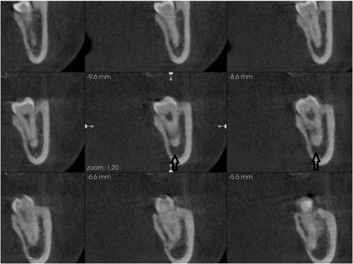
Figure 2. The MC is visible on six slices (open arrow), therefore, was given a score of 2 (very good visibility).
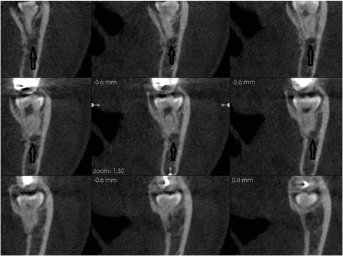
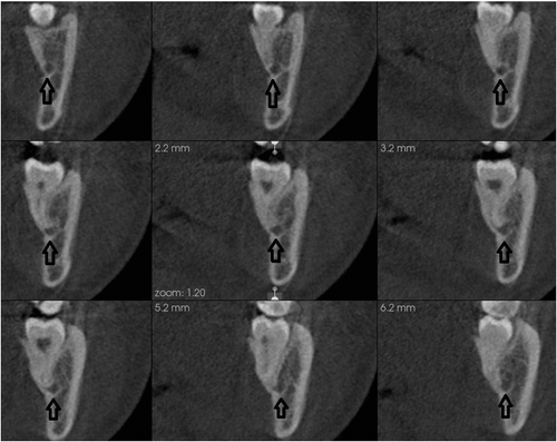
Figure 3. The MC is visible on nine slices (open arrow), therefore, was given a score of 3 (excellent visibility).
All images were evaluated on LCD monitor with installed CS 3D imaging viewer (CS 3D imaging viewer, 3.2.9 Carestream, Rochester, NY, USA). When necessary, the window settings were adjusted to optimize the images for evaluation.
Statistical analysis
Intra-class correlation coefficient (ICC) was used to asses inter-observer agreement depending on the following criteria: a value of less than 0.40 was considered to indicate poor agreement, from 0.40 to 0.60 fair agreement, from 0.61 to 0.80 good agreement and higher than 0.80 excellent agreement. Kruskal–Wallis test was used to examine the differences in visibility of the canal according to its location and the type of tooth impaction. Statistical significance was set at a level of 0.05.
All statistical analyses were performed by using a statistical software package (SPSS version 16; Chicago, IL, USA).
Results and discussion
The inter-observer agreement for the visibility scores of the MC was excellent; ICC for average measures was 0.90. Between the two observers, disagreement was present in five scores and consensus was reached after discussion.
The visibility scores of the MC were good, very good and excellent at 3, 25 and 122 IMTM sites, respectively. The distribution of the scores in relation to the type of tooth impaction and its location (on pseudo-panoramic and cross-sectional images) are shown in –. The most frequent type of tooth impaction was mesial, and the MC was most frequently observed in superimposed and inferior locations on pseudo-panoramic and cross-sectional images, respectively. According to Kruskal–Wallis test, there were no statistically significant differences in the visibility scores of the MC according to its location or the type of tooth impaction.
Table 1. Distribution of the visibility scores in relation to the type of tooth impaction on pseudo-panoramic images.
Table 2. Distribution of the visibility scores in relation to the location of MC on pseudo-panoramic images.
Table 3. Distribution of the visibility scores in relation to the location of MC on cross-sectional images.
In the current study, we evaluated the visibility of the MC on cross-sectional CBCT images at IMTM sites. At most sites, the visibility score was very good to excellent, meaning that the MC was visible on most of the CBCT images. Such findings are in agreement with Jung et al.[Citation15] and Oliveira-Santos et al.[Citation23] studies. In these studies, the highest visibility of the MC was at the third molar area. In our study, the canal could be identified and considered visible even if it was not corticated, and the usage of small voxel size (0.2 mm) could also be a contributing factor to the attained excellent visibility of the canal.
In a study done by Lofthag-Hansen et al.,[Citation25] the MC was visible only in one-third of the cross-sectional CBCT images, and visibility was improved when other CBCT images were included in evaluation. Similarly, in another study done by Takahashi et al.,[Citation26] the MC was visible in two-thirds of the CT cross-sectional images and visibility was improved when the MC was outlined in reformatted panoramic images. In both of these studies, the assessment of the MC visibility was not confined to the third molar area, and the poor visibility of the MC on cross-sectional images could be due to using large image thickness (1 mm). Furthermore, using large inter-slice interval (1 mm) and CT images rather than CBCT images, might also be the cause for poor visibility of the MC in the study of Takahashi et al.[Citation26]
In contrast to the previous CBCT studies,[Citation15,Citation23,Citation25] our cross-sectional images were parallel with the long axis of the impacted teeth, which enabled us to see the impacted teeth with complete roots in upright position, meaning that the location of the MC in relation to the impacted teeth could be assessed with easiness. Moreover, this could be another contributing factor in the achieved excellent visibility of the MC. Changing slice angulation may lead to different orientation of the canal in relation to bone marrow spaces and impacted teeth.[Citation30] This, in turn, may affect the visibility of the canal, especially if it was not corticated. In our current study, we did not attempt to compare the visibility of the MC on cross-sectional images generated with different slice angulations; therefore, further studies will be needed to resolve this issue.
Some studies evaluated the visibility of the MC on digital panoramic, reformatted panoramic and CT or CBCT cross-sectional images.[Citation15,Citation19,Citation21,Citation26] When visibility was compared between CBCT or CT cross-sectional images and digital panoramic images, CBCT and CT cross-sectional images outperformed digital panoramic images in identification of the MC.[Citation15,Citation21] In contrast with these results, the visibility of the MC was significantly higher in panoramic views when reformatted from CT and compared with cross-sectional CT views.[Citation26] This could be due to the high quality and minimum thickness of reformatted CT panoramic views when compared with conventional digital panoramic radiographs.[Citation19]
Although the depiction of the MC is consistent and comparable between CT and CBCT,[Citation17,Citation18] CBCT is recommended and preferred over CT for the reasons mentioned previously.[Citation17,Citation18,Citation27] Therefore, a drawback can be noticed in the previous studies which used CT images for visualizing the MC for dental tasks.[Citation21,Citation26] Moreover, rather than comparing the visibility of the MC on reformatted panoramic views with cross-sectional views,[Citation26] it is necessary to identify the MC on both of the views,[Citation9] since the MC has different locations in both.
IMTMs can be classified according to their inclination into mesial, distal, vertical and horizontal.[Citation31] Different types of tooth impaction are associated with different anatomical relations with the MC.[Citation32,Citation33] Therefore, we were under the impression that the visibility of the MC might be affected by the type of tooth impaction, especially if they are in contact on panoramic radiograph. Our results are in line with previous studies.[Citation32,Citation33] However, the differences in visibility of the MC were not statistically significant according to different types of tooth impaction.
Conclusions
In conclusion, regardless of the different types of tooth impaction and different locations of the MC, the visibility of the MC is excellent on most of the cross-sectional CBCT images at IMTM sites. CBCT is considered an excellent diagnostic tool for achieving these results.
Disclosure statement
The authors deny any conflicts of interest. The authors declare that they have no competing interests.
References
- de Melo Albert DG, Gomes AC, do Egito Vasconcelos BC, et al. Comparison of orthopantomographs and conventional tomography images for assessing the relationship between impacted lower third molars and the mandibular canal. J Oral Maxillofac Surg. 2006;64:1030–1037.
- Flygare L, Ohman A. Preoperative imaging procedures for lower wisdom teeth removal. Clin Oral Investig. 2008;12:291–302.
- Roeder F, Wachtlin D, Schulze R. Necessity of 3D visualization for the removal of lower wisdom teeth: required sample size to prove non-inferiority of panoramic radiography compared to CBCT. Clin Oral Investig. 2012;16:699–706.
- Sanmartí-Garcia G, Valmaseda-Castellón E, Gay-Escoda C. Does computed tomography prevent inferior alveolar nerve injuries caused by lower third molar removal? J Oral Maxillofac Surg. 2012;70:5–11.
- Guerrero ME, Botetano R, Beltran J, et al. Can preoperative imaging help to predict postoperative outcome after wisdom tooth removal? A randomized controlled trial using panoramic radiography versus cone-beam CT. Clin Oral Investig. 2014;18:335–342.
- Kositbowornchai S, Densiri-aksorn W, Piumthanaroj P. Ability of two radiographic methods to identify the closeness between the mandibular third molar root and the inferior alveolar canal: a pilot study. Dentomaxillofac Radiol. 2010;39:79–84.
- Pawelzik J, Cohnen M, Willers R, et al. A comparison of conventional panoramic radiographs with volumetric computed tomography images in the preoperative assessment of impacted mandibular third molars. J Oral Maxillofac Surg. 2002;60:979–984.
- Sivolella S, Boccuzzo G, Gasparini E, et al. Assessing the need for computed tomography for lower-third-molar extraction: a survey among 322 dentists. Radiol Med. 2012;117:112–124.
- Valmaseda-Castellón E, Berini-Aytés L, Gay-Escoda C. Inferior alveolar nerve damage after lower third molar surgical extraction: a prospective study of 1117 surgical extractions. Oral Surg Oral Med Oral Pathol Oral Radiol Endod. 2001;92:377–383.
- Szalma J, Lempel E, Jeges S, et al. The prognostic value of panoramic radiography of inferior alveolar nerve damage after mandibular third molar removal: retrospective study of 400 cases. Oral Surg Oral Med Oral Pathol Oral Radiol Endod. 2010;109:294–302.
- Jhamb A, Dolas RS, Pandilwar PK, et al. Comparative efficacy of spiral computed tomography and orthopantomography in preoperative detection of relation of inferior alveolar neurovascular bundle to the impacted mandibular third molar. J Oral Maxillofac Surg. 2009;67:58–66.
- Hasegawa T, Ri S, Shigeta T, et al. Risk factors associated with inferior alveolar nerve injury after extraction of the mandibular third molar – a comparative study of preoperative images by panoramic radiography and computed tomography. Int J Oral Maxillofac Surg. 2013;42:843–851.
- Ghaeminia H, Meijer GJ, Soehardi A, et al. Position of the impacted third molar in relation to the mandibular canal. Diagnostic accuracy of cone beam computed tomography compared with panoramic radiography. Int J Oral Maxillofac Surg. 2009;38:964–971.
- Ghaeminia H, Meijer GJ, Soehardi A, et al. The use of cone beam CT for the removal of wisdom teeth changes the surgical approach compared with panoramic radiography: a pilot study. Int J Oral Maxillofac Surg. 2011;40:834–839.
- Jung YH, Cho BH. Radiographic evaluation of the course and visibility of the mandibular canal. Imaging Sci Dent. 2014;44:273–278.
- de Oliveira-Santos C, Souza PH, de Azambuja Berti-Couto S, et al. Assessment of variations of the mandibular canal through cone beam computed tomography. Clin Oral Investig. 2012;16:387–393.
- Liang X, Jacobs R, Hassan B, et al. A comparative evaluation of Cone Beam Computed Tomography (CBCT) and Multi-Slice CT (MSCT). Part I. On subjective image quality. Eur J Radiol. 2010;75:265–269.
- Naitoh M, Nakahara K, Suenaga Y, et al. Comparison between cone-beam and multislice computed tomography depicting mandibular neurovascular canal structures. Oral Surg Oral Med Oral Pathol Oral Radiol Endod. 2010;109:25–31.
- Angelopoulos C, Thomas SL, Hechler S, et al. Comparison between digital panoramic radiography and cone-beam computed tomography for the identification of the mandibular canal as part of presurgical dental implant assessment. J Oral Maxillofac Surg. 2008;66:2130–2135.
- Gerlach NL, Meijer GJ, Maal TJ, et al. Reproducibility of 3 different tracing methods based on cone beam computed tomography in determining the anatomical position of the mandibular canal. J Oral Maxillofac Surg. 2010;68:811–817.
- Kamrun N, Tetsumura A, Nomura Y, et al. Visualization of the superior and inferior borders of the mandibular canal: a comparative study using digital panoramic radiographs and cross-sectional computed tomography images. Oral Surg Oral Med Oral Pathol Oral Radiol. 2013;115:550–557.
- Naitoh M, Katsumata A, Kubota Y, et al. The role of objective plane angulation on the mandibular image using cross-sectional tomography. J Oral Implantol. 2006;32:117–121.
- Oliveira-Santos C, Capelozza AL, Dezzoti MS, et al. Visibility of the mandibular canal on CBCT cross-sectional images. J Appl Oral Sci. 2011;19:240–243.
- Shokri A, Shakibaei Z, Langaroodi AJ, et al. Evaluation of the mandibular canal visibility on cone-beam computed tomography images of the mandible. J Craniofac Surg. 2014;25:273–277.
- Lofthag-Hansen S, Gröndahl K, Ekestubbe A. Cone-beam CT for preoperative implant planning in the posterior mandible: visibility of anatomic landmarks. Clin Implant Dent Relat Res. 2009;11:246–255.
- Takahashi A, Watanabe H, Kamiyama Y, et al. Localizing the mandibular canal on dental CT reformatted images: usefulness of panoramic views. Surg Radiol Anat. 2013;35:803–809.
- Hashimoto K, Arai Y, Iwai K, et al. A comparison of a new limited cone beam computed tomography machine for dental use with a multidetector row helical CT machine. Oral Surg Oral Med Oral Pathol Oral Radiol Endod. 2003;95:371–377.
- Neves FS, Vasconcelos TV, Oenning AC, et al. Oblique or orthoradial CBCT slices for preoperative implant planning: which one is more accurate? Braz J Oral Sci. 2014;13:104–108.
- Chadwick JW, Lam EW. The effects of slice thickness and interslice interval on reconstructed cone beam computed tomographic images. Oral Surg Oral Med Oral Pathol Oral Radiol Endod. 2010;110:37–42.
- Lübbers HT, Kruse AL, Obwegeser JA, et al. Oblique high resolution tomography: the ideal plane for visualization of the gonial section of the mandibular canal and its related structures? J Healthc Eng. 2012;3:87–104.
- Almendros-Marqués N, Berini-Aytés L, Gay-Escoda C. Evaluation of intraexaminer and interexaminer agreement on classifying lower third molars according to the systems of Pell and Gregory and of Winter. J Oral Maxillofac Surg. 2008;66:893–899.
- Schneider T, Filo K, Kruse AL, et al. Variations in the anatomical positioning of impacted mandibular wisdom teeth and their practical implications. Swiss Dent J. 2014;124:520–538.
- Lübbers HT, Matthews F, Damerau G, et al. Anatomy of impacted lower third molars evaluated by computerized tomography: is there an indication for 3-dimensional imaging? Oral Surg Oral Med Oral Pathol Oral Radiol Endod. 2011;111:547–550.