
Duygu Çapar G, Sapmaz-Metin M, Kütan E, et al. Preventive effect of doxycycline sponge against bisphosphonate-related osteonecrosis of the jaws: an animal study. BIOTECHNOL BIOTEC EQ. 2016; 30 [ePub ahead of print].
http://dx.doi.org/10.1080/13102818.2016.1174078
When the above article was first published online, the following mistakes were published.
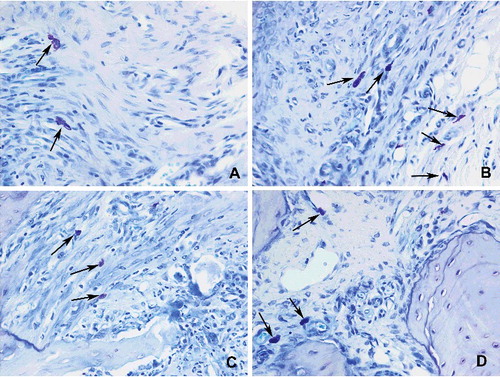
1. The caption for figure 4 was incorrectly published as:
Figure 4. Histopathological view showing the osteoblastic activity: cuboidal and dense stained osteoblasts (arrow) reflected osteoid formation. Basophilic reversal lines (arrowhead) were considered to identify the bone remodelling (A) control, moderate osteoblastic activity,(B and C) Groups I and II, mild to moderate activities along with small amounts of reversal lines, (D) Group III, an increased osteoblastic activity (PAS +HL 200x).
The correct version is:
Figure 4. Histopathological view showing the osteoblastic activity: cuboidal and dense stained osteoblasts (arrow) reflected osteoid formation. Basophilic reversal lines (arrowhead) were considered to identify the bone remodelling (A) control, moderate osteoblastic activity,(B and C) Groups I and II, mild to moderate activities along with small amounts of reversal lines, (D) Group III, an increased osteoblastic activity (Toluidin blue 400x).
2. The image for Figure 5 was incorrectly published as:
Incorrect:
Correct:
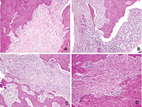
3. The Caption for Figure 6 was incorrectly published as:
Figure 6. Histopathological view showing the osteoclast density: (A) control, osteoclasts (arrowhead) were shown associated with bone surface in their active pole in the control group. (B) Detached (arrowhead) and apoptotic (star) osteoclasts were marked in Group I. (C and D) Osteoclast number with normal appearance was increased in Groups II and III. Giant type osteoclasts were characteristics of that groups (PAS + HL 200x).
The correct version is:
Figure 6. Histopathological view showing the osteoclast density: (A) control, osteoclasts (arrowhead) were shown associated with bone surface in their active pole in the control group. (B) Detached (arrowhead) and apoptotic (star) osteoclasts were marked in Group I. (C and D) Osteoclast number with normal appearance was increased in Groups II and III. Giant type osteoclasts were characteristics of that groups (Mallory Triple 400x).
The authors would like to apologise for any inconvenience caused.