
ABSTRACT
Following root canal treatment, sealers may contact periapical tissue. The purpose of this study was to evaluate the systemic toxic effects of epoxy resin-based sealers (AH Plus and Obtuseal). Inductively coupled plasma-mass spectrometry (ICP-MS) was used to measure levels of trace elements (beryllium, magnesium, aluminium, calcium, chromium, arsenic and lead) in the brain, kidney and liver of rats. Twenty sterilized polyethylene tubes were then filled with AH Plus and Obtuseal and implanted into the dorsal subcutaneous tissue of 10 rats; three unoperated animals were used as a control group. After 45 days, the rats were sacrificed by cervical dislocation following anaesthesia, and brains, kidneys and livers were removed from all experimental animals. ICP-MS analysis was used to determine levels of trace elements. Data were analysed using Kruskal–Wallis and Connover post hoc tests. No significant differences were found in aluminium and calcium levels, but brains, kidneys and livers showed significantly higher amounts of magnesium and chromium than the corresponding controls. In the kidney and liver samples, arsenic levels were found to be higher than in the control group. Lead was detected at higher levels only in liver samples from the AH Plus group. Beryllium was not detected in any organ. It was concluded that AH Plus and Obtuseal release minimal quantities of trace elements when in contact with subcutaneous tissue, and further studies are needed to understand the systemic effects of these materials.
Introduction
Sealers may be classified according to their main chemical content, which usually includes calcium hydroxide (CaOH) or an epoxy resin [Citation1]. Because of the physical limitations of core materials, sealers play an important role in root canal obturation, as they fill all voids and spaces within the tooth [Citation2].
Materials such as AH Plus (Dentsply De Trey, Germany), which is based on an epoxy resin, and Obtuseal (A.T.O., Zizine, France), based on an epoxy resin and CaOH, have recently become available for endodontic use.
Sealers containing CaOH have shown good biological results [Citation3]. AH Plus contains bisphenol A, F epoxy resin, calcium tungstate, zirconium oxide, silica, iron oxide pigments, dibenzyldiamine, aminoadamantane, tricyclodecane-diamine (TCD-diamine) and silicone oil but not DGEBA or, more importantly, CaOH. Obtuseal's base tube contains TCD-diamine, a radiopaque excipient; the catalyst tube contains CaOH, diglycidyl ether of bisphenol A (DGEBA) and radiopaque excipient.
Root canal sealers are intended for use within the root canal system [Citation4] but may inadvertently extrude into the periradicular tissue, causing tissue irritation and delayed healing [Citation5]. Similarly, destruction and corrosion products from root canal sealers may reach periradicular tissues through dentinal tubules, lateral and accessory canals or apical foramina [Citation6]. Products that reach the periradicular tissues may even access distant tissues through the circulatory or lymphatic systems [Citation7].
Concentrations of trace elements affect a range of organs in both humans and animals [Citation8]. While elements such as cobalt, copper, chromium and nickel are essential components of biological structures, they can also be toxic at concentrations beyond functional limits. Other elements such as arsenic, lead, cadmium, magnesium and calcium are known for their toxic role in certain biochemical reactions [Citation9,Citation10]. In studies examining implants [Citation11] and dental applications [Citation12,Citation13] in animals, AH Plus has been found to have biocompatible properties, but the effects of Obtuseal remain unknown.
Inductively coupled plasma-mass spectrometry (ICP-MS) is widely used for estimating trace metals in biological fluids because of its multi-element analysis capabilities and limits of detection that are generally lower than for other systems such as inductively coupled plasma-optical emission spectrometry (ICP-OES) or graphite furnace atomic absorption spectrometry (GF-AAS). The well-documented advantages of ICP-MS also include greater sensitivity and selectivity [Citation14–16].
Sealers are often in direct contact with pulpal and periradicular tissues, which may allow trace metals in AH Plus and Obtuseal to leach out into the body. The aim of this study was to measure concentrations of beryllium (Be), magnesium (Mg), aluminium (Al), calcium (Ca), chromium (Cr), arsenic (As) and lead (Pb) in rat organs following implantation of AH Plus or Obtuseal.
Materials and methods
Experimental animals
Thirteen Wistar albino rats aged 3–4 months and weighing 240–280 g were used in this study. The animals were provided with food and water ad libitum while housed in plastic cages (40 × 32 × 17 cm) in ventilated rooms (12 h light/dark cycles; room temperature 21–25 °C). The study protocol was reviewed and approved by the Experimental Animal Ethics Committee of the Inonu University of Health Sciences (2013/A-16).
Study design and experimental procedures
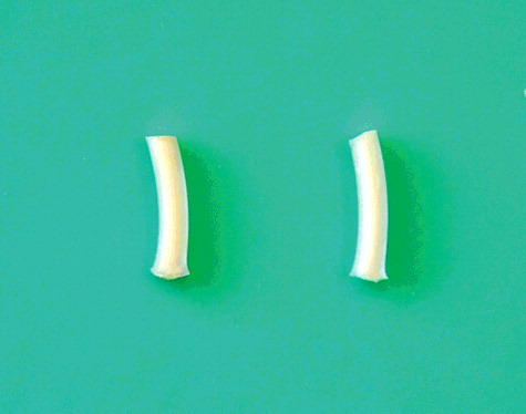
Twenty sterilized polyethylene tubes (nontoxic Scalp Vein 19G; 1.3 mm internal diameter, 10 mm long) were prepared, with one side opened and the other side closed. AH Plus and Obtuseal sealers were prepared according to the manufacturer's recommendations. The tubes were separated into two experimental groups, and sealers were inserted into the tubes using a Lentulo spiral (Maillefer Dentsply, Tulsa, OK, USA) (). All tubes were sterilized using ethylene oxide gas. Five animals were assigned to each experimental group, and two tubes were inserted in the back of each animal. A control group of three animals remained unoperated.
Figure 1. The test tubes filled with AH Plus and Obtuseal sealer.
Surgical procedure
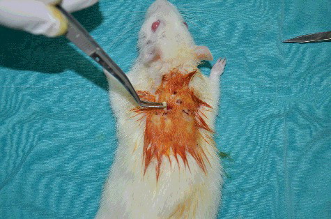
All surgery was performed under sterile conditions. Before the procedure, rats were anaesthetized using intraperitoneal 10% ketamine (0.1 mL/100 g) and 2% xylazine (0.1 mL/100 g). Once an anaesthetized rat became unresponsive, the surgical sites on the dorsal skin were shaved and disinfected with povidone–iodine. Using a scalpel, two 5-mm incisions were made in the skin, without opening the peritoneum. With the aid of presel, the test tubes were carefully placed at the incision site (). The area around the wound was then surgically stitched with a single 4/0 silk suture and disinfected with povidone-iodine. At 45 days, the rats were sacrificed by cervical dislocation following anaesthesia, and brain, kidneys and liver were removed from each animal.
Figure 2. The insertion of test tubes into the incision site with the aid of a presel.
Sample preparation and inductively coupled plasma-mass spectrometry
Using the Berghof microwave digestion system, tissue samples were digested in an acid solution – first in a microwave inside a closed Teflon vessel for 1 h 45 min, and then in a microwave oven (Berghof Speedwave, Germany). Prior to analysis, samples were isolated from metallic materials and dust to avoid contamination. A 250 mg sample was placed in the digestion vessel, and after adding 5 mL of nitric acid (65%), the mixture was carefully shaken. After waiting for at least 20 min, the vessels were closed, and samples were heated in the microwave oven in accordance with the digestion program [Citation17].
Digestion solutions were diluted and analysed on an Agilent 7500a series ICP/MS. For quality control, the concentration of internal standards was 200 ppb (9Be, 45Sc, 103Rh, 208Bi); reference materials were run with the samples, and tuning parameters () were controlled before the analyses. The axis cut point of the calibration line was used to obtain the detection limit for each element. It was also important to assure linear rank of the methodology by analysing different standards with lower and higher known concentrations of each element (0, 1, 5, 10, 20, 30, 40 and 50 ppb). At least five different reference materials were used to encompass all the elements used. Duplicate samples were also used to determine the precision of the analysis. For each element, at least three standards were used to take account of the instrument's analytical working range. Ultrapure water was used to prepare calibration standards and blanks, and three replicate determinations were performed for each sample. A reagent blank was subtracted from all sample results, and detection limits were calculated as three times the standard deviation for reagent blanks [Citation18].
Table 1. Work specification resume for ICP-MS Agilent 7500a and measurement parameters.
Statistical analysis
Each variable was reported as mean and standard error (SE). Normality of continuous variables in groups was determined by the Shapiro–Wilk test. Results were statistically analysed using the Kruskal–Wallis and Connover post hoc tests; a value of P < 0.05 was considered to be statistically significant. Data were evaluated using SPSS for Windows (version 15.0, SPSS Inc., Chicago, IL, USA).
Results and discussion
The chemical effect of root canal sealers used adjacent to periapical tissues is a predictor of their physical, chemical and biological properties [Citation19,Citation20]. In the case of a sealer that has extended beyond the root canal space, corrosion of the sealer surface causes in vivo release of various elements over time. The elements released from root canal sealers may be retained by cells or transferred to other organs for elimination, but migration of metallic trace elements from root canal sealers to distant organs has not been widely studied. Knowledge of their chemical effects can guide practitioners in selecting the optimal material for given clinical conditions.
The present study investigated concentrations of seven trace elements (Be, Al, As, Ca, Cr, Mg and Pb) in the brain, kidney and liver of rats. Along with the kidneys, the liver organizes homeostasis and is one of the body's fundamental biochemical workplaces, metabolizing many chemicals, as well as trace elements. For this reason, concentrations of major and trace elements in the liver have been studied in both animals [Citation21] and humans [Citation22].
Pilot in vivo studies to evaluate biocompatibility commonly involve subcutaneous insertion of the material to be analysed into small animals. The rat is most commonly used because, in addition to its suitability as an experimental model for mammals, its size permits safe and easy administration of materials. Additionally, rats’ more accelerated metabolism facilitates acceptable results in a shorter period of time [Citation23].
The introduction of new root canal sealers to the market means that their characteristics must be tested and compared with commonly used materials [Citation24]. The present study compared AH Plus, a commonly used root canal sealer, with Obtuseal, an epoxy resin-based sealer that also contains CaOH. Because chemical products such as AH Plus may vary in composition, the study analysed alterations in their trace metal content.
shows a comparison of element concentrations in brains, kidneys and livers. Al was present in similar levels in all tissue samples when compared to the control group (p = 0.339). Extreme exposure to Al in close contact with human tissues increases risk of Alzheimer's disease [Citation25]. Using EDX, Borges et al. [Citation26] found that AH Plus was composed of carbon, oxygen, calcium, chlorine, tungsten and zirconium. Thus, there is no statistically significant difference between experimental and control groups.
Table 2. Concentrations (µg⋅kg−1) of six elements in rat organs (n = 5).
In the present study, AH Plus and Obtuseal showed similar levels of Cr in brain, kidney and liver. There was no statistically significant difference between the two sealers, but when compared with the control group, all organ levels were found to be significantly elevated (brain: p = 0.030; kidney: p = 0.027; liver: p = 0.037). Decreased Cr levels in humans are associated with impaired glucose tolerance, glycosuria, fasting hyperglycaemia and elevated levels of circulating insulin and glucagon [Citation27]. Chromium toxicity is rare, and the only reported adverse effects in humans present as liver or kidney problems [Citation28,Citation29].
Levels of Mg in the experimental rats’ organs increased significantly by comparison with the control group (brain: p = 0.009; kidney: p = 0.039; liver: p = 0.031). Magnesium plays a significant role in the actions of numerous enzymes involved in glucose oxidation and may also play a role in the release of insulin [Citation30]. Increased magnesium levels in humans lead to clinical manifestation of hypermagnesemia, which is associated with neuromuscular and cardiovascular toxicity and metabolic disturbance [Citation31].
Arsenic is known as a carcinogenic chemical. There is substantial evidence of an association between As and inorganic As compounds and lung, skin and urinary bladder carcinogens in humans, as well as some evidence for carcinogenicity in the kidneys, liver and prostate [Citation32]. In the present study, significantly higher levels of As were found in rat kidneys (p = 0.026) and liver (p = 0.037). For both root canal sealers, As was detected in the kidneys and livers, respectively, at concentrations of 0.0071 and 0.0122 ppb (for AH Plus) and 0.0154 and 0.02 ppb (for Obtuseal). However, As2O3 (trivalent inorganic arsenic) is known to inhibit cellular functions and disrupt intracellular microstructures at concentrations of 1 mmol L−1 in eukaryotic cells [Citation33,Citation34] the observed values are below the risk limits.
According to the International Agency for Research on Cancer (IARC), beryllium is known to be a Category 1A carcinogen [Citation34]. In this study, Be was not detected in either AH Plus or Obtuseal. As Beryllium is an element with a low atomic number, quantitative analysis of this element may be imprecise.
Lead is known to cause adverse health effects that include neurotoxicity, nephrotoxicity and damage to the haematological and cardiovascular systems, and the IARC has classified lead as a possible human carcinogen (group 2B) [Citation35]. In the present study, small amounts of lead (1 ppb) were detected in the livers of the AH plus group. Although there was a statistically significant difference (p = 0.020), this value was close to the control group.
The methods used to determine trace element concentrations include ICP-OES, ICP-MS and GF-AAS. For this purpose, ICP-MS technology is more sensitive than ICP-OES [Citation36]. The benefits of ICP-MS technique include its high accuracy and lower detection limits as compared to GF-AAS, and it also has multi-element analysis capabilities [Citation37]. On that basis, ICP-MS technology was chosen for the present study, and the distribution of trace elements (Be, Al, As, Ca, Cr, Mg and Pb) and their potential toxicity in rat organs were successfully assessed using ICP-MS.
According to their manufacturers’ descriptions, neither AH Plus nor Obtuseal contain Mg, Cr or As. Both AH Plus and Obtuseal contain Ca [Citation38], but no significant difference was found when compared with the control groups. Additionally, as that study found traces of Mg in AH Plus [Citation38], there is a need to explain the increase in these elements in the organs. These results may be a consequence of contamination during manufacture or of some industrial secret [Citation38]. Another possible explanation is that the body naturally acquires these trace elements and uses them in the metabolic process. However, AH Plus and Obtuseal might alter kidney, liver or brain metabolism, so altering the body's ability to eliminate these substances.
Conclusions
In this study, the accumulation of trace elements (Mg, Al, Ca, Cr, As and Pb) in different organs was measured by ICP-MS, with a calculated detection limit sensitivity of 0.2 ppb. When in contact with subcutaneous tissue, AH Plus and Obtuseal release minimal quantities of trace elements. Although elevated levels of certain elements were observed in rat organs, they were below toxic levels in all cases, indicating that AH Plus and Obtuseal are nontoxic. The results also indicate the occurrence of sealer dissolution occurs. The trick of this study filling materials should be kept within root canal boundaries. This study is clearly of interdisciplinary relevance. It may contribute to overcoming some current difficulties related to sealer quality and dissolution in biological tissues and fluids. Further studies should examine these sealers effects on organ metabolism and blood contamination.
Ethical approval
Ethical approval was given by Animal Ethics Research Committee of Inonu University of Health Science (2013/A-16).
Disclosure statement
None declared.
Additional information
Funding
Related Research Data
References
- Kim RJ, Shin JH. Cytotoxicity of a novel mineral trioxide aggregate-based root canal sealer [corrected]. Dent Mater J. 2014;33:313–318.
- Farhad AR, Hasheminia S, Razavi S, et al. Histopathologic evaluation of subcutaneous tissue response to three endodontic sealers in rats. J Oral Sci. 2011;53:15–21.
- Gomes-Filho JE, Bernabe PF, Nery MJ, et al. Reaction of rat connective tissue to a new calcium hydroxide-based sealer. Oral Surg Oral Med Oral Pathol Oral Radiol Endod. 2008;106:e71–76.
- Bernath M, Szabo J. Tissue reaction initiated by different sealers. Int Endod J. 2003;36:256–261.
- Sari S, Duruturk L. Radiographic evaluation of periapical healing of permanent teeth with periapical lesions after extrusion of AH Plus sealer. Oral Surg Oral Med Oral Pathol Oral Radiol Endod. 2007;104:e54–59.
- Geurtsen W, Leyhausen G. Biological aspects of root canal filling materials–histocompatibility, cytotoxicity, and mutagenicity. Clin Oral Invest. 1997;1:5–11.
- Sarmiento-González A, Encinar JR, Marchante-Gayon JM, et al. Titanium levels in the organs and blood of rats with a titanium implant, in the absence of wear, as determined by double-focusing ICP-MS. Anal Bioanal Chem. 2009;393:335–343.
- Kuriwaki J, Nishijo M, Honda R, et al. Effects of cadmium exposure during pregnancy on trace elements in fetal rat liver and kidney. Toxicol Lett. 2005;156:369–376.
- Khan N, Choi JY, Nho EY, et al. Determination of minor and trace elements in aromatic spices by micro-wave assisted digestion and inductively coupled plasma-mass spectrometry. Food Chem. 2014;158:200–206.
- Alonso ML, Montana FP, Miranda M, et al. Interactions between toxic (As, Cd, Hg and Pb) and nutritional essential (Ca, Co, Cr, Cu, Fe, Mn, Mo, Ni, Se, Zn) elements in the tissues of cattle from NW Spain. Biol Met. 2004;17:389–397.
- Sousa CJ, Montes CR, Pascon EA, et al. Comparison of the intraosseous biocompatibility of AH Plus, EndoREZ, and epiphany root canal sealers. J Endod. 2006;32:656–662.
- Tanomaru Filho M, Leonardo MR, Silva LA, et al. Effect of different root canal sealers on periapical repair of teeth with chronic periradicular periodontitis. Int Endod J. 1998;31:85–89.
- Leonardo MR, Salgado AA, da Silva LA, et al. Apical and periapical repair of dogs' teeth with periapical lesions after endodontic treatment with different root canal sealers. Pesqui Odontol Bras. 2003;17:69–74.
- Goullé JP, Mahieu L, Castermant J, et al. Metal and metalloid multi-elementary ICP-MS validation in whole blood, plasma, urine and hair. Forensic Sci Int. 2005;153:39–44.
- Bazzi A, Nriagu JO, Linder AM. Determination of toxic and essential elements in children's blood with inductively coupled plasma-mass spectrometry. J Environ Monit. 2008;10:1226–1232.
- Heitland P, Koster HD. Biomonitoring of 37 trace elements in blood samples from inhabitants of northern Germany by ICP-MS. J Trace Elem Med Biol. 2006;20:253–262.
- Rattanachongkiat S, Millward GE, Foulkes ME. Determination of arsenic species in fish, crustacean and sediment samples from Thailand using high performance liquid chromatography (HPLC) coupled with inductively coupled plasma mass spectrometry (ICP-MS). J Environ Monit. 2004;6:254–261.
- De Blas Bravo I, Sanz Castro R, Lopez Riquelme N, et al. Optimization of the trace element determination by ICP-MS in human blood serum. J Trace Elem Med Biol. 2007;21(Suppl 1):14–17.
- Dammaschke T, Gerth HU, Zuchner H, et al. Chemical and physical surface and bulk material characterization of white ProRoot MTA and two Portland cements. Dent Mater. 2005;21:731–738.
- Estrela C, Sousa-Neto MD, Guedes OA, et al. Characterization of calcium oxide in root perforation sealer materials. Braz Dent J. 2012;23:539–546.
- Seixas TG, Kehrig Hdo A, Fillmann G, et al. Ecological and biological determinants of trace elements accumulation in liver and kidney of Pontoporia blainvillei. Sci Total Environ. 2007;385:208–220.
- Varga I, Szebeni A, Szoboszlai N, et al. Determination of trace elements in human liver biopsy samples by ICP-MS and TXRF: hepatic steatosis and nickel accumulation. Anal Bioanal Chem. 2005;383:476–482.
- Zmener O. Tissue response to a new methacrylate-based root canal sealer: preliminary observations in the subcutaneous connective tissue of rats. J Endod. 2004;30:348–351.
- Ricucci D, Langeland K. Apical limit of root canal instrumentation and obturation, part 2. A histological study. Int Endod J. 2002;31:394–409.
- Forbes WF, Gentleman JF. Risk factors, causality, and policy initiatives: the case of aluminum and mental impairment. Exp Geront. 1998;33:141–154.
- Borges AH, Guedes OA, Orçati Dorilêo MGC, et al. Analysis of chemical elements and heavy metals in MTA Fillapex and AH Plus. Oral Health Dent Manag. 2014;13:1007–1012.
- Goldhaber SB. Trace element risk assessment: essentiality vs. toxicity. Regul Toxicol Pharmacol. 2003;38:232–242.
- Loubières Y, de Lassence A, Bernier M, et al. Acute, fatal, oral chromic acid poisoning. J Toxicol Clin Toxicol. 1999;37:333–336.
- Cerulli J, Grabe DW, Gauthier I, et al. Chromium picolinate toxicity. Ann Pharmacother. 1998;32:428–431.
- Paolisso G, Barbagallo M. Hypertension, diabetes mellitus, and insulin resistance: the role of intracellular magnesium. Am J Hypertens. 1997;10:346–355.
- Topf JM, Murray PT. Hypomagnesemia and hypermagnesemia. Rev Endocr Metab Disord. 2003;4:195–206.
- Tokar EJ, Benbrahim-Tallaa L, Ward JM, et al. Cancer in experimental animals exposed to arsenic and arsenic compounds. Crit Rev Toxicol. 2010;40:912–927.
- Pan X, Reissman S, Douglas NR, et al. Trivalent arsenic inhibits the functions of chaperonin complex. Genetics. 2010;186:725–734.
- Kum KY, Kim EC, Yoo YJ, et al. Trace metal contents of three tricalcium silicate materials: MTA Angelus, Micro Mega MTA and Bioaggregate. Int Endod J. 2014;47:704–710.
- García-Lestón J, Mendez J, Pasaro E, et al. Genotoxic effects of lead: an updated review. Environ Int. 2010;36:623–636.
- Khan N, Jeong IS, Hwang IM, et al. Method validation for simultaneous determination of chromium, molybdenum and selenium in infant formulas by ICP-OES and ICP-MS. Food Chem. 2013;141:3566–3570.
- Janasik B, Trzcinka-Ochocka M, Brodzka R. [Selenium determination in plasma/serum by inductively coupled plasma mass spectrometry (ICP-MS): comparison with graphite furnace atomic absorption spectrometry (GF-AAS)]. Med Pr. 2011;62:489–498.
- Sampaio FC, Alencar AH, Guedes OA, et al. Chemical elements characterization of root canal sealers using scanning electron microscopy and energy dispersive X-ray analysis. Oral Health Dent Manag. 2014;13:27–34.