Abstract
In this study, the case of a patient who developed artistic ability following a traumatic brain injury is reported. The subject was a 49-year-old male who suffered brain injury at the age of 44 due to an accidental fall. At age 48, he began drawing with great enthusiasm and quickly developed a personal style with his own biomorphic iconography. At first, his drawing was restricted to realistic reproductions of photographs of buildings, but his style of drawing changed and became more personal and expressionistic over the following 6 months.
A number of reports have examined the development of visual artistic ability following degenerative or other types of brain injury (Chatterjee, Citation2006; Zaidel, Citation2005). However, the emergence of de novo artistic ability is rarely seen in brain-damaged patients (Pollak, Mulvenna, & Lythgoe, Citation2007); this is particularly true for patients with traumatic brain injury (Schott, Citation2012). Here, the case of a patient who began drawing following a traumatic brain injury and quickly developed a personal style is reported.
Methods
Case report
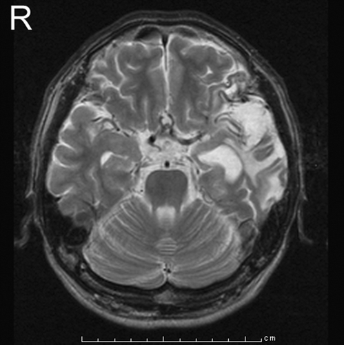
In December 2008, a 47-year-old male former truck driver was presented at Showa University Hospital with an unidentified complaint. Approximately 3 years earlier, in March 2005, he had fallen from the loading deck of a truck, suffered a closed head injury, and was admitted to a neurosurgical unit for the period of a month. After discharge from the hospital, the patient moved to a rehabilitation facility, where he stayed for 6 months and attended private cognitive training once a week and received outpatient treatment in the neurosurgery, ophthalmology, otolaryngology, and rehabilitation units once every 2 or 3 months. During the initial phase of his injury, the patient had several neurological deficits, including hearing loss (no hearing in the left ear and poor hearing in the right ear), a taste disorder, an olfactory disturbance, and a vestibular nerve balance disorder in addition to neuropsychological disturbances that included amnesia, executive dysfunction, irritability, and attention problems. Three years after his accident, he and his wife were aware that his symptoms were getting worse. He noticed palsy in both his upper and lower extremities, he could not change his clothes by himself, and an ophthalmologist found a gradual loss of visual acuity in the left eye. In July 2006, the visual acuity in the left eye was 20/20, but it was 20/40 by November 2008. In March 2010, a magnetic resonance imaging (MRI) scan revealed a high-intensity area in the left temporofrontal lobe (), and a neuropsychological examination in January 2011 found preserved visuospatial abilities but disturbed verbal functions, as measured with the Wechsler Adult Intelligence Scale-Revised (WAIS-R) subscales (Information, 8; Picture Completion, 12; Block Design, 16). During examination using the Cognistat tests in May 2013, the patient displayed verbal and memory deterioration but visuospatial function was well preserved (orientation: 9; attention span: 0; comprehension: 10; repetition: 5; naming: 7; constructional ability: 11; memory: 7, calculation skills: 10; reasoning: 9; judgment: 10).
Artistic expression
In the summer of 2010 (5 years post-injury), the patient started to attend a day treatment center where he became very interested in drawing, an interest that extended to his free time as well as to his time at the center. All works were drawn in colored pencils on paper in art sketchbooks (Japanese standard F4 size; 330 × 216 mm), and his artistic style changed significantly over several months. In the first phase (Summer 2010), he copied from photographs primarily depicting buildings, landscapes, or portraits, and his work was characterized by attention to detail (). In the second phase (Autumn 2010), the work was more tile-like and regular (), whereas the work became more florid and decorative in the third phase (Winter 2010) (). In the fourth phase (Spring 2011), the pictures became more biomorphic with regular motifs, such as an eye ().
Figure 2. In the first stage, the patient copied from photographs of buildings, landscapes, and portraits, and his drawing style was characterized by observational accuracy (A). In the second stage, he became interested in regular, tile-like patterns (B). In the third stage, he began to draw looser, more ornate shapes (C). In the fourth stage, his pictures were iconographic and biomorphic using, for example, eye shape as a motif (D). With kind permission from FUMIO. [To view this figure in color, please see the online version of this article.]
![Figure 2. In the first stage, the patient copied from photographs of buildings, landscapes, and portraits, and his drawing style was characterized by observational accuracy (A). In the second stage, he became interested in regular, tile-like patterns (B). In the third stage, he began to draw looser, more ornate shapes (C). In the fourth stage, his pictures were iconographic and biomorphic using, for example, eye shape as a motif (D). With kind permission from FUMIO. [To view this figure in color, please see the online version of this article.]](/cms/asset/4211150a-e5de-4315-b8b3-befbefddfa12/nncs_a_873058_f0002_oc.jpg)
When one of the current authors (AM) asked him about his usual painting style and the reasons for his work, he answered that he drew pictures without a ruler, worked on several pictures at once, and was influenced by internal images. The patient also remarked that he tended to avoid visiting art exhibitions, both before and after the accident, because he was hesitant to be influenced by other artists. Following the accident, his only visit to a museum was to an exhibit of the work of Taro Okamoto, a Japanese abstract and avant-garde painter and sculptor. Following this experience, the patient drew , after which his painting style became abstract, a style that he has maintained to this day (). His wife reported that the patient generally remained in his own room and was obsessively devoted to drawing throughout the day, except for sometimes eating. As a result, she became concerned about his physical condition but not his drawing.
Procedure
After obtaining informed consent from the patient and his wife, 12 paintings created by the patient between 2010 and 2011 were collected. Two paintings were from Summer 2011 (Time 1), three from Autumn 2011 (Time 2), four from Winter 2011 (Time 3), and three from Spring 2012 (Time 4). Sixteen naïve college students (10 females and six males; age range: 20–23 years, mean: 20.9, SD: 0.86) were recruited to examine these pieces. The students were blind to the background of the paintings, including the profile of the painter and the purpose of the research. The students were asked to rate each painting in random order using a semantic differential method, as previously described (Okada & Inoue, Citation1991). The questionnaire was constructed with 23 pairs of adjectives using a 7-point scale.
Results
A principal component analysis (PCA) and a multiple analysis of variance (MANOVA) were performed. The PCA indicated four main axes of variation (eigenvalues > 1.0) and explained 89.8% of the total variance; the first component explained 58.2% of the variance, and the second, third, and fourth components explained 15.6%, 9.4%, and 6.4% of the variance, respectively. The interpretive labels given to the four factors were activity, evaluation, intensity, and unsteadiness. The MANOVA was performed with these four factors as the dependent variables and with the individual time periods (Time 1, Time 2, Time 3, and Time 4) as four levels of the independent variable. We found a significant effect of time on the combined dependent variables (F[12, 13.5] = 3.76, p < .05; Wilks’ Lambda = .02; partial ƞ2 = .011), but analyses of each individual dependent variable using a Bonferroni-adjusted alpha level of .012 found that only the factor of activity differed according to time period (F[3,8] = 26.38, p < .001, partial ƞ2 = .908).
Discussion
In the present study, the case of a patient who developed de novo painting ability following a traumatic brain injury is reported. Currently, a number of reports have examined the development of artistic ability after brain damage (Chatterjee, Citation2006; Zaidel, Citation2005); however, this case has several distinct features. First, the damage to the current patient was caused by a traumatic brain injury, whereas previous cases have generally involved neural degeneration such as frontotemporal dementia (FTD; Midorikawa, Fukutake, & Kawamura, Citation2008; Miller et al., Citation1998), Alzheimer’s disease (AD; Chakravarty, Citation2011), Parkinson’s disease (PD; Walker, Warwick, & Cercy, Citation2006), or cerebrovascular disease (Lythgoe, Pollak, Kalmus, de Haan, & Chong, Citation2005). The emergence of artistic ability is rarely seen in patients following traumatic brain injury (Schott, Citation2012). To our knowledge, only a single report mentions artistic style subsequent to such an injury (Bogousslavsky, Citation2005), but the patient, Guillaume Apollinaire, was not a de novo artist. Rather, prior to his injury, this patient was already a poet and a critic. In the current case, the main lesion was in the left anterior temporal cortex in an area consistent with previous reports of patients who developed artistic ability after the onset of disease (Midorikawa et al., Citation2008; Miller et al., Citation1998; Miller, Boone, Cummings, Read, & Mishkin, Citation2000). These patients all suffered from degenerative disease rather than a traumatic brain injury, suggesting that the lesion or site of damage is more important to de novo artistic expression than is the pathological background of the insult. It has been proposed that temporal lesions and mild frontal lesions may play an integral role in the development of artistic creativity (Pollak et al., Citation2007). In previous cases, many brain-damaged patients created paintings in a realistic style (Miller et al., Citation1998), but in this case, the presence of a frontal lesion was not confirmed. Therefore, it is plausible that the preserved frontal lobe function of the current patient was a crucial underlying factor in his characteristic painting style.
Second, a period of 5 years separated the injury from the initiation of drawing activity, which began when he started going to the day-care center. However, there was no special training program for painting at this center, indicating that his improved painting skills were not due to training but were developed of his own accord. This further suggests that his painting techniques and style likely emerged from his own internal drive and volition.
Third, this investigation demonstrated that the patient’s drawing style and the perceptions underpinning his paintings rapidly changed in just 6 months and that he was very prolific (nearly 24 pictures per month) in a compulsive way. Therefore, it is possible that the compulsiveness of the current patient was crucial to the development of his skills and perceptions.
Fourth, the painting style of the patient changed from a form of realism to a form of iconographic and biomorphic abstraction. Patients with FTD who develop artistic ability after the onset of their disease tend to suffer from left temporal lesions, but their paintings are realistic or surrealistic without a significant symbolic or abstract component (Miller & Hou, Citation2004). During the initial period (Time 1), the current patient painted in a realistic style that was compatible with that of previous FTD patients. However, the subsequent development of his abstract painting style is inexplicable. It has recently been shown that many artistic patients exhibit a hectic urge to create or demonstrate compulsive behaviors related to their painting activity (Schott, Citation2012). The patient in this case displayed compulsive behaviors such as the repetition of identical drawings and ceaselessly painting for hours or even days. Moreover, because several de novo artists exhibited a compulsive nature in their painting activity after suffering from various diseases such as stroke (Lythgoe et al., Citation2005; Thomas-Anterion et al., Citation2010), FTD (Miller & Hou, Citation2004), or PD (Walker et al., Citation2006), it appears that it is not the etiology of the insult but rather the compulsive nature of the behavior that is crucial for a de novo artist. Therefore, the urge to create may be a central factor during the development of drawing activity.
Even though many de novo artists did not change their painting style during the course of their disease, some established artists have changed their style. Several abstract artists developed a realistic style (Seeley et al., Citation2008; Tanabe et al., Citation1996), whereas others changed from a realistic to an abstract style (Crutch & Rossor, Citation2006; Mell, Howard, & Miller, Citation2003). The current patient was a de novo artist, but he fits into the latter pattern of change. However, this type of change in an established artist has been observed following various insults such as AD (Crutch & Rossor, Citation2006) and FTD (Mell et al., Citation2003), and therefore, we could not confirm a relationship between the lesion and the pattern of change.
Unfortunately, some debate remains. In the chronic phase, the patient exhibited several symptoms of neurological deterioration, suffered from palsy in both his upper and lower extremities, and experienced a gradual loss of visual acuity. Therefore, it is possible that some type of neural degeneration might have affected his painting style. However, after treatment, there was not any evidence of a continued gradual loss of neurological function or any apparent changes in his painting style, suggesting there was not a progressive disease concomitant with his traumatic brain injury.
In conclusion, the post-injury ability of untutored artists such as the current patient suggests the likelihood that artistic ability is not restricted to a particular sort of person and may emerge in anyone.
Funding
AM was supported by a Chuo University Grant for Special Research, and AM and MK were supported by a Grant-in-Aid for Scientific Research from the Ministry of Education, Culture, Sports, Science and Technology (MEXT, 22730587(AM), 23591283(MK)). MK was supported by the Tamagawa University Center of Excellence under the Ministry of Education, Culture, Sports, Science, and Technology (MEXT).
References
- Bogousslavsky, J. (2005). Artistic creativity, style and brain disorders. European Neurology, 54(2), 103–111. doi:10.1159/000088645
- Chakravarty, A. (2011). De novo development of artistic creativity in Alzheimer’s disease. Annals of Indian Academy of Neurology, 14(4), 291–294. doi:10.4103/0972-2327.91953
- Chatterjee, A. (2006). The neuropsychology of visual art: Conferring capacity. International Review of Neurobiology, 74, 39–49. doi:10.1016/S0074-7742(06)74003-X
- Crutch, S. J., & Rossor, M. N. (2006). Artistic changes in Alzheimer’s disease. International Review of Neurobiology, 74, 147–161. doi:10.1016/S0074-7742(06)74012-0
- Lythgoe, M. F., Pollak, T. A., Kalmus, M., de Haan, M., & Chong, W. K. (2005). Obsessive, prolific artistic output following subarachnoid hemorrhage. Neurology, 64(2), 397–398. doi:10.1212/01.WNL.0000150526.09499.3E
- Mell, J. C., Howard, S. M., & Miller, B. L. (2003). Art and the brain: The influence of frontotemporal dementia on an accomplished artist. Neurology, 60(10), 1707–1710.
- Midorikawa, A., Fukutake, T., & Kawamura, M. (2008). Dementia and painting in patients from different cultural backgrounds. European Neurology, 60(5), 224–229.
- Miller, B. L., Boone, K., Cummings, J. L., Read, S. L., & Mishkin, F. (2000). Functional correlates of musical and visual ability in frontotemporal dementia. The British Journal of Psychiatry, 176, 458–463.
- Miller, B. L., Cummings, J., Mishkin, F., Boone, K., Prince, F., Ponton, M., & Cotman, C. (1998). Emergence of artistic talent in frontotemporal dementia. Neurology, 51(4), 978–982.
- Miller, B. L., & Hou, C. E. (2004). Portraits of artists: Emergence of visual creativity in dementia. Archives of Neurology, 61(6), 842–844.
- Okada, M., & Inoue, J. (1991). A psychological analysis about the elements of artistic evaluation on viewing paintings. Journal of the Yokohama National University, Educational Sciences, 31, 45–66.
- Pollak, T. A., Mulvenna, C. M., & Lythgoe, M. F. (2007). De novo artistic behaviour following brain injury. Frontiers of Neurology and Neuroscience, 22, 75–88. doi:10.1159/0000102873
- Schott, G. D. (2012). Pictures as a neurological tool: Lessons from enhanced and emergent artistry in brain disease. Brain: A Journal of Neurology, 135(6), 1947–1963. doi:10.1093/brain/awr314
- Seeley, W. W., Matthews, B. R., Crawford, R. K., Gorno-Tempini, M. L., Foti, D., Mackenzie, I. R., & Miller, B. L. (2008). Unravelling Bolero: Progressive aphasia, transmodal creativity and the right posterior neocortex. Brain: A Journal of Neurology, 131(1), 39–49. doi:10.1093/brain/awm270
- Tanabe, H., Nakagawa, Y., Ikeda, M., Hashimoto, M., Yamada, N., Kazui, H., … Okuda, J. (1996). Selective loss of semantic memory for words. In K. Ishikawa, J. L. McGaugh, & H. Sakata (Eds.), Brain processes and memory (pp. 141–152). Amsterdam: Elsevier.
- Thomas-Anterion, C., Creach, C., Dionet, E., Borg, C., Extier, C., Faillenot, I., & Peyron, R. (2010). De novo artistic activity following insular-SII ischemia. Pain, 150(1), 121–127. doi:10.1016/j.pain.2010.04.010
- Walker, R. H., Warwick, R., & Cercy, S. P. (2006). Augmentation of artistic productivity in Parkinson’s disease. Movement Disorders: Official Journal of the Movement Disorder Society, 21(2), 285–286. doi:10.1002/mds.20758
- Zaidel, D. W. (2005). Neuropsychology of art: Neurological, cognitive and evolutionary perspectives (1st ed.). Hove: Psychology Press.


![Figure 3. One of the most recent pictures by the patient (Winter 2013); after Time 4, he did not draw using a motif. With kind permission from FUMIO. [To view this figure in color, please see the online version of this article.]](/cms/asset/35a6773e-0473-4ffa-aaaa-f86477cea614/nncs_a_873058_f0003_oc.jpg)