ABSTRACT
A variety of marine benthic algae belonging to the Chlorophyta, Phaeophyta, and Rhodophyta were collected from different coastal areas of Karachi (Pakistan), and several biological tests were conducted on their methanol extracts in order to investigate antibacterial, antifungal, phytotoxic, and insecticidal activities. Brown seaweeds showed greater antibacterial activity than the green and red ones. Botryocladia leptopoda. (J.Ag.) Kylin exhibited the greatest antifungal activity and the least was exhibited by Codium shameelii. Nizam. The highest phytotoxic activity was displayed by Ulva intestinalis. L., and Osmundea pinnatifida. (Huds.) Stack. showed the greatest insecticidal activity as compared to the other investigated species. Furthermore, a number of species were analyzed for their elemental composition with the help of a Perkin-Elmer 3100 atomic absorption spectrometer. Elements such as Ca, Cd, Co, Cr, Cu, Fe, K, Mg, Na, Pb, and Zn were determined quantitatively. Among them, Ca, Cr, and Pb were found to occur in the highest proportion in green seaweeds, Co, Cu, Fe, and Zn in highest proportion in brown seaweeds, and Cd, K, Mg, and Na were in highest proportion in the investigated red seaweeds.
Introduction
The sea offers a reservoir of useful seaweeds with biodynamic activities. In recent years, a number of marine plant extracts have exhibited a variety of antimicrobial activities (Baslow, Citation1969; Naqvi et al., Citation1980; Rao et al., Citation1991; Usmani et al. Citation1991, Alam et al., Citation1994). Among seaweeds growing at the coast of Karachi, extracts of Codium dwarkense. Børg. and Bryopsis plumosa. J. Ag. exhibited antifungal activity (Aliya & Shameel, Citation1999), the methanol extract of Caulerpa veravalensis. Thivy et Chauhan showed activity against Candida tropicalis. Castellani et Berkhout, and that of Ulva prolifera. (Mull.) J. Ag. displayed activity against Aspergillus niger. van Tieghem (Anonymous, Citation1985). Seaweeds concentrate minerals and trace elements from marine water and convert them in organic forms as they grow in a mineral-rich medium (Chapman & Chapman, Citation1980). The numerous elements coming from the sea are Ca, Cl, Cu, I, Mg, Mn, Na, P, S, and Zn (Jarvis, Citation1976). They selectively absorb elements like Na, K, Ca, Mg, I, and Br from the seawater and accumulate them in their thalli. The accumulated elements vary from species to species. For example, large quantities of K and I are taken up by many brown seaweeds and Ca and Br by red algae. Marine algae generally contain Na, K, Ca, Mg, and Fe in large quantities, up to 15–25% of dry weight. The inorganic content appears very high when compared with 5–6% in hay or nearly 4% in cereals. Seaweeds are known as alkaline food, as their inorganic components play a very important role in preventing blood acidosis (Kaur, Citation1997).
The Karachi Coast (100 km) is located on the Arabian Sea. It includes beaches and numerous islands. The coastal waters around Manora, Sandspit, Hawkesbay, Buleji, Paradise Point, Pacha, Nathiagali, and Cape Monze are inhabited by a variety of marine benthic algae (Shameel & Tanaka, Citation1992). Although a lot of work has been done on their taxonomy and distribution, as well as morpho-ecological and phycochemistry studies (Anand, Citation1940, Citation1943; Nizamuddin, Citation1963, Citation1964; Saifullah, Citation1973; Shameel, Citation1987; Afaq-Husain et al., Citation1991; Shameel et al., Citation1996, Citation2000; Usmanghani & Shameel, Citation1996; Hameed et al., Citation2000, Citation2001), little data are available in the literature related to their bioactivity and elemental composition (Rizvi & Shameel, Citation2001). Therefore, this work was undertaken to examine specimens from the coast of Karachi, Pakistan.
Materials and Methods
Collection of marine algae
The healthy and mature specimens of different species of marine algae were collected in bulk quantity from sandy bays, large and shallow sand-bottom flats, small and large pools with rocky and sandy bottom at the ledges along various coastal areas of Karachi, Pakistan (e.g., Manora, Buleji, Sandspit, and Paradise Point) during 1997–2001 (). The sublittoral algae were picked up as drift material. The collected seaweeds were brought to the laboratory, washed immediately with running water to remove epiphytes and attached debris, and then soaked in MeOH for biological testing. A part of the specimens were washed with distilled water for elemental composition. Species were identified by one of us (M.S.), and voucher specimens (KUH-SW) were placed in the Seaweed Herbarium, MAH Qadri Biological Research Centre, University of Karachi.
Table 1. Investigated seaweeds and the area of their collection.
Antibacterial bioassay
The antibacterial bioassay was performed against a variety of Gram-positive and Gram-negative bacteria using the agar well diffusion technique (Carron et al., Citation1987). The details of test organisms are given in . The 6 mg sample of each crude methanol extract was used for this test. All pathogenic microbes were clinical isolates and kindly provided by the Department of Microbiology, University of Karachi, except Staphylococcus aureus. Rosenbach and Candida albicans. Robin (Berkhout), which were generously given by Liaquat National Hospital, Karachi. The pure bacterial cultures were inoculated in nutrient broth and incubated at 37°C for 2–8 h until turbidity developed. The turbidity of nutrient broth (NB) in the test tube was compared with the McFarland turbidity standard (Oxoid Uni Path Ltd., Hampshire, UK). Test samples at a concentration of 2 µg/100 ml as well as dimethyl sulfoxide (DMSO) were added in their respective wells (Atta-ur-Rahman et al., Citation2001). The zones of inhibition were measured in millimeters and compared with a reference antibacterial drug, tetracycline.
Table 2. Test organisms used for bioassays.
Antifungal bioassay
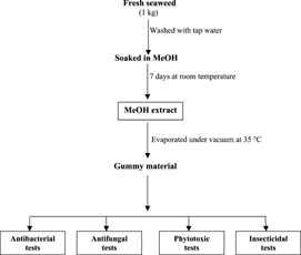
The fresh algal material (1 kg each) was soaked in MeOH for 7 days at room temperature (). The MeOH extract was filtered through Whatman filter paper and concentrated under reduced pressure at 35°C in a rotary evaporator. The crude gummy methanol extract (24 mg) was dissolved in 1 ml of sterile DMSO serving as stock solution. Sabouraud dextrose agar (SDA; Merck, Darmstadt, Germany) was prepared in screw-capped tubes and autoclaved at 121°C for 15 min (Paxton, Citation1991). Tubes were allowed to cool to 50°C, and nonsolidified SDA medium was mixed with 66.6 µl of stock solution giving a final concentration of 400 µg/ml of SDA. Tubes were then allowed to solidify in a slanting position at room temperature. Each tube was inoculated with a 4-mm-diameter piece of inoculum removed from a 7-day-old culture of fungi. For nonmycelial growth, an agar surface streak was employed. The various fungal organisms used are given in . The tubes were incubated at 27–29°C for 7–10 days (Atta-ur-Rahman et al., Citation2001). Growth inhibition was calculated in percentage and compared with standard antibiotic drugs, miconazole and ketoconazole, sometimes amphotericin B and benlate.
Figure 1 Scheme for algal extraction.
Lemna. bioassay
This bioassay was used to study the phytotoxic activity of MeOH extracts of the seaweeds on the plant Lemna aequinoctialis. Welv. The stock solutions were prepared by dissolving 30 mg of the crude extract in 1.5 ml of methanol. Nine flasks (three for each concentration) were inoculated with 1000, 100, and 10 µl of the stock solution for 1000, 100, and 10 µg/ml. The solvent was evaporated overnight under sterile conditions. To each flask, 20 ml of E-medium at pH of 5.5–6.0 was added. Then, 10 plants of Lemna aequinoctialis. having a rosette of three fronds were added to each flask. Two other flasks were supplemented with solvent and reference plant growth inhibitor as well as promoter serving as negative and positive controls, respectively. For a positive control, paraquat (ICI Pak. Ltd, Karachi, Pakistan) was used. The flasks were plugged with cotton and placed in a growth cabinet for 7 days. On the seventh day, the number of fronds per flask were counted (Atta-ur-Rahman, Citation1991). Interpretation of the result was made by analyzing growth regulation in percentage, which was calculated with reference to the negative control.
Insecticidal assay
This simple test was used to assess the insecticidal activity of each MeOH extract (200 mg) of the seaweeds. The insects were exposed to the test extracts by a direct contact method using filter paper impregnated with test sample (1571.33 µg/cm2, pyrethroid and permethrin, 1:1). Afterwards, 10 adult insects of different types and of the same age were transferred to Petri dishes. A negative control was treated with solvent for the determination of solvent effects. Another batch supplemented with reference insecticides (i.e., Mortein Coopex; Reckitt Benckiser Pak. Ltd., Karachi, Pakistan) was used. All these were kept without food for 24 h at 27–30°C. Mortality counts were carried out after a 24 h exposure period (Farhana, Citation2000).
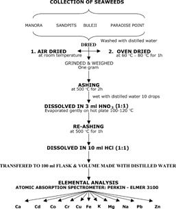
Ashing and digestion of the seaweeds
The algal material was initially dried under shade at room temperature and later in an oven at 60–80°C for 1 h. It was then powdered through a grinder, 1 g of the ground sample was accurately weighed in a porcelain crucible, and ashed at 500°C in an oven to constant weight for 2 h (). The ash was cooled at room temperature, wet with 10 drops of distilled water, and carefully dissolved in 3 ml of HNO3 (1:1). The acid solution of the sample was then heated gently on a hot plate at 100–120°C until it was nearly dry. The crucible was returned to a muffle furnace and ashed again for 1 h at 500°C. It was then cooled and dissolved in 10 ml of HCl (1: 1), and the solution was filtered through Whatman filter paper no. 42 (Schleir & Schuell, Dassel, Germany) into a 100 mL volumetric flask. The solution was then diluted to final volume with distilled water, mixed well, and prepared for AAS analysis (Jones, Citation1984).
Figure 2 Scheme for ashing and digestion of seaweeds.
Elemental assay
Flame atomic absorption spectrometry (AAS; model 3100, Perkin-Elmer, Norwalk, CT, USA) was performed at Hamdard University, Karachi, for the purpose of estimating Ca, Cd, Co, Cr, Cu, Fe, K, Mg, Na, Pb, and Zn. The various instrument parameters are presented in . Instructions for instrument setting, calibration, and assay for specific elements as described in the operational manual were strictly followed.
Table 3. Instrument parameters.
Results and Discussion
The results of antibacterial tests obtained on 14 seaweeds are shown in the , expressed in terms of zones of inhibition in millimeters. They indicate that 10 algal extracts inhibited the growth of Gram-positive and Gram-negative bacteria. The values higher than 12 were replicated two times, and an average was obtained. Extracts from Ulva intestinalis., Stoechospermum polypodioides., and Osmundea pinnatifida. could prevent the growth of only one bacterium, and extracts from Sargassum ilicifolium. and Champia compressa. inhibited only two bacteria. Extracts from Colpomenia sinuosa. and Iyengaria stellata. showed positive activity against six bacteria including three Gram-positive and three Gram-negative bacterial strains that appeared to be the most active, and the extracts from Cystoseria indica. exhibited positive activity against four bacteria, indicating that brown seaweeds have more active antibacterial components than do the green and red ones. Extracts from Codium iyengarii., Ulva fasciata., and Padina antillarum. showed no inhibition against all nine of the bacteria that were investigated. Similar observations have been made on a variety of algae such as Ulva compressa., Padina gymnospora., Sargassum wightii., and Gracillaria corticata. that were active against Gram-positive cultures of Bacillus. (Rao et al., Citation1991). It has also been reported that extracts from Gracilaria corticata. and Padina gymnospora. show no antibacterial activity against Bacillus megaterium. and Staphylococcus aureus. Rosenbach (Ahmad & Perveen, Citation1993).
Table 4. Antibacterial activity of crude methanol extract of different seaweeds shown as zone of inhibition in millimeters.
Twenty-three species of marine benthic algae belonging to the Chlorophyta, Phaeophyta, and Rhodophyta were tested against seven species of human, three species of animal, and three species of plant pathogens for in vitro. fungicidal activity (). Treatments were replicated two times, and an average value was obtained. Only five species of marine algae exhibited moderate activity (45–50%). Extracts from Botryocladia leptopoda. appeared to be the most active, whereas extracts from Codium shameelii. were the least active. Extracts from Codium iyengarii. displayed good activity, and in other observations this extract showed significant antifungal activity against a variety of pathogens (Ali et al., Citation2000). The crude methanol extract of Stoechospermum polypodioides. has been found to inhibit the growth of Micrococcus pyrogenes. Lehman et Neumann var. aureus. Hucker (Anonymous, Citation2000). The ethanol extracts derived from seven seaweed species showed no detectable antifungal activity against Epidermophyton floccose. (Harz) Langeron et Milochevitch, Microsporum canis. Bodin, and Trichophyton rubrum. (Castellani) Sabouraud (Alam et al., Citation1994). It appears that different seaweed extracts behave variably against different fungal species.
Table 5. Antifungal activity of crude MeOH extract of different seaweeds shown as % inhibition.
The results of the Lemna. bioassay of methanol extracts of the investigated seaweeds at concentrations of 10, 100, and 1000 µg/ml are presented in , and an average phytotoxic activity of the three mentioned concentrations is graphically expressed in . The extracts from all the seaweeds inhibited the growth of Lemna aequinoctialis.. The extract from Ulva intestinalis. displayed the highest activity, followed by the extract from Champia compressa.. The extracts from Codium shameelii. and Sargassum boveanum. exhibited poor activity, and in another observation, the ethanol extract from S. tenerrimum. showed 100% inhibition of the fronds at a concentration of 500 µg/ml (Ali et al., Citation2000). It appears that different species of the same genus act variably. Probably they accumulate different natural products, which may be responsible for phytotoxic activity. As compared to the extracts from green seaweeds displaying great fluctuations in their activity, the extracts from the investigated brown seaweeds exhibited uniformity in their phytotoxic activities.
Figure 3 Average phytotoxic activity: 1 = Codium shameelii., 2 = Ulva fasciata., 3 = Ulva intestinalis., 4 = Colpomenia sinuosa., 5 = Cystoseira indica., 6 = Dictyota hauckiana., 7 = Iyengaria stellata., 8 = Sargassum boveanum., 9 = Botryocladia leptopoda., 10 = Champia compressa., 11 = Hypnea musciformis., 12 = Osmundea pinnatifida..
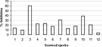
Table 6. Phytotoxic activity of seaweeds in % inhibition.
For insecticidal bioassays, eight marine algae were chosen, and their crude extracts were tested against five different insects, Callosobruchus analis. Fabricius, Rhyzopertha dominica. Fabricius, Sitophilus oryzae. L. Fabricius, Tribolium castaneum. Herbst, and Trogoderma granarium. Everts (). Treatments were replicated two times, and an average value was obtained. Ulva intestinalis, Sargassum boveanum., and Osmundea pinnatifida. exhibited moderate activity, whereas Ulva fasciata., Sargassum ilicifolium., and Champia compressa. showed a very poor activity against these insects. Botryocladia lepdopoda. and Hypnea musciformis. displayed no activity. Osmundea pinnatifida. appeared to be the most active seaweed: it was found to contain a large variety of natural products (Ali et al., Citation2000), which might be responsible for such activity.
Table 7. Insecticidal test for seaweeds in % mortality.
Twenty-six species of seaweeds were analyzed for the composition of 11 elements (). Among these elements, Ca, Fe, K, Mg, and Na were found in large amounts (on the average 2411.38–76714.25 ppm), Cr, Cu, Pb, and Zn were present in small amounts (on the average 5.88–53.28 ppm), and Cd and Cr were detected in extremely small amounts (on the average 1.61–5.13 ppm). Iron has been found in large quantity, Cr and Zn in medium quantity, and Co in small quantity in several brown seaweeds of the Saronic Gulf, Greece (Kanias et al., Citation1991). The intestinal absorption of seaweed minerals (Ca, Fe, and Zn) exhibited an interesting result when observed in vivo. using a perfused intestinal loop in the rat (Bougle et al., Citation1996). The average quantity of Na was found to be the highest among investigated algae (76714.25 ppm) followed by K (69535.17 ppm) and Ca (26746.67 ppm). The average amounts of Co (5.88 ppm) and Cr (5.13 ppm) were quite low; Cd was detected in the lowest quantity (1.61 ppm). However, Cd was present in smallest amount in Chlorophyta (1.38) as compared to Phaeophyta (1.18) as well as Rhodophyta (2.27 ppm). It was also detected in very small quantity in several other species of green, brown, and red seaweeds (Hasni & Sarwar, Citation1985; Rizvi & Shameel, Citation2001). In general, Ca, Cr, and Pb were found to occur in highest proportion in green seaweeds, Co, Cu, Fe, and Zn in highest proportion in brown seaweeds, and Cd, K, Mg, and Na in highest proportion in the investigated red seaweeds. When collectively considered, the average quantity of the detected 11 elements was highest (20,645.31 ppm) in the red seaweeds and lowest (12,276.43 ppm) in the brown seaweeds, and the average amount of green seaweeds was in-between (15,544.42). In many studies, the quantity of different essential elements was higher in red seaweeds as compared to other groups (Ganesan et al., Citation1991; Munda & Hudnick, Citation1991; Rajendran et al., Citation1993).
Table 8. Elemental composition in seaweeds from the Karachi coast.
Kanias et al. (Citation1991) determined trace elements in the dry matter of brown algae of the Saronic Gulf. Some species of marine algae from the coast of Goa, India, have been analyzed for Co, Cu, Fe, Mn, Ni, Pb, and Zn (Forsk) C. Ag. (Hoppe & Levring, Citation1982). Ulva lactuca. (Forsk) C. Ag. from China and Southeast Asia, has been reported to be rich in iron (Ahmad et al., Citation1989). The edible green alga Codium intricatum. (Mosure-miru) was found to contain a considerable quantity of iodine (0.13–0.16% of the dry weight), and red algae such as Gelidium. and Grateloupia. contained a medium amount (Chapman & Champan, Citation1980). The trace metal distribution in seaweeds of the Indian Coast has also been well documented. Metal concentration in the seaweeds were in the order Fe > Mn > Zn > Cu with the exception of a few seaweeds (e.g., Ulva. reticulata., Sargassum wightii Grev.., and Sarconema. sp.), which concentrated more Zn than Mn. Seaweeds from other tropical areas exhibit a similar trend. It seems that the tropical seaweeds tend to accumulate more Fe than Mn, Zn, and Cu (Ganesan et al., Citation1991).
This type of research work is to be continued in order to know the proportion of those elements in algal thalli that are beneficial for the human body, such as Ca, Fe, I, K, Na, and Zn, and also to investigate the distribution of various elements in different thallus parts of marine benthic algae for the sake of comparison. Some seaweeds are used as food because they are not poisonous, usually have soft tissues, and as such have many indirect medicinal effects.
Acknowledgments
The Authors are greatly indebted to the Vice Chancellor, Director, Hamdard Institute of Advanced Studies & Research, Hamdard University, and to Prof. Dr. M. Iqbal Choudhary, H.E.J. Research Institute of Chemistry, University of Karachi, for his keen interest. This was supported by a research grant from the U.S. Office of Naval Research (ONR Res. Proj. No. NP-3), for which we are grateful to Prof. Dr. Viqar Uddin Ahmad, P. I. of the ONR Research Projects.
References
- Afaq-Hussain S, Nizamuddin M, Shameel M (1991): The structure and reproduction of a new taxon Dermonema abbottiae. (Nemaliales-Rhodophyta) from the coast of Pakistan. Pak J Sci Ind Res 34: 75–82.
- Ahmad J, Ganapathy SN, Siddiqi TO, Hamdard ME (1989): The distribution of elements in some plant species of the botanical kingdom. In: Said M, Rahman MA, D'Silva LA, eds., Elements in Health and Disease. Karachi, Hamdard University Press, pp. 143–167.
- Ahmad VU, Perveen S (1993): Medicinal significance of marine algae. Hamd Med 36: 33–35.
- Alam K, Agua T, Maven R, Taie R, Rao KS, Burrows I, Huber ME, Rali T (1994): Preliminary screening of seaweeds, seagrass and lemongrass oil from Papupa New Guinea for antimicrobial and antifungal activity. Int J Pharmacog 32: 396–399.
- Ali MS, Mazhar F, Saleem M, Jahangir M, Pervez K, Usmanghani K, Ahmad VU (2000): Chemistry and biology of algae from sea coasts of Karachi. In: Ahmad VU, ed., Proceedings of the National ONR Symposium on Arabian Sea as a Resource of Biological Diversity. HEJ Res Ins Chem, Karachi University, pp. 3–44.
- Aliya R, Shameel M (1999): Phytochemical evaluation of four coenocytic green seaweeds from the coast of Karachi. Pak J Mar Biol (Mar. Res.) 5: 65–76.
- Anand PL (1940): Marine Algae from Karachi. I. Chlorophyceae. Lahore, Punjab University Botany Publications, 52 pp.
- Anand PL (1943): Marine Algae from Karachi. II. Rhodophyceae. Lahore, Punjab University Botany Publications, 76 pp.
- Anonymous (1985): The Wealth of India (Raw Materials). Vol. 1 A. New Delhi, CSIR, 138–159.
- Anonymous (2000): The Wealth of India (Raw Materials). Vol. 1 A (Revised). New Delhi, CSIR, 38–40.
- Atta-ur-Rahman (1991): Studies in Natural Product Chemistry, Vol. 9 (Part-B). Amsterdam, Elsevier Science Publications, 383 pp.
- Atta-ur-Rahman, Choudhary MI, Thomsen WJ (2001): Bioassay Techniques for Drug Development. Amsterdam, The Netherlands, Harwood Academy Publications, 222 pp.
- Baslow, MH (1969): Marine Pharmacology. Baltimore, William and Wilkins, pp. 56–85.
- Bougle D, Boudey M, Arhan P, Bureau F, Neuville D, Drosdowsky M (1996): In vivo. study of the absorption of seaweed mineral by perfused rat intestine. Phytother Res 10: 325–326. [CROSSREF], [CROSSREF]
- Carron R, Maran A, Montero JM, Fernandozlago, Derminguez A (1987): Plantes. Med Phytotherap 21: 195.
- Chapman VJ, Chapman DJ (1980): Seaweeds and Their Uses, 3rd ed. New York: Chapman and Hall, pp. 62–96.
- Farhana K (2000): Screening of insecticides by impregnation method on storage pests. In: Manual of Bioassay Techniques. Workshop on techniques for product development from medicinal plants, HEJ Research Institute of Chemistry, Pakistan and Inter-Islamic Network on Tropical Medicine, Malaysia, pp. 15–17.
- Ganesan M, Kannan R, Rajendran K, Govindasamy C, Sampathkumar P, Kannan L (1991): Trace metals distribution in seaweeds of the Gulf of Mannar, Bay of Bengal. Mar Pollut Bull 22: 205–207. [CROSSREF], [CROSSREF], [CSA]
- Hameed S, Ahmad M, Shameel M (2000): An ecological study on the tide pools of the rocky ledge at Pacha, near Karachi (Pakistan). Pak J Mar Biol 6: 179–197. [CSA]
- Hameed S, Ahmad M, Shameel M (2001): Common structure and species composition of macroorganisms at the rocky bench at Pacha, near Karachi, Pakistan. Pak J Mar Biol 7: 135–146. [CSA]
- Hasni S, Sarwar M (1985): Nutritional analysis of few seaweeds found at Karachi coast. J Pharm Univ Kar 4: 21–29. [CSA]
- Hoppe HA, Levring T (1982): Marine Algae in Pharmaceutical Sciences, Vol. 2, Berlin, Germany, Walter de Gruyter, pp. 3–48.
- Jarvis DC (1976): Folk Medicine. London, Pan Books, pp. 120–132.
- Jones JB (1984): Plants. In: William S, ed, An Official Method of Analysis. Association of official Analytical Chemists, pp. 38–64.
- Kanias G, Skaltsa H, Tsitsa E, Loukis A, Bitis J (1991): Comparative phytochemical and pharmacological study of eight brown algae from the Saronic Gulf (Greece). Planta Med 57: 399–590.
- Kaur I (1997): Potentials and future prospects. In: Vijayaraghavan MR, Kaur I, eds., Brown Algae. New Delhi, India, APH Publishing, pp. 277–294.
- Munda IM, Hudnik V (1991): Trace metal content in some seaweeds from northern Adriatic. Bot Mar 34: 241–249. [CSA]
- Naqvi SWA, Solimabi S, Kamat SY, Fernandez L, Reddy CVG, Bhakuni DS, Dhawan BN (1980): Screening of some marine plants from the Indian coast for biological activity. Bot Mar 24: 51–55. [CSA]
- Nizamuddin M (1963): Studies on the green alga, Udotea indica. A. & E. S. Gepp 1911. Pac Sci 17: 243–245.
- Nizamuddin M (1964): Studies on the genus Caulerpa. from Karachi. Bot Mar 6: 204–223.
- Paxton JD (1991): Assay for antifungal activity. In: Dey PM, Harborne JB, Hostettmann K, eds., Methods in Plants Biochemistry. London, Academic Press, pp. 33–46.
- Rajendran K, Sampathkumar P, Govindasamy C, Ganesan M, Kannan R, Kannan L (1993): Levels of trace metals (Mn, Fe, Cu, and Zn) in some Indian seaweeds. Mar Pollut Bull 26: 283–285. [CROSSREF], [CROSSREF], [CSA]
- Rao DS, Girijavallabhan KG, Muthusamy S, Chandrika V, Gopinathan CP, Kalimuthu S. Najamuddin M (1991): Bio-acivity of marine algae. In: Thompson M-F, Sarojini R, Nagabhushanam R, eds., Bioactive Compounds from Marine Organisms with Emphasis on the Indian Ocean. New Delhi, India, Oxford & IBH Publishing, pp. 373–377.
- Rizvi MA, Shameel M (2001): Distribution of elements in marine algae of Karachi coast. Pak J Bot 33: 357–363.
- Saifullah SM (1973): A preliminary survey of the standing crop of seaweeds from Karachi coast. Bot Mar 16: 139–144. [CSA]
- Shameel M (1987): A preliminary survey of seaweeds from the coast of Lasbela, Pakistan. Bot Mar 30: 511–515. [CSA]
- Shameel M, Aisha K, Khan SH (1996): A preliminary survey of seaweeds from the coast of Makran, Pakistan. Bot Mar 39: 223–230. [CSA]
- Shameel M, Khan SH, Afaq-Husain S (2000): Biodiversity of marine benthic algae along the coast of Balochistan, Pakistan. Pak J Mar Biol 6: 69–100. [CSA]
- Shameel M, Tanaka J (1992): A preliminary checklist of marine algae from the coast and inshore waters of Pakistan. In: Nakaike T, Malik S, eds., Cryptogamic Flora of Pakistan, Vol. I. National Science Museum, Tokyo, pp. 1–64.
- Usmanghani K, Shameel M (1996): Fatty acid composition of seaweeds of Pakistan. Pak J Pharm Sci 9: 53–68. [CSA]
- Usmani, JN, Mahboob A, Kalhoro and Ismail S (1991) Biological activities in seaweeds. Pak J Sci Ind Res 34: 247–248.