Abstract
Seven pure flavonolignans were isolated from an extract of milk thistle [Silybum marianum.(L.) Gaertn (Asteraceae)], by semipreparative reverse-phase HPLC, and identified based on spectroscopic and LC-MS/IT-TOF data. All seven compounds were screened as potential antitumor-promoting agents by using the in vitro. short-term 12-O.-tetradecanoylphorbol-13-acetate (TPA)-induced Epstein-Barr virus early antigen (EBV-EA) activation assay in Raji cells. They showed good inhibitory activity (87.7–94.9%) at 1000 mol ratio/TPA. Silychristin A (1) and silychristin B (2) were slightly more potent than the well-known antitumor promoter β.-carotene. Silychristins A (1) and B (2) and isosilybins A (6) and B (7) were more active than the clinically proven cancer prevention components silybins A (4) and B (5).
Introduction
Milk thistle, Silybum marianum. (L.) Gaertn (Asteraceae), is native to the Mediterranean area and was grown in southern Europe as a vegetable (Ball & Kowdley, Citation2005). Extracts of S. marinaum. have been used as traditional medicine since the time of ancient Greece (Flora et al., Citation1998; Dhiman & Chawla, Citation2005; Rambaldi et al., Citation2005). Silymarin is the collective name of several flavonolignans, including silybinin, silydianin, and silychristin, extracted from S. marianum. (Luper, Citation1998; Crocenzi & Roma, Citation2006). For more than 25 years, silymarin has been used clinically in Europe as an antihepatotoxic agent (Bhatia et al., Citation1999), and in recent years, milk thistle has become one of the most popular herbal remedies used by patients with liver disease and ranked 10th in U.S. sales among all supplements sold in 2000 (Jacobs et al., Citation2002).
Previous studies have related the hepatoprotective effects of silymarin to its antioxidant and strong free radical scavenging effects (Luper et al., Citation1998) and more recently to anticholestatic properties (Crocenzi & Roma, Citation2006). In addition, other studies have reported its effects in the prevention and therapy of cancer as summarized recently by Aggarwal and Shishodia (Citation2006). The anticancer effect of silymarin has been linked to the main constituent, silybin, in milk thistle. Silybin consists of two isomers, silybin A and silybin B, which have shown antineoplastic activity in several cancer models, and a phase I clinical study in prostate cancer patients was reported recently (Flaig et al., Citation2007). Additional studies of silybin in cancer prevention were also summarized (Katiyar, Citation2005; Singh & Agrawal, Citation2005). In our continuing investigation of antitumor-promoting agents, an extract of milk thistle was subjected to high-performance liquid chromatography (HPLC). Seven flavonolignans were collected and identified based on spectroscopic and LC-MS/IT-TOF data. All seven compounds were screened for antitumor-promoting effects. The results showed that these compounds are potent inhibitors of skin tumor promotion.
Materials and Methods
Chemistry
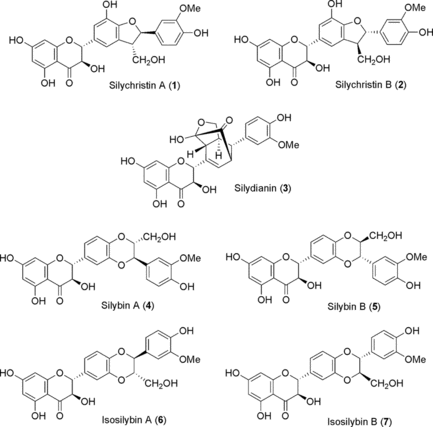
Milk thistle extract was dissolved in methanol and separated on a semipreparative HPLC system (Shimadzu SLC 10A system controller, Shimadzu LC-20A pump) with a photodiode array detector (Shimadzu SPD-M10A detector). The separation was performed on a Alltech Alltima C18-5 22 i.d. × 250 mm column. Seven major peaks were collected at UV absorption 254 nm with an isocratic methanol-water (52:48) solvent system. At a flow rate of 4.5 mL/min, the retention times for each compound were 34.8, 38.8, 44.5, 87.9, 97.6, 126.2, and 135.0 min, respectively. From UV, 1H NMR, 13C NMR, and LC-MS/IT-TOF data, these compounds were identified as silychristin A (1), silychristin B (2), silydianin (3), silybin A (4), silybin B (5), isosilybin A (6) and isosilybin B (7) (), and these assignments were confirmed by comparing their spectroscopic data with prior literature (Lee & Liu, Citation2003; Smith et al., Citation2005).
In vitro. EBV-EA activation assay
Epstein-Barr virus early antigen (EBV-EA) positive serum from a patient with nasopharyngeal cancer (NPC) was provided by Professor H. Hattori (Department of Otorhinolaryngology, Kobe University). The EBV genome-carrying human lymphoblastoid cell (Rahi cells derived from Burkitt lymphoma) were cultivated in 10% fetal bovine serum (FBS) RPMI 1640 medium (Sigma R8758; Sigma, St. Louis, MO, USA). Spontaneous activation of EBV-EA in a subline of Raji cells was less than 0.1%. The inhibition of EBV-EA activation was assayed using Raji cells (virus nonproducer type) incubated at 37°C for 48 h in 1 mL of medium containing n.-butyric acid (4 mM, inducer), TPA (32 pM = 20 ng in 2 µL DMSO), and various amounts of the test compound (1–7) dissolved in 5 µL DMSO. Smears were made from the cell suspension. The EBV-EA inducing cells were stained with high-titer EBV-EA positive serum from NPC patients and detected by an indirect immunofluorescence technique. In each assay, 500 cells were counted, and the number of stained cells (positive cells) was recorded. Triplicate assays were performed for each data point. The average EBV-EA induction of the test compound was expressed as a ratio relative to the control experiment (100%), which was carried out with n.-butyric acid (4 mM) plus TPA (32 pM). EBV induction was ordinarily around 35%, and the viability of treated Raji cells was assayed by Trypan blue staining method. The cell viability of the TPA positive control was greater than 80%. Only compounds that induced less than 80% (% of control) of the EBV-active cells (those with a cell viability of more than 60%) were considered able to inhibit the activation caused by promoter substances.
Cytotoxicity determination
For the determination of the cytotoxicity or cell viability of surviving cells, the Trypan blue staining method was used. After EBV-EA activating assays, 0.1 mL of treated cells (suspended in PBS) was stained with 0.1 mL of 0.25% Trypan blue solution. Dead cells were stained blue. Viable unstained cells were counted.
Results and Discussion
The biological screening was carried out by using a short-term in vitro. synergistic assay on EBV-EA activation induced by TPA (Henle & Henle, Citation1966; Takasaki et al., Citation1990). In this assay, we used three different reference compounds, β.-carotene, curcumin, and glyzyrrhizin, which are all widely studied natural products and known to be active in this widely used test for cancer prevention using animal models (Suzuki et al., Citation2006; Wang et al., Citation2006). In this test system, all seven compounds showed potent inhibition of EBV activation (), which was comparable or better than that of the reference compounds. In addition, all of the test compounds showed low cytotoxicity toward Raji cells (IC50 398–482 µM). The results in show that the isomeric compounds 1 and 2 had significant EBV-EA inhibition effects (92.7% and 64.1% for 1; 94.9% and 66.7% for 2) at test concentrations of 1000 and 500 mol ratio/TPA, respectively, and were the two most potent compounds among all test samples. Compounds 6 and 7 showed similar inhibition effects to those of β.-carotene (91.4% inhibition at 1000 mol ratio/TPA, 65.8% at 500 mol ratio/TPA). Moreover, at concentrations of 100 and 10 mol ratio/TPA, 6 and 7 displayed greater inhibition than β.-carotene. Compounds 3, 4, and 5 also showed good inhibition effects at concentrations of 1000, 500, and 100 mol ratio/TPA.
Table 1.. Inhibitory effects of compounds 1–7 on TPA-mediated EBV-EA induction.
Silybins A and B (4 and 5) are the major components in the active extract of milk thistle (silymarin) and have been linked previously to skin cancer prevention effects via anti-inflammatory, antioxidant, and immunomodulatory mechanisms (Katiyar, Citation2005). Our study results now reveal that all seven major compounds in silymarin show good EBV-EA inhibition activities. In our testing model, silychristins A and B (1 and 2) and isosilybins A and B (6 and 7) had better inhibition effects compared with β.-carotene, and silybins A and B (4 and 5) showed lower activity compared with the other flavonolignans in silymarin. Further in vivo. testing on mouse skin papillomas is ongoing.
In conclusion, seven pure compounds silychristin A (1), silychristin B (2), silydianin (3), silybin A (4), silybin B (5), isosilybin A (6), and isosilybin B (7) were isolated from an extract of milk thistle. Evaluation with an in vitro. EBV-EA activation assay showed that silychristin B (2) was the most active compound with 94.9% EBV-EA inhibition at 1000 mol ratio/TPA. As it also showed low cytotoxicity, 2 could be valuable as an antitumor promoter or as a lead compound for new cancer preventive drug development.
Acknowledgments
This investigation was supported in part by grant CA-17625 from the National Cancer Institute and GM-076152 from National Institute of General Medial Sciences, NIH, awarded to K.H.L. This study was also supported in part by a grant from the Ministry of Education, Sciences, Sports and Culture, and the Ministry of Health and Welfare, Japan (H.T.). Thanks are due to Dr. Takashi Tatsuzaki of Tokiwa Phytochemical Co., Ltd, for the gift of Silybum marianum. extract.
References
- Aggarwal BB, Shishodia S (2006): Molecular targets of dietary agents for prevention and therapy of cancer. Biochem Pharmacol 71: 1397–1421.
- Ball KR, Kowdley KV (2005): A review of Silybum marianum. (milk thistle) as a treatment for alcoholic liver disease. J Clin Gastroenterol 39: 520–528.
- Bhatia N, Zhao J, Wolf DM, Agarwal R (1999): Inhibition of human carcinoma cell growth and DNA synthesis by silibinin, an active constituent of milk thistle: Comparison with silymarin. Cancer Lett 147: 77–84.
- Crocenzi FA, Roma MG (2006): Silymarin as a new hepatoprotective agent in experimental cholestasis: New possibilities for an ancient medication. Curr Med Chem 13: 1055–1074.
- Dhiman RK, Chawla YK (2005): Herbal medicines for liver diseases. Dig Dis Sci 50: 1807–1812.
- Flaig TW, Gustafson DL, Su L-J, Zirrolli JA, Crighton F, Harrison GS, Pierson AS, Agarwal R, Glode LM (2007): A phase I and pharmacokinetic study of silybin-phytosome in prostate cancer patients. Invest New Drugs 25: 139–146.
- Flora K, Hahn M, Rosen H, Benner K (1998): Milk thistle (Silybum marianum.) for the therapy of liver disease. Am J Gastroenterol 93: 140–143.
- Henle G, Henle W (1966): Immunofluorescence in cells derived from Burkitt's lymphoma. J Bacteriol 91: 1248–1256.
- Jacobs BP, Dennehy C, Ramirez G, Sapp J, Lawrence V (2002): Milk thistle for the treatment of liver disease: A systematic review and meta-analysis. Am J Med 113: 506–515.
- Katiyar S (2005): Silymarin and skin cancer prevention: Anti-inflammatory, antioxidant and immunomodulatory effects (review). Int J Oncol 26: 169–176.
- Lee DY-W, Liu Y (2003): Molecular structure and stereochemistry of silybin A, silybin B, isosilybin A, and isosilybin B, isolated from Silybum marianum. (milk thistle). J Nat Prod 66: 1171–1174.
- Luper S (1998): A review of plants used in the treatment of liver disease: Part 1. Altern Med Rev 3: 410–421.
- Rambaldi A, Jacobs BP, Iaquinto G, Gluud C (2005): Milk thistle for alcoholic and/or hepatitis B or C liver diseases—a systematic Cochrane hepato-biliary group review with meta-analyses of randomized clinical trials. Am J Gastroenterol 100: 2583–2591.
- Singh RP, Agarwal R (2005): Mechanisms and preclinical efficacy of silibinin in preventing skin cancer. Eur J Cancer 41: 1969–1979.
- Smith WA, Lauren DR, Burgrss EJ, Perry NB, Martin RJ (2005): A silychristin isomer and variation of flavonolignan levels in milk thistle (Silybum marianum.) fruits. Planta Med 71: 877–880.
- Suzuki M, Nakagawa-Goto K, Nakamura S, Tokuda H, Morris-Natschke SL, Kozuka M, Nishino H, Lee K-H (2006): Cancer preventive agents. Part 5. Anti-tumor-promoting effects of coumarins and related compounds on Epstein-Barr virus activation and two-stage mouse skin carcinogenesis. Pharm Biol 44: 178–182.
- Takasaki M, Konoshima T, Fujitani K, Yoshida A, Nishimura H, Tokuda H, Nishino H, Iwashima A, Kozuka M (1990): Inhibitors of skin-tumor promotion. VIII. Inhibitory effects of euglobals and their related compounds on Epstein-Barr virus activation. (1). Chem Pharm Bull 38: 2737–2739.
- Wang X, Nakagawa-Goto K, Kozuka M, Tokuda H, Nishino H, Lee K-H (2006): Cancer preventive agents. Part 6: Chemopreventive potential of furanocoumarins and related compounds. Pharm Biol 44: 116–120.
