
Abstract
Context
Shen-Shi-Jiang-Zhuo formula (SSJZF) exhibits a definite curative effect in the clinical treatment of non-alcoholic fatty liver disease (NAFLD).
Objective
To explore the therapeutic effect and mechanism of SSJZF on NAFLD.
Materials and methods
Sprague Dawley rats were randomly divided into control, NAFLD, positive drug (12 mg/kg/day), SSJZF high-dose (200 mg/kg/day), SSJZF middle-dose (100 mg/kg/day), and SSJZF low-dose (50 mg/kg/day) groups. After daily intragastric administration of NAFLD rats for 8 weeks, lipid metabolism and hepatic fibrosis were evaluated by biochemical indices and histopathology. Then we uncovered the main active compounds and mechanism of SSJZF against NAFLD by integrating RNA-sequencing and network pharmacology, and PI3K/AKT pathway activity was verified by western blot.
Results
High dose SSJZF had the best inhibitory effect on hepatic lipid accumulation and fibrosis in rats with NAFLD, which significantly down-regulated total triglycerides (58%), cholesterol (62%), aspartate aminotransferase (57%), alanine aminotransferase (41%) andγ-glutamyl transpeptidase (36%), as well as the expression of ACC (5.3-fold), FAS (12.1-fold), SREBP1C (2.3-fold), and CD36 (4.4-fold), and significantly reduced collagen deposition (67%). Then we identified 23 compounds of SSJZF that acted on 25 key therapeutic targets of NAFLD by integrating RNA-sequencing and network pharmacology. Finally, we also confirmed that high dose SSJZF increased p-PI3K/PI3K (1.6-fold) and p-AKT/AKT (1.6-fold) in NAFLD rats.
Discussion and conclusion
We found for first time that SSJZF improved NAFLD in rats by activating the PI3K/Akt pathway. These findings provide scientific support for SSJZF in the clinical treatment of NAFLD and contribute to the development of new NAFLD drugs.
Introduction
Non-alcoholic fatty liver disease (NAFLD) is the most common chronic liver disease characterised by excess lipid accumulation in liver without alcohol consumption (Brunt et al. Citation2015). NAFLD includes benign non-alcoholic fatty liver disease and the more severe form of non-alcoholic steatohepatitis (NASH), which can develop into cirrhosis and hepatocellular carcinoma (Cobbina and Akhlaghi Citation2017). NAFLD has rapidly become the most prevalent cause of liver dysfunction around the world, as lifestyles become increasingly sedentary and dietary patterns change (Zeng et al. Citation2020). Lifestyle modification, including weight loss and physical exercise, is still the key of NAFLD prevention and treatment (Younossi et al. Citation2019; Zeng et al. Citation2020). The current medications of NAFLD include metformin, antihyperlipidemic drugs, antioxidants, etc., which may exert serious side effects (Riley et al. Citation2008; Chalasani et al. Citation2018; Zsori et al. Citation2019). Thus, it is of great significance to explore more effective interventions for the treatment of NAFLD (Cobbina and Akhlaghi Citation2017).
Traditional Chinese medicine (TCM), as an important complementary and alternative medicine approach, has been shown efficacy and safety in treating complex diseases, including NAFLD (Qin et al. Citation2020; Sun et al. Citation2021). In TCM, the treatment of NAFLD mainly focussed on the holism of hepatoprotection, which manifests as various forms and features in mechanism, including lipid metabolism modulation and anti-fibrosis, etc. (Dai et al. Citation2021). As evidenced by animal and cellular studies indicated that quercetin exerts hepatoprotection against NAFLD by antihepatic steatosis or antifibrosis (Marcolin et al. Citation2012; Chen et al. Citation2021). Studies have shown that resveratrol can inhibit the accumulation of hepatic lipids and improve hepatic fibrosis for treatment of NAFLD (Izzo et al. Citation2021; Tejada et al. Citation2021). Shen-Shi-Jiang-Zhuo Formula (SSJZF) is a TCM formula consisting of Atractylodes lancea (Thunb.) DC. (Compositae), Poria cocos (Schwein) F.A. Wolf (Polyporaceae), Magnolia officinalis (Rehder & E. H. Wilson) N. H. Xia & C. Y. Wu (Magnoliaceae), Rhizoma Pinelliae (Thunb.) Breit. (Araceae), Talcum (Silicates), Tetrapanax papyriferus (Hook.) K. Koch (Araliaceae), Radix Bupleuri (Umbelliferae), Citrus aurantium L. (Rutaceae), Radix Paeoniae rubra (Ranunculaceae), Panax notoginseng (Burkill) F.H. Chen (Araliaceae), Rubia cordifolia Linn (Rubiaceae), Fructus Broussonetiae (Moraceae), and Pinellia ternate (Thunb.) Ten. ex Breitenb (Araceae). Our previous research found that SSJZF could alleviate the symptoms of NAFLD patients through improving liver function, as well as reducing the lipid levels of NAFLD patients (Lu et al. Citation2018). However, the underlying mechanisms and active compounds of SSJZF in the treatment of NAFLD remained unknown.
Over the past decades, with the rapid development of high-throughput analysis technologies, mRNA expression profiling has been widely used as an effective tool to explore molecular mechanisms of drug treatment of disease and uncover new therapeutic targets. For example, Cheng et al. (Citation2019) determined that SAHA is a new treatment method for NAFLD by drug screening based on mRNA expression profile. Zhu, Li, et al. (2019) used high-throughput RNA sequencing to explore mRNA expression profiling by methionine-and-choline-deficient-induced NAFLD with or without Qinggang formula and found the protective effect of Qinggan formula on NFLDA was related to immune function. In addition, network pharmacology is a newly-developed discipline based on the large number of database resources. Network pharmacology has been widely used to explore therapeutic targets and underlying mechanisms of TCM treatment, such as discovery of TCM-derived target, prediction of pharmacological action, new multicomponent and formula discovery, etc. (Zhang et al. Citation2013; Jiang et al. Citation2015; Sun et al. Citation2020; Zhou et al. Citation2020). In brief, network pharmacology is an attractive modality to explore the association of multi-targets and multi compounds on the disease (Hopkins Citation2007, Citation2008). Network pharmacology can identify some key active ingredients and protein targets of TCM formulas involved with the disease (Chen et al. Citation2018; Lee et al. Citation2018). Furthermore, Li et al. (2021) proved that the multi-target regulation and compatibility mechanism of Gegen Qinlian Decoction on the Wnt signalling pathway by using an integrated strategy of network pharmacology and RNA-sequencing. Zhou et al. (2021) showed that base on network pharmacology combination with transcriptomics analysis to develop a novel anti-liver fibrosis new formula from Xiaoyaosan decoction, and its anti-liver fibrosis function may be related to the regulation of PI3K/AKT and JAK/STAT signalling.
In the present study, we used the rat model of high-fat-diet (HFD)-induced NAFLD to explore whether the protective effect of SSJZF on NAFLD was realised by improving hepatic lipid accumulation in clinical practice. Further, we used comprehensive analysis of mRNA-sequencing data mining, network pharmacology and bio-informatics to investigate the underlying mechanisms and effective compounds SSJZF against NAFLD, which provided theoretical basis for the application of SSJZF in clinical treatment of NAFLD.
Methods and methods
Drug preparation
SSJZF consists of Atractylodes lancea (Guangdong YuanSenTai Pharmaceutical Co., Ltd., No.190801), Poria cocos (Anhui XieHe ChengYao Decoction Pieces Co., Ltd., No.19111902), Magnolia officinalis (Traditional Chinese Medicine Decoction Pieces Factory of Guangdong Medicine Company, No.H4920711), Rhizoma Pinelliae (Sichuan Qianfang Traditional Chinese Medicine Co., Ltd., No.202007105), Talcum (Sichuan Qianfang Traditional Chinese Medicine Co., Ltd., No.20200701), Tetrapanax papyriferus (Bozhou Yonggang Decoction Pieces Factory Co., Ltd., No.200618), Radix Bupleuri (Traditional Chinese Medicine Decoction Pieces Factory of Guangdong Medicine Company, No.B5720214), Citrus aurantium (Guangdong YuanSenTai Pharmaceutical Co., Ltd., No.191001), Radix paeoniae rubra (Sinopharm Holdings Shenzhen Medicinal Materials Co., Ltd., No.200401), Panax notoginseng (Traditional Chinese Medicine Decoction Pieces Factory of Guangdong Medicine Company, No.S4420712), Rubia cordifolia (Inner Mongolia Yikang Pharmaceutical Co., Ltd., No19102301), and Fructus Broussonetiae (Anhui Boyao Qiancao Decoction Pieces Co., Ltd., No.1906054).
In accordance with the dose ratio of SSJZF (), the mixtures of crude herbs were immersed in 6 volumes of distilled water (v/w) for 60 min. After heating and refluxing for 2 h, the decoction was collected. The decoction was filtered, and the residue with 6 volumes of water refluxed again for 2 h. Finally, the water extract solution was concentrated to dryness by a rotary evaporator and stored at 4 °C for further study. The solid drugs obtained in the previous step were diluted gradient into 40, 20, and 10 mg/mL by using normal saline, and then the NAFLD rats were treated by intragastric administration according to low- (50 mg/kg/day), medium- (100 mg/kg/day), and high-dose (200 mg/kg/day). In addition, polyene phosphatidylcholine capsule group (positive drug control, PC) were diluted into 2.4 mg/mL by using normal saline, and then the NAFLD rats were treated by intragastric administration according to administration dosage (12 mg/kg/day). All drugs for intragastric administration are then diluted to the appropriate concentration when it will be used.
Table 1. The dosage ratio of each herb used in preparing solid SSZJF.
NAFLD rat model and drug intervention
Epidemiological and animal model studies have shown that female sex hormone oestrogen is protective against NAFLD/NASH (Hamada et al. Citation2000; Klair et al. Citation2016). To avoid the protective effects of oestrogen in female rats, male rats were chosen for the study. All the animal procedures were performed in accordance with the ‘Guide for the Care and Use of Laboratory Animals’ published by the National Institutes of Health. This study has been approved by the Commission for Bioethics and Research Ethics of Shenzhen Bao’an TCM Hospital Group (permit number: 2019013).
The NAFLD model rats were established by feeding with HFD for 4 weeks to induce NAFLD as previous described (Sheng et al. Citation2019). Six-week-old male SPF Sprague Dawley (SD) rats (n = 40) were purchased from Guangdong Medical Laboratory Animal Centre (Guangzhou, China). The animals were housed in cages with the temperature of 21–25 °C, humidity of 55–65%, and a 12 h dark/light cycle. All rats were allowed free access to water and food. After acclimated for 1 week, all rats were randomly divided into the following six groups (n = 6 per group): normal control group (NC), fed with normal diet; NAFLD group (NAFLD), fed with HFD for 16 weeks; NAFLD + polyene phosphatidylcholine capsule group (positive drug control, PC), after 8 weeks of HFD feeding, rats were orally treated with polyene phosphatidylcholine (Sigma, St. Louis, MO, USA) at a dose of 12 mg/kg/day via a lavage needle and fed on HFD feeding for 8 weeks; NAFLD + low dose SSJZF group (L-SSJZF), after 8 weeks of HFD feeding, rats were orally treated with solid SSJZF at a dose of 50 mg/kg/day via a lavage needle and fed on HFD feeding for 8 weeks; NAFLD + middle dose SSJZF group (M-SSJZF), after 8 weeks of HFD feeding, rats were orally treated with solid SSJZF at a dose of 100 mg/kg/day via a lavage needle and fed on HFD feeding for 8 weeks; NAFLD + high dose SSJZF group (H-SSJZF), after 8 weeks of HFD feeding, rats were orally treated with solid SSJZF at a dose of 200 mg/kg/day via a lavage needle and fed on HFD feeding for 8 weeks. Raw drug dose of rats was obtained according to the conversion of body surface area and man-mouse dose, and the medium dose of crude drug given to rats was the clinical dose of human. After 4 weeks intervention, rats were sacrificed to collect blood samples and live tissues samples according to previously described protocols (Feng et al. Citation2019) for further analysis.
Haematoxylin and eosin (H&E) staining
For histopathological analysis, fresh liver tissues were fixed in 4% paraformaldehyde (Sigma-Aldrich, Germany) for 24 h at 4 °C, following by dehydration in alcohol gradients. And then fixed samples were processed into paraffin-embedded 4 μm sections and stained with H&E (Thermo Fisher Scientific, USA).
Oil red O staining
For hepatic lipogenesis observation, fresh liver tissues were dehydrated with a sucrose gradient and then frozen in OCT embedding medium (Sigma-Aldrich, Germany) using liquid nitrogen. After freezing, the frozen liver tissues were sectioned at 10 μm in a freezing microtome and stained with Oil Red O (Sigma-Aldrich, Germany) in dark. Subsequently, the sections were differentiated and washed. The nucleus was stained for 3 min with haematoxylin, and sealed with glycerol gelatine for observation.
Masson staining
The degree of hepatic fibrosis was assessed by Masson staining. Briefly, fresh liver tissue was fixed in 4% paraformaldehyde overnight, dehydrated, paraffin-embedded, and sectioned. The sections were stained using Masson kits (Service Biological Technology, Wuhan, China) according to the instructions. The collagen deposition was observed using a light microscope and analysed by the Image-Pro Plus software for relative quantification.
Hydroxyproline (HYP) content analysis
HYP content was used to analyse by Hydroxyproline Content Assay Kit (Solarbio, Beijing, China) following the instructions. In brief, the liver samples (0.2 g) were digested with 2 mL Extract solution in 110 °C oven for 6 h. After cooling, adjust the PH to 7 with 10 mol/L NaOH constant volume to 4 mL with distilled water and then centrifuged at 16,000 rpm for 20 min. After centrifugation, the supernatant was taken and measured by visible spectrophotometry. Hydroxyproline content was corrected to liver weight and expressed as µg hydroxyproline/mg liver.
Immunohistochemical (IHC) analysis
Immunostaining was performed to determine the subcellular localisation and expression pattern of fibrosis related protein (α-SMA, collagen I, collagen IV, or fibronectin) in HCC tissues of mice in each group. Briefly, the paraffin-embedded sections were dewaxed, rehydrated, and incubated with 3% H2O2 for blocking endogenous peroxidase. Then antigen retrieval was performed by microwave heating. After blocking non-specific antigen binding with 5% BSA at 37 °C for 1 h, the sections were incubated with a specific primary antibody against α-SMA, collagen I, collagen IV, and fibronectin (1:100 dilution, Abcam, USA) at 4 °C overnight. After incubating with the corresponding secondary antibodies at 37 °C for 1 h, the sections were stained with diaminobenzidine and counterstained with haematoxylin. Representative images were taken using an Olympus light microscope and analysed by the Image-Pro Plus software for relative quantification.
Lipid metabolism and liver function analysis
The levels of total cholesterol (TC), triglyceride (TG), aspartate aminotransferase (AST), alanine aminotransferase (ALT), and γ-glutamyl transpeptidase (γ-GT) in serum were accessed by an automatic biochemistry analyser (Hitachi, Japan) according to the manufacturer’s instructions. In addition, the levels of TC, TG, AST, ALT, and γ-GT in liver tissues were detected using Total cholesterol assay kit (Nanjing Jiancheng Bioengineering Institute, China), Triglyceride assay kit (Nanjing Jiancheng Bioengineering Institute, China), Aspartate aminotransferase Assay Kit (Nanjing Jiancheng Bioengineering Institute, China), Alanine aminotransferase Assay Kit (Nanjing Jiancheng Bioengineering Institute, China), and γ-glutamyl transferase Assay Kit (Nanjing Jiancheng Bioengineering Institute, China), respectively.
Real-time quantitative PCR (RT-qPCR)
Total RNA from liver tissues was extracted with Trizol (Invitrogen) according to the manufacturer’s instructions. Then cDNA synthesis was performed using PrimeScript™ RT reagent Kit with gDNA Eraser (Takara, Dalian, China). RT-qPCR was performed to determine the relative mRNA level using the TB Green® Fast qPCR Mix (Takara, Dalian, China) on the ABI 7500 Fast Real-Time PCR system (Applied Biosystems). GAPDH was used as an internal control for mRNAs. The relative mRNA expression of genes was calculated through the 2−ΔΔCt method. All the primer sequences used in this study are shown in .
Table 2. All the primer sequences used in this study.
mRNAs-sequencing and differential mRNA analysis
The next generation sequencing experiments were performed by Shanghai Biotechnology (Shanghai, China). Briefly, total RNAs from rat livers were extracted by Trizol (Thermo Scientific, USA), and the purity and integrity of RNA were detected by agarose gel electrophoresis analysis. Then, the purity of RNA (OD 260/280) was detected using Nanodrop (Thermo Scientific, USA), and the RNA concentration was accurately quantified using Qubit 2.0 (Thermo Scientific, USA). Finally, the integrity of RNA was accurately detected by Agilent 2100 (Agilent, USA). The cDNA library was constructed using NEBNext1 UltraTM RNA Library Prep Kit for Illumina (E7530, NEB, USA), and high-throughput sequencing was then performed using the HiSeqTM 2500 system (Illumina). Clean data was aligned to the rat reference genome by TopHat software and gene expression levels were assessed using fragments per kilobase of transcript per million mapped fragments (FPKM). The limma package in R was used to analyse the difference of expression matrix of sequencing data. Differentially expressed genes were identified with the threshold of p-value < 0.05 and |log2 FC| > 0.5. Unsupervised hierarchical clustering and the visualisation of heatmap were performed using the R platform (http://www.r-project.org/).
Network pharmacology
Screening compounds and target genes of SSJZF
The Traditional Chinese Medicine Systems Pharmacology (TCMSP, https://tcmspw.com/tcmsp.php) database was used to identify the compounds in SSJZF. The screened the potential active compounds of the formula with the characteristics of oral bioavailability (OB) ≥ 30% and drug-likeness (DL) ≥ 0.18. OB indicates the drug-like nature of molecules as therapeutic agents and represents the relative amount of orally administered drug that reaches the blood circulation, shown by the convergence of the adsorption, distribution, metabolism, and excretion (ADME) process. ‘DL’ is a qualitative concept used in drug design to determine how drug-like a prospective compound is to describe and optimise pharmacokinetic and pharmaceutical properties (Zhu et al. Citation2019). The target genes of potential active compounds were further screened from TCMSP, and the network of potential active compounds with their target genes were constructed and visualised using Cytoscape software (version 3.7.2.).
Identifying the potential targets of SSJZF in NAFLD
First, the NAFLD-related target genes were gathered by the databases, including NCBI gene (https://www.ncbi.nlm.nih.gov/gene/), GeneCards (https://www.genecards.org), MalaCards (https://www.malacards.org/pages/info) (Paolacci et al. Citation2019), and DisGeNET (http://www.disg.enet.org) (Yu et al. Citation2019). Then, the overlapped genes both in NAFLD-related target genes and SSJZF target genes were displayed by Venn diagrams, which were compound-disease common targets.
Compound-target network construction
To study the anti-NAFLD mechanism and active compound of SSJZF, the active compounds and their potential target proteins were integrated to construct compound-target network and then were visualised using Cytoscape software (version 3.7.2.). In the network, the nodes represented the compounds and targets, and the edges represented the interactions between the targets and compounds.
Function enrichment analysis
Based on the clusterProfiler R package (version 3.6.1; https://www.r-project.org/) installed on the Bioconductor, KEGG pathway analysis can be conducted, which can be used to explore the functional spectrum of genes or gene clusters (Chen et al. Citation2017). KEGG is a bioinformatics repository that contains comprehensive information related to biological pathways. The enrichment analysis results with statistical significance (p < 0.05) were screened out and visualised.
Western blot analysis
Total proteins were extracted by RIPA buffer (i.e., 50 mM Tris-HCl pH 7.4, 150 mM NaCl, 2 mM EDTA, 1% NP-40, and 0.1% SDS, Thermo Fisher Scientific, USA) containing proteinase inhibitor cocktail (Roche, Germany) and phosSTOP phosphatase inhibitor cocktail (Roche, Germany) from homogenised liver tissues. Then the proteins were separated by SDS - PAGE electrophoresis, and were transferred to polyvinylidene fluoride (PVDF) membranes (Millipore, Germany). The membranes were blocked with 5% slim milk and incubated with primary antibodies including p-PI3K (ab245781, Abcam, USA; 1:1,000), PI3K (ab191606, Abcam, USA; 1:1,000), p-Akt (ab81283, Abcam, USA; 1:1,000), Akt (ab8805, Abcam, USA; 1:1,000) and GAPDH (ab8245, Abcam, USA; 1:1,000). After incubation with secondary antibodies, protein bands were visualised using an ECL detection kit (Thermo Fisher Scientific, USA). Protein expression levels were quantified by densitometry using ImageJ software (National Institutes of Health, Bethesda, MD, USA) and normalised to GAPDH (ab8245, Abcam, USA; 1:1000).
Statistical analysis
The data were represented as the mean ± SD. All statistical analysis and plotted images were performed using GraphPad Prism 6.0 (GraphPad Software, CA). The paired or unpaired Student’s t-test was used for comparison between two groups and one-way ANOVA for more than two groups. The p-values < 0.05 were considered as statistically significant.
Results
SSJZF ameliorated hepatic lipid accumulation, hepatic function injury, and hepatic fibrosis in rat with NAFLD
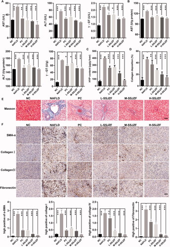
To confirm the efficiency of SSJZF for NAFLD treatment, rats were fed with high-fat diet (HFD) for 8 weeks to induce NAFLD and then subjected to treatment with or without SSJZF or PC by gavage for 8 weeks. The pathological changes of rat liver tissues were detected by H&E staining and Oil Red O staining (). Compared with NC group, cellular lipid was accumulated in the liver tissues of NAFLD rats. Low dose of SSJZF slightly reduced liver lipid deposits in the hepatocytes, while the middle and high doses of SSJZF intervention reduced lipogenesis notably in the hepatocytes. In addition, the levels of TC and TG in the serum and liver tissues of NAFLD rat models were detected by an automatic biochemistry analyser (). The results showed that the levels of TC and TG in rat with NAFLD were significantly increased compared with that of in control rats, and the effect was reduced in SSJZF-treated rats. Further, the results of RT-qPCR indicated that mRNA expression of lipid synthesis and uptake-relate genes (ACC, FAS, SREBP1C, and CD36) was significantly up-regulated in NAFLD rat model, and up-regulation of these genes were reversed by SSJZF or PC treatment (). To further investigate whether SSJZF relieved hepatic function in rats with NAFLD, we estimated the contents of AST, ALT, and γ-GT in the serum and liver tissues (). The results showed that the contents of AST, ALT, and γ-GT were significantly increased in NAFLD rats compared with that in control rats, and the relative indexes were reversed by middle dose or high dose of SSJZF treatment, suggesting SSJZF diminished hepatic function damage in rats with NAFLD. Finally, the degree of liver fibrosis was evaluated by Masson staining, HYP content detection kit, and immunohistochemistry. The liver fibrosis marker HYP content was significantly enhanced in liver tissues of the NAFLD rats, whereas SSJZF treatment could resist the elevated HYP content in liver tissues of the NAFLD rats (). Consistent with HYP content, Masson staining also showed that the accumulation of collagen significantly increased in liver tissues of the NAFLD rats, and the enhancement was inhibited by SSJZF treatment (). In addition, the results of immunohistochemistry also showed that the protein expression level of fibrosis markers (α-SMA, collagen I, collagen IV, and fibronectin) was significantly up-regulated in the NAFLD rat model, and up-regulation of these protein were reversed by SSJZF or PC treatment (), suggesting that SSJZF ameliorated liver fibrosis in rats with NAFLD. Altogether, our data suggested that SSJZF attenuated hepatic lipid accumulation, function damage, and fibrosis in rats with NAFLD.
Figure 1. SSJZF treatment reduced hepatic lipid accumulation in rats with NAFLD (n = 6 per group). NAFLD: non-alcoholic fatty liver disease; SSJZF: Shen-Shi-Jiang-Zhuo formula; NC: normal control; PC: positive control; L-SSJZF: NAFLD rats treated with low dose SSJZF group; M-SSJZF: NAFLD rats treated with middle dose SSJZF group; H-SSJZF: NAFLD rats treated with high dose SSJZF group. (A) HE staining of rat liver tissues. Bar = 100 μm. (B) Oil red staining of rat liver tissues. Bar = 100 μm. (C) The content levels of TC and TG in serum and liver samples of rats were detected by an automatic biochemistry analyser. *p < 0.05, **p < 0.01, ***p < 0.001. TC, Total cholesterol; TG, Triglycerides. (D) The mRNA expression of lipid synthesis and uptake-relate genes (ACC/FAS/SREBP1C/CD36) in liver tissues were detected by RT-PCR. *p < 0.05, **p < 0.01, ***p < 0.001.
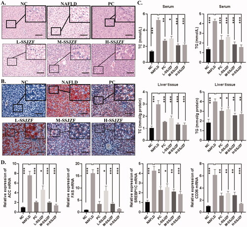
Figure 2. SSJZF treatment relieved hepatic function damage and fibrosis in rats with NAFLD (n = 6 per group). NAFLD: non-alcoholic fatty liver disease; SSJZF: Shen-Shi-Jiang-Zhuo formula; NC: normal control; PC: positive control; L-SSJZF: NAFLD rats treated with low dose SSJZF group; M-SSJZF: NAFLD rats treated with middle dose SSJZF group; H-SSJZF: NAFLD rats treated with high dose SSJZF group; AST: Aspartate aminotransferase; ALT: Alanine aminotransferase; γ-GT: γ-glutamyl transpeptidase. (A) The content levels of AST, ALT, and γ-GT in serum of rats were analysis by an automatic biochemistry analyser. *p < 0.05, **p < 0.01, ***p < 0.001. (B) The content levels of AST, ALT, and γ-GT in liver tissues of rats were analysis by an automatic biochemistry analyser. *p < 0.05, **p < 0.01, ***p < 0.001. (C) The liver fibrosis marker hydroxyproline content was measured by Hydroxyproline Content assay kit. (D) The collagen relative deposition was evaluated by Masson staining. (E) The representative Masson stain diagrams. Bar = 50 μm. (F) Immunohistochemical results showed that the reduced expressions of hepatic fibrosis by SSJZ treatment corresponded to a reduction in hepatic gene expression of fibrosis markers (α-SMA, collagen I, collagen IV, and fibronectin). Bar = 50 μm.
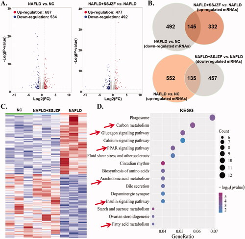
Differential mRNA expression profile of liver tissues in NAFLD rats treated with or without SSJZF
To elucidate the therapeutic mechanism of SSJZF against NAFLD, we performed mRNAs - sequencing on livers tissues obtained from control rats, NAFLD rats, and NAFLD rats treated with SSJZF. Differentially mRNA expression was obtained with the threshold of P-value < 0.05 and |log FC| > 1. A total of 1224 differentially expressed genes were identified by comparing the NAFLD rats with the control rats, of which 687 were up-regulated and 537 were down-regulated (). A total of 969 differentially expressed genes were identified by comparing the SSJZF-treated NAFLD rats with the NAFLD rats, of which 477 were up-regulated and 492 were down-regulated (). Importantly, we found that 280 mRNAs of significantly dysregulation in NAFLD were reversed by SSJZF treatment (), which were enriched in 10 KEGG pathways such as fatty acid metabolism, glucagon signalling pathway, PPAR signalling pathway and arachidonic acid metabolism (). Taken together, the data suggested that a potent effect of SSJZF on a global regulation of hepatic gene expression in rats with NAFLD trending towards a normal profile, and the therapeutic effect of SSJZF on NAFLD rats might be mainly related to the regulation of lipid metabolism.
Figure 3. Differential mRNA expression profiles of liver tissues in NAFLD rats treated with or without SSJZF (n = 3 per group). NAFLD: non-alcoholic fatty liver disease; SSJZF: NAFLD rats treated with Shen-Shi-Jiang-Zhuo formula; NC: normal control. (A) Volcano plot of mRNA expression profiles between NC group and NAFLD group or NAFLD group and NAFLD + SSJZF group. (B) Venn diagram showed the intersection of differential genes between NAFLD group vs. NC group and differential genes between NAFLD + SSJZF group vs. NAFLD group. (C) Heatmap showed that 280 mRNAs of significant dysregulation in NAFLD were reversed by SSJZF treatment. (D) KEGG pathway enrichment for the 280 mRNAs of significant dysregulation.
Reconstruction and verification of SSJZF effect network based on pharmacodynamic difference gene for NAFLD
SSJZF consists of complexity of compounds and TCMSP was used to retrieve the potential compounds. With the limitation of OB ≥ 30% and DL ≥ 0.18, a total of 159 potential compounds of SSJZF were collected after deleting the duplicates (). The target genes of these potential compounds were further screened from TCMSP, a total of 227 target genes of these potential compounds were collected after eliminating the repetitive genes (). The SSJZF active compounds-target genes network was constructed and visualised by Cytoscape software and the compounds with the most target genes were β-sitosterol, baicalein, hederagenin, kaempferol, luteolin, oleic acid, quercetin, stigmasterol, and wogonin (). We further screened the NAFLD-related genes using NCBI-gene, GeneCards, MalaCards and DisGeNET databases, and 1323 NAFLD-related genes were collected after eliminating the repetitive genes. Furthermore, the Venn diagram was established to clarify the relationship between SSJZF and NAFLD-related genes, and 121 overlapping target genes were obtained to be associated with NAFLD and SSJZF (). The predictive empirical compound-target network model (ECT network model) of SSJZF in the treatment of NAFLD was constructed by Cytoscape visualisation software and found that a total of 95 potential compounds in SSJZF and 121 related targets of NAFLD were collected (). On the basis this model, 969 differential genes obtained from SSJZF pharmacodynamic experiments were mapped into the ECT network model, and to reliably screen and identify 23 potential compounds of SSJZF and 25 potential target genes (). Finally, a validated compound-target network model (VCT network model) of SSJZFZ in the treatment of NAFLD was extracted and reconstructed, and the plot of the VCT network model was shown in . Among the 8 compounds (quercetin, kaempferol, baicalein, ellagic acid, linolenic acid, luteolin, stigmasterol, and naringenin) were connected to more than three genes, and 7 genes (GSTEM1, MYC, ICAM, BAX, MAPK14, MMP9 and VEGFA) were regulated by more than or equal to three compounds in this network (), indicating that these compounds and genes might be key nodes in this network.
Figure 4. The general compounds-target network of SSJZF was constructed and visualised with Cytoscape visualisation software. The orange circles indicate the herbs of SSJZF. The green circles indicate the compounds in the herbs. The red circles indicate the potential targets of the compounds. SSJZF: Shen-Shi-Jiang-Zhuo formula.
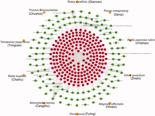
Figure 5. The predictive empirical compound-target network model (ECT network model) of SSJZFZ in the treatment of NAFLD. (A) 121 overlapping target genes within NAFLD-related genes and target genes of SSJZF were obtained by Venn diagram. (B) The predictive empirical compound-target network model (ECT network model) of SSJZFZ in the treatment of NAFLD was constructed by Cytoscape visualisation software. The orange circles indicate the herbs of SSJZF. The green circles indicate the compounds in the herbs. The red circles indicate the potential targets of the compounds.
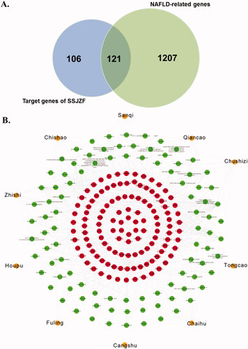
Figure 6. The validated compound-target network model (VCT network model) of SSJZFZ in the treatment of NAFLD. (A) Venn diagram showed 25 overlapping target genes within potential SSJZF targets according to network pharmacology and based on pharmacodynamic difference gene for NAFLD. (B) The validated compound-target network model (VCT network model) of SSJZFZ in the treatment of NAFLD was constructed by Cytoscape visualisation software. The blue rhombuses indicate the herbs of SSJZF. The cyan rectangles indicate the compounds in the herbs. The green circles indicate the potential targets of the compounds.
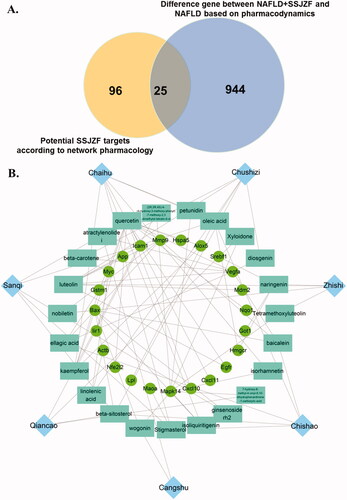
Table 3. Active compounds of SSJZF.
Table 4. Target genes of SSJZF active compounds.
Table 5. The validated compound-target network model (VCT network model) of SSJZFZ in the treatment of NAFLD was identified 23 compounds of SSJZF and 25 potential target genes.
To further examine the signalling pathways and functions of these target genes, we performed functional enrichment analysis using KEGG pathways. The result indicated that 25 genes were mainly enriched in PI3K/Akt signalling pathway, endocrine resistance, TNF signalling pathway, lipid and atherosclerosis, Rap1 signalling pathway, MAPK signalling pathway, etc. (). Taken together, our data suggested that the treatment of SSJZF on NAFLD might be achieved through multi-compounds, multi-targets, and multi-pathways.
Figure 7. SSJZF enhanced PI3K/Akt signalling pathway activation in rat with NAFLD. NAFLD: non-alcoholic fatty liver disease; SSJZF: Shen-Shi-Jiang-Zhuo formula; NC: normal control; PC: positive control; L-SSJZF: NAFLD rats treated with low dose SSJZF group; M-SSJZF: NAFLD rats treated with middle dose SSJZF group; H-SSJZF: NAFLD rats treated with high dose SSJZF group. A. KEGG pathway enrichment of the intersection 25 different genes. B/C. The expression of total PI3K (t-PI3K), phosphorylated PI3K (p-PI3K), total Akt (t-Akt) and phosphorylated Akt (p-Akt) in liver tissues was detected by Western blots. *p < 0.05, **p < 0.01.
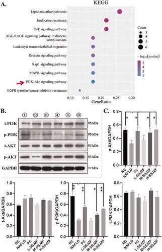
SSJZF enhanced PI3K/Akt signalling pathway activity in liver tissues of NAFLD rats
PI3K/Akt signalling pathway has also been demonstrated to play a critical role in the regulation of liver lipid metabolism in NAFLD (Wang et al. Citation2014; Zeng et al. Citation2014; Wang et al. Citation2019). The comprehensive analysis of network pharmacology and sequencing in the early stage found that PI3K signalling pathway was involved in the therapeutic effect of SSJZF on NAFLD (). Here, in order to confirm whether SSJZF regulates PI3K/Akt signalling pathway in liver tissue of NAFLD rats, western blot analyses were performed to verify the PI3K/Akt signalling pathway activation in NAFLD rats with or without SSJZF treatment (). Compared with the control group, the phosphorylation levels of PI3K and AKT was significantly decreased in liver tissues of NAFLD rats, but the total protein of PI3K and AKT was not remarkably altered, indicating PI3K/Akt signalling pathway was restrained in liver tissues of NAFLD rats. While the restraint of PI3K/Akt signalling pathway in liver tissues of NAFLD rats was reversed by treatment of PC or SSJZF, indicating SSJZF enhanced PI3K/Akt signalling pathway activity in liver tissues of NAFLD rats. And the efficiency of high dose of SSJZF is more significant than PC in PI3K/Akt signalling pathway activation. Taken together, our data indicated that SSJZF might alleviate hepatic lipid accumulation in NAFLD rat model via activating the PI3K/Akt signalling pathway.
Discussion
NAFLD, currently most common form of chronic liver disease in both adults and children worldwide, is associated with hepatic lipids metabolism (Ipsen et al. Citation2018; Gjorgjieva et al. Citation2019; Reimer et al. Citation2020). Over the last decades, great studies have been dedicated to the understanding of the pathogenesis of NAFLD, but there are currently no effective drugs to treat NAFLD (Gao et al. Citation2020). In this study, our results revealed that SSJZF ameliorated high-fat diet-induced NAFLD in rats. Further, the comprehensive analysis of mRNA-sequencing and network pharmacology was used to uncover the main active ingredients and protective mechanisms of SSJZF in NAFLD.
The traditional Chinese medicine SSJZF has been clinically used to treat NAFLD for a long time in China. In our previous study, we indicated that SSJZF had higher clinical therapeutic efficacy on NAFLD (Lu et al. Citation2018). However, preclinical experimental evidence supporting its efficacy is lacking. Our results revealed for first time that SSJZF improve abnormal lipid metabolism, liver function injury, and hepatic fibrosis in rats with NAFLD, suggesting that SSJZF ameliorated HFD-induced NAFLD in rats. The results of this study are consistent with our preliminary clinical study. Liver fibrosis, also one of the important characteristics of NAFLD, is a necessary progression for NAFLD to develop into non-alcoholic steatohepatitis, cirrhosis or hepatocellular carcinoma (HCC), and is also a reversible pathological stage that can effectively intervene and prevent NAFLD (Campos-Murguía et al. Citation2020). Animal experiment showed that apoptosis signal-regulating kinase 1 (ASK1) depletion increased hepatic steatosis and liver fibrosis, and ASK1 may be a novel therapeutic target to the hepatic fibrosis of NAFLD (Challa et al. Citation2019). In addition, studies have been carried out on the therapeutic effect of Traditional Chinese medicine in NAFLD fibrosis. For example, polydatin alleviate liver fibrosis and relieves hepatic fat accumulation in rats with NAFLD (Li et al. Citation2018). Silybin prevented hepatic steatosis and fibrosis in NASH mice by inhibiting Nf-κB Pathway (Takatani et al. Citation2020). Our results indicated that SSJZF reduced the hepatic fibrosis in rats with NAFLD. Studies have shown that quercetin can ameliorate tissue fibrosis in a variety of diseases, including hepatic fibrosis (Wu et al. Citation2017), and this study indirectly supports our findings.
To further explore the protective mechanism of SSJZF in the treatment of HFD-induced NAFLD in rats, we performed mRNA-sequencing analysis. Our data indicated that 280 mRNAs of significantly dysregulation in NAFLD were reversed by SSJZF treatment, which were enriched 10 KEGG, such as arachidonic acid metabolism and fatty acid metabolism. Recent studies have shown that the abnormality of hepatic lipid metabolism (such as fatty acid metabolism and arachidonic acid metabolism) is a key factor leading to the occurrence and development of NAFLD (Pei et al. Citation2020). Sztolsztener et al. (Citation2020) suggested that arachidonic acid is an early indicator of inflammation in the development of NAFLD. Furthermore, we identified 23 active compounds of SSJZF in NAFLD base on mRNAs-sequencing combined with network pharmacology. Among the active compounds of SSJZF, quercetin, β-carotene, luteolin, stigmasterol, and diosgenin have been proved to alleviate NAFLD. For example, quercetin is a flavonoid which takes part in various processes, such as anti-inflammatory and antioxidative (Wang et al. Citation2020). It is proved that the combination of quercetin and DHA-rich fish oil may partly alleviate NAFLD by reducing oxidative stress, suppressing fatty acid synthesis, ameliorating inflammation (Xu et al. Citation2019; Yang et al. Citation2019). A cross-sectional study showed that β-carotene to retinol ratio is associated with the severity of NAFLD (Kimura et al. Citation2020). It is reported that diosgenin ameliorated free fatty acid-induced NAFLD via AMPK/ACC pathway (Fang et al. Citation2019). Many studies have shown that luteolin alone and combination therapy may be the treatment of NAFLD development (Sagawa et al. Citation2015; Zhu et al. Citation2020; Sun et al. Citation2021). Moreover, Studies have shown that stigmasterol alters lipid metabolism and alleviates NAFLD in mice fed a high-fat western-style diet (Feng, Dai, et al. Citation2018; Feng, Gan, et al. Citation2018). Furthermore, our previous research has proved that SSJZF significantly reduces the process of NAFLD clinically (Lu et al. Citation2018). Taking the results into consideration, we suggested that SSJZF may reduce NAFLD syndromes through quercetin, β-carotene and diosgenin.
Further, we identified 23 compounds of SSJZF which acted on 25 key therapeutic targets of NAFLD, such as Mapk14, MMP9, Myc, Bax. Xiao et al. (Citation2017) reported that Mapk14 restricted the hepatic fat accumulation by antagonising JNK activation. Hwang et al. (Citation2020) reported that hepatocyte-specific deletion of Mapk14 in HFD-induced fatty liver exacerbated steatosis and liver injury, indicating Mapk14 plays distinct roles in NAFLD. Zhang et al. (Citation2019) indicated that macrophage Mapk14 promotes nutritional steatohepatitis through M1 polarisation. Recent studies have shown that MMP9 levels have been identified as a predictor of poor prognosis in patients with NAFLD (Coilly et al. Citation2019). Our data indicated MMP9 was a key target of SSJZF in the treatment of NAFLD. In addition, studies have shown that Myc activity was repressed by SIRT7 to suppress ER stress and prevent fatty liver disease (Shin et al. Citation2013), suggesting that Myc played an important role in NAFLD. A previous study showed that increasing the activation of BAX accelerates apoptotic death in severe hepatic steatosis (Liu et al. Citation2013). Blueberry combined with probiotics alleviates NAFLD via activation of JAK1/STAT3 signalling and inhibition of the apoptosis factor BAX (Zhu et al. Citation2018). Studies reported that transmembrane BAX inhibitor motif-containing 1 and Bax inhibitor-1 attenuated non-alcoholic steatohepatitis by a novel anti-inflammatory approach or limiting IREα signalling (Lebeaupin et al. Citation2018; Wree et al. Citation2018). Our results identified these hub genes as SSJZF potential targets on NAFLD were in accordance with previous studies to some extent. In-depth investigation of these targets may provide us novel therapies of NAFLD.
Numerous signalling pathways were screened based on the network pharmacology as well as RNA sequencing, including MAPK signalling pathway, TNF signalling pathway, and PI3K/Akt signalling pathway. Tumour necrosis factor (TNF) is a pivotal modulating component of the immune system, which regulates innate and adaptive immunity and implicates different inflammatory diseases such as tumour and inflammatory bowel disease (Lopetuso et al. Citation2018; Fischer et al. Citation2020). It has been reported that TNF played an important role in NAFLD progression, and NASH patients exhibited higher levels of TNF (Handa et al. Citation2017). Anti-TNF agents have been reported protective effects in NAFLD animal models including reduction of AST, ALT, hepatic inflammation and fibrosis (Koca et al. Citation2008). Another study has revealed that curcumin, an active ingredient of turmeric [Curcuma longa Linn, (Zingiberaceae)], could decrease TNF level in serum and improve NASH in patients (Saadati et al. Citation2019). These results suggested that TNF might be a potential target for NAFLD treatment. Moreover, the PI3K/Akt signalling pathway has been conserved to regulate and maintain appropriate cell growth, survival and metabolism (Barker et al. Citation2020), which is an important signalling pathway of insulin action and related to NAFLD progression (Zeng et al. Citation2014). We initially determined whether the PI3K/Akt signalling pathway was involved in the efficiency of SSJZF for NAFLD treatment, as previous studies have emphasised the significant role the PI3K/AKT pathway plays in process of NAFLD. The PI3K/Akt signalling pathway might modulate NAFLD progression (Liu et al. Citation2012; Matsuda et al. Citation2013). Akt can reduce the expression of fatty acid oxidation genes to regulate the process of hepatic glucose and lipid metabolism (Zeng et al. Citation2014). Previous studies also indicated the role the PI3K/Akt signalling pathway played in NAFLD treated with TCM. For example, Tangganjian decoction decreased triglyceride and total cholesterol, and increased the expression of PI3K and Akt in liver (Fan et al. Citation2018), as well as Shengmai-Yin and Ganmaidazao decoction, which could improve liver metabolism partially by activiting the PI3K/Akt pathway in type 2 diabetes mellitus with NAFLD (Sun et al. Citation2019). Our results were consistent with previous studies to some extent. It proved that SSJZF promoted activation of PI3K/Akt signalling, correlating with the evidence that the pathway alleviated hepatic lipogenesis and regulated lipid metabolism (Wang et al. Citation2014; Liao et al. Citation2018). Therefore, activation of the PI3K/Akt signalling pathway might be the mechanism of amelioration of NAFLD treated by SSJZF.
However, this study has several limitations. First, a lack of rescue experiment was performed to verify SSJZF improve hepatic lipid accumulation, hepatic fibrosis, and hepatic function injury in rats with NAFLD model by regulating PI3K/Akt pathway signalling. Second, the molecular mechanism of SSJZF regulation of PI3K/AKT pathway signalling has not been further clarified. Third, we screened out 23 chemical compounds as the potential effective ingredients of SSJZF by the comprehensive analysis of network pharmacology and mRNA-sequencing data mining, but high-performance liquid chromatography coupled with quadrupole time-of-flight mass spectrometry technique (HPLC-QTOF-MS) was not used to further identify these compounds in SSJZF and blood-absorbed compounds. In future study, these parts will be included in our experiments.
Conclusions
In the present study, our data revealed that SSJZF ameliorated HLF-induced NAFLD via activating the PI3K/AKT pathway signalling. Importantly, we screened out 23 chemical compounds as the potential effective ingredients of SSJZF which acted on 25 targets of NAFLD by the comprehensive analysis of network pharmacology, mRNA-sequencing data mining and bioinformatics. This study not only provided theoretical basis for the application of SSJZF in clinical treatment of NAFLD, but also screened out some major compounds of SSJZF and their potential targets which will facilitate the development of new drugs for NAFLD.
Author contributions
All authors made substantial contributions to the conception, study design, acquisition of data, analysis and interpretation of data; took part in drafting the article or revising it critically for important intellectual content; agreed to submit to the current journal; gave final approval of the version to be published; and agree to take responsibility and be accountable for the contents of the work.
Disclosure statement
No potential conflict of interest was reported by the author(s).
Additional information
Funding
References
- Barker RM, Holly JMP, Biernacka KM, Allen-Birt SJ, Perks CM. 2020. Mini review: opposing pathologies in cancer and Alzheimer’s disease: does the PI3K/Akt pathway provide clues? Front Endocrinol (Lausanne). 11:403.
- Brunt EM, Wong VW, Nobili V, Day CP, Sookoian S, Maher JJ, Bugianesi E, Sirlin CB, Neuschwander-Tetri BA, Rinella ME. 2015. Nonalcoholic fatty liver disease. Nat Rev Dis Primers. 1:15080.
- Campos-Murguía A, Ruiz-Margáin A, González-Regueiro JA, Macías-Rodríguez RU. 2020. Clinical assessment and management of liver fibrosis in non-alcoholic fatty liver disease. World J Gastroenterol. 26(39):5919–5943.
- Chalasani N, Younossi Z, Lavine JE, Charlton M, Cusi K, Rinella M, Harrison SA, Brunt EM, Sanyal AJ. 2018. The diagnosis and management of nonalcoholic fatty liver disease: practice guidance from the American Association for the Study of Liver Diseases. Hepatology. 67(1):328–357.
- Challa TD, Wueest S, Lucchini FC, Dedual M, Modica S, Borsigova M, Wolfrum C, Blüher M, Konrad D. 2019. Liver ASK1 protects from non-alcoholic fatty liver disease and fibrosis. EMBO Mol Med. 11(10):e10124.
- Chen L, Cao Y, Zhang H, Lv D, Zhao Y, Liu Y, Ye G, Chai Y. 2018. Network pharmacology-based strategy for predicting active ingredients and potential targets of Yangxinshi tablet for treating heart failure. J Ethnopharmacol. 219:359–368.
- Chen L, Liu J, Mei G, Chen H, Peng S, Zhao Y, Yao P, Tang Y. 2021. Quercetin and non-alcoholic fatty liver disease: a review based on experimental data and bioinformatic analysis. Food Chem Toxicol. 154:112314.
- Chen L, Zhang YH, Wang S, Zhang Y, Huang T, Cai YD. 2017. Prediction and analysis of essential genes using the enrichments of gene ontology and KEGG pathways. PLoS One. 12(9):e0184129.
- Cheng Z, Wen Y, Liang B, Chen S, Liu Y, Wang Z, Cheng J, Tang X, Xin H, Deng L. 2019. Gene expression profile-based drug screen identifies SAHA as a novel treatment for NAFLD. Mol Omics. 15(1):50–58.
- Cobbina E, Akhlaghi F. 2017. Non-alcoholic fatty liver disease (NAFLD) - pathogenesis, classification, and effect on drug metabolizing enzymes and transporters. Drug Metab Rev. 49(2):197–211.
- Coilly A, Desterke C, Guettier C, Samuel D, Chiappini F. 2019. FABP4 and MMP9 levels identified as predictive factors for poor prognosis in patients with nonalcoholic fatty liver using data mining approaches and gene expression analysis. Sci Rep. 9(1):19785.
- Dai X, Feng J, Chen Y, Huang S, Shi X, Liu X, Sun Y. 2021. Traditional Chinese Medicine in nonalcoholic fatty liver disease: molecular insights and therapeutic perspectives. Chin Med. 16(1):68.
- Fan Y, He Z, Wang W, Li J, Hu A, Li L, Yan L, Li Z, Yin Q. 2018. Tangganjian decoction ameliorates type 2 diabetes mellitus and nonalcoholic fatty liver disease in rats by activating the IRS/PI3K/AKT signaling pathway. Biomed Pharmacother. 106:733–737.
- Fang K, Wu F, Chen G, Dong H, Li J, Zhao Y, Xu L, Zou X, Lu F. 2019. Diosgenin ameliorates palmitic acid-induced lipid accumulation via AMPK/ACC/CPT-1A and SREBP-1c/FAS signaling pathways in LO2 cells. BMC Complement Altern Med. 19(1):255.
- Feng S, Dai Z, Liu AB, Huang J, Narsipur N, Guo G, Kong B, Reuhl K, Lu W, Luo Z, et al. 2018. Intake of stigmasterol and beta-sitosterol alters lipid metabolism and alleviates NAFLD in mice fed a high-fat western-style diet. Biochim Biophys Acta Mol Cell Biol Lipids. 1863(10):1274–1284.
- Feng S, Gan L, Yang CS, Liu AB, Lu W, Shao P, Dai Z, Sun P, Luo Z. 2018. Effects of stigmasterol and beta-sitosterol on nonalcoholic fatty liver disease in a mouse model: a lipidomic analysis. J Agric Food Chem. 66(13):3417–3425.
- Feng Y, Chen Y, Yang B, Lan Q, Wang T, Cui G, Ren Z, Choi IC, Leung GP, Yan F, et al. 2019. Hepatoprotective effect of jianpi huoxue formula on nonalcoholic fatty liver disease induced by methionine-choline-deficient diet in rat. Biomed Res Int. 2019:1–12. 2019:
- Fischer R, Kontermann RE, Pfizenmaier K. 2020. Selective targeting of TNF receptors as a novel therapeutic approach. Front Cell Dev Biol. 8:401.
- Gao Y, Zhang W, Zeng LQ, Bai H, Li J, Zhou J, Zhou GY, Fang CW, Wang F, Qin XJ. 2020. Exercise and dietary intervention ameliorate high-fat diet-induced NAFLD and liver aging by inducing lipophagy. Redox Biol. 36:101635.
- Gjorgjieva M, Sobolewski C, Dolicka D, Correia de Sousa M, Foti M. 2019. miRNAs and NAFLD: from pathophysiology to therapy. Gut. 68(11):2065–2079.
- Hamada N, Ogawa Y, Saibara T, Murata Y, Kariya S, Nishioka A, Terashima M, Inomata T, Yoshida S. 2000. Toremifene-induced fatty liver and NASH in breast cancer patients with breast-conservation treatment. Int J Oncol. 17(6):1119–1123.
- Handa P, Vemulakonda AL, Maliken BD, Morgan-Stevenson V, Nelson JE, Dhillon BK, Hennessey KA, Gupta R, Yeh MM, Kowdley KV. 2017. Differences in hepatic expression of iron, inflammation and stress-related genes in patients with nonalcoholic steatohepatitis. Ann Hepatol. 16(1):77–85.
- Hopkins AL. 2007. Network pharmacology. Nat Biotechnol. 25(10):1110–1111.
- Hopkins AL. 2008. Network pharmacology: the next paradigm in drug discovery. Nat Chem Biol. 4(11):682–690.
- Hwang S, Wang X, Rodrigues RM, Ma J, He Y, Seo W, Park SH, Kim SJ, Feng D, Gao B. 2020. Protective and detrimental roles of p38alpha mitogen-activated protein kinase in different stages of nonalcoholic fatty liver disease. Hepatology. 72(3):873–891.
- Ipsen DH, Lykkesfeldt J, Tveden-Nyborg P. 2018. Molecular mechanisms of hepatic lipid accumulation in non-alcoholic fatty liver disease. Cell Mol Life Sci. 75(18):3313–3327.
- Izzo C, Annunziata M, Melara G, Sciorio R, Dallio M, Masarone M, Federico A, Persico M. 2021. The role of resveratrol in liver disease: a comprehensive review from in vitro to clinical trials. Nutrients. 13(3):933.
- Jiang X, Lv B, Li P, Ma X, Wang T, Zhou Q, Wang X, Gao X. 2015. Bioactivity-integrated UPLC/Q-TOF-MS of Danhong injection to identify NF-kappaB inhibitors and anti-inflammatory targets based on endothelial cell culture and network pharmacology. J Ethnopharmacol. 174:270–276.
- Kimura M, Mikami K, Endo T, Matsuzaka M, Sawada N, Igarashi G, Iino C, Hasegawa T, Sawada K, Ando M, et al. 2020. Association between serum β-carotene-to-retinol ratio and severity of hepatic steatosis in non-alcoholic fatty liver disease in Japan: A cross-sectional study. Nutrition. 79–80:110984.
- Klair JS, Yang JD, Abdelmalek MF, Guy CD, Gill RM, Yates K, Unalp-Arida A, Lavine JE, Clark JM, Diehl AM, et al. 2016. A longer duration of estrogen deficiency increases fibrosis risk among postmenopausal women with nonalcoholic fatty liver disease. Hepatology. 64(1):85–91.
- Koca SS, Bahcecioglu IH, Poyrazoglu OK, Ozercan IH, Sahin K, Ustundag B. 2008. The treatment with antibody of TNF-alpha reduces the inflammation, necrosis and fibrosis in the non-alcoholic steatohepatitis induced by methionine- and choline-deficient diet. Inflammation. 31(2):91–98.
- Lebeaupin C, Vallee D, Rousseau D, Patouraux S, Bonnafous S, Adam G, Luciano F, Luci C, Anty R, Iannelli A, et al. 2018. Bax inhibitor-1 protects from nonalcoholic steatohepatitis by limiting inositol-requiring enzyme 1 alpha signaling in mice. Hepatology. 68(2):515–532.
- Lee AY, Park W, Kang TW, Cha MH, Chun JM. 2018. Network pharmacology-based prediction of active compounds and molecular targets in Yijin-Tang acting on hyperlipidaemia and atherosclerosis. J Ethnopharmacol. 221:151–159.
- Li R, Li J, Huang Y, Li H, Yan S, Lin J, Chen Y, Wu L, Liu B, Wang G, et al. 2018. Polydatin attenuates diet-induced nonalcoholic steatohepatitis and fibrosis in mice. Int J Biol Sci. 14(11):1411–1425.
- Li Y, Gong Y, Zhang X, Wang J, Cheng Y, Liu F, Shi X, Xu W, Dong L. 2021. Exploring the synergistic mechanism of gegen qinlian decoction on the WNT signaling pathway using an integrated strategy of network pharmacology and RNA-seq. J Ethnopharmacol. 278:114283.
- Liao X, Song L, Zhang L, Wang H, Tong Q, Xu J, Yang G, Yang S, Zheng H. 2018. LAMP3 regulates hepatic lipid metabolism through activating PI3K/Akt pathway. Mol Cell Endocrinol. 470:160–167.
- Liu K, Lou J, Wen T, Yin J, Xu B, Ding W, Wang A, Liu D, Zhang C, Chen D, et al. 2013. Depending on the stage of hepatosteatosis, p53 causes apoptosis primarily through either DRAM-induced autophagy or BAX. Liver Int. 33(10):1566–1574.
- Liu Y, Han X, Bian Z, Peng Y, You Z, Wang Q, Chen X, Qiu D, Ma X. 2012. Activation of liver X receptors attenuates endotoxin-induced liver injury in mice with nonalcoholic fatty liver disease. Dig Dis Sci. 57(2):390–398.
- Lopetuso L, Mocci G, Marzo M, D’Aversa F, Rapaccini G, Guidi L, Armuzzi A, Gasbarrini A, Papa A. 2018. Harmful effects and potential benefits of bnti-tumor necrosis factor (TNF)-alpha on the liver. IJMS. 19(8):2199.
- Lu Y, Hu J, Wu F, Zheng J, Yang C, Chen H. 2018. Clinical effect of shenshijiangzhuo formula of non-alcoholic fatty liver disease. Heilongjiang J Trad Chinese Med. 47:9–10.
- Marcolin E, San-Miguel B, Vallejo D, Tieppo J, Marroni N, González-Gallego J, Tuñón MJ. 2012. Quercetin treatment ameliorates inflammation and fibrosis in mice with nonalcoholic steatohepatitis. J Nutr. 142(10):1821–1828.
- Matsuda S, Kobayashi M, Kitagishi Y. 2013. Roles for PI3K/AKT/PTEN pathway in cell signaling of nonalcoholic fatty liver disease. ISRN Endocrinol. 2013:472432.
- Paolacci S, Precone V, Acquaviva F, Chiurazzi P, Fulcheri E, Pinelli M, Buffelli F, Michelini S, Herbst KL, Unfer V, et al. 2019. Genetics of lipedema: new perspectives on genetic research and molecular diagnoses. Eur Rev Med Pharmacol Sci. 23(13):5581–5594.
- Pei K, Gui T, Kan D, Feng H, Jin Y, Yang Y, Zhang Q, Du Z, Gai Z, Wu J, et al. 2020. An overview of lipid metabolism and nonalcoholic fatty liver disease. Biomed Res Int. 2020:4020249.
- Qin M, Yuan F, Ren J, Chi Z. 2020. Effectiveness and safety of traditional Chinese medicines for non-alcoholic fatty liver disease: protocol for systematic review and meta-analysis. Medicine (Baltimore). 99(25):e20699.
- Reimer KC, Wree A, Roderburg C, Tacke F. 2020. New drugs for NAFLD: lessons from basic models to the clinic. Hepatol Int. 14(1):8–23.
- Riley P, Sudarshi D, Johal M, Benedict A, Panteli J, Crook M, O’Donohue J. 2008. Weight loss, dietary advice and statin therapy in non-alcoholic fatty liver disease: a retrospective study. Int J Clin Pract. 62(3):374–381.
- Saadati S, Sadeghi A, Mansour A, Yari Z, Poustchi H, Hedayati M, Hatami B, Hekmatdoost A. 2019. Curcumin and inflammation in non-alcoholic fatty liver disease: a randomized, placebo controlled clinical trial. BMC Gastroenterol. 19(1):133.
- Sagawa H, Naiki-Ito A, Kato H, Naiki T, Yamashita Y, Suzuki S, Sato S, Shiomi K, Kato A, Kuno T, et al. 2015. Connexin 32 and luteolin play protective roles in non-alcoholic steatohepatitis development and its related hepatocarcinogenesis in rats. Carcinogenesis. 36(12):1539–1549.
- Sheng D, Zhao S, Gao L, Zheng H, Liu W, Hou J, Jin Y, Ye F, Zhao Q, Li R, et al. 2019. BabaoDan attenuates high-fat diet-induced non-alcoholic fatty liver disease via activation of AMPK signaling. Cell Biosci. 9:77.
- Shin J, He M, Liu Y, Paredes S, Villanova L, Brown K, Qiu X, Nabavi N, Mohrin M, Wojnoonski K, et al. 2013. SIRT7 represses Myc activity to suppress ER stress and prevent fatty liver disease. Cell Rep. 5(3):654–665.
- Sun C, Zhang H, Wang X, Liu X. 2020. Ligamentum flavum fibrosis and hypertrophy: molecular pathways, cellular mechanisms, and future directions. FASEB J. 34(8):9854–9868.
- Sun L, Gu X, Pan Z, Guo X, Liu J, Atianjoh FE, Wu S, Mo K, Xu B, Liang L, et al. 2019. Contribution of dnmt1 to neuropathic pain genesis partially through epigenetically repressing kcna2 in primary afferent neurons. J Neurosci. 39(33):6595–6607.
- Sun WL, Yang JW, Dou HY, Li GQ, Li XY, Shen L, Ji HF. 2021. Anti-inflammatory effect of luteolin is related to the changes in the gut microbiota and contributes to preventing the progression from simple steatosis to nonalcoholic steatohepatitis. Bioorg Chem. 112:104966.
- Sun Y, Tan Z, Jiang Z, Li M, Wang W, Huang Y, Sun J. 2021. Comparative efficacy and safety of traditional Chinese patent medicine for NAFLD in childhood or adolescence: A protocol for a Bayesian network meta analysis. Medicine (Baltimore). 100(3):e24277.
- Sztolsztener K, Chabowski A, Harasim-Symbor E, Bielawiec P, Konstantynowicz-Nowicka K. 2020. Arachidonic acid as an early indicator of inflammation during non-alcoholic fatty liver disease development. Biomolecules. 10(8):1133.
- Takatani N, Kono Y, Beppu F, Okamatsu-Ogura Y, Yamano Y, Miyashita K, Hosokawa M. 2020. Fucoxanthin inhibits hepatic oxidative stress, inflammation, and fibrosis in diet-induced nonalcoholic steatohepatitis model mice. Biochem Biophys Res Commun. 528(2):305–310.
- Tejada S, Capó X, Mascaró CM, Monserrat-Mesquida M, Quetglas-Llabrés MM, Pons A, Tur JA, Sureda A. 2021. Hepatoprotective effects of resveratrol in non-alcoholic fatty live disease. Curr Pharm Des. 27(22):2558–2570.
- Wang C, Chi Y, Li J, Miao Y, Li S, Su W, Jia S, Chen Z, Du S, Zhang X, et al. 2014. FAM3A activates PI3K p110alpha/Akt signaling to ameliorate hepatic gluconeogenesis and lipogenesis. Hepatology. 59(5):1779–1790.
- Wang S, Yang FJ, Shang LC, Zhang YH, Zhou Y, Shi XL. 2019. Puerarin protects against high-fat high-sucrose diet-induced non-alcoholic fatty liver disease by modulating PARP-1/PI3K/AKT signaling pathway and facilitating mitochondrial homeostasis. Phytother Res. 33(9):2347–2359.
- Wang YY, Chang CY, Lin SY, Wang JD, Wu CC, Chen WY, Kuan YH, Liao SL, Wang WY, Chen CJ. 2020. Quercetin protects against cerebral ischemia/reperfusion and oxygen glucose deprivation/reoxygenation neurotoxicity. J Nutr Biochem. 83:108436.
- Wree A, Inzaugarat ME, Feldstein AE. 2018. Transmembrane BAX Inhibitor motif-containing 1, a novel anti-inflammatory approach for nonalcoholic steatohepatitis treatment. Hepatology. 67(1):438–441.
- Wu L, Zhang Q, Mo W, Feng J, Li S, Li J, Liu T, Xu S, Wang W, Lu X, et al. 2017. Quercetin prevents hepatic fibrosis by inhibiting hepatic stellate cell activation and reducing autophagy via the TGF-β1/Smads and PI3K/Akt pathways. Sci Rep. 7(1):9289.
- Xiao Y, Wang J, Yan W, Zhou K, Cao Y, Cai W. 2017. p38alpha MAPK antagonizing JNK to control the hepatic fat accumulation in pediatric patients onset intestinal failure. Cell Death Dis. 8(10):e3110.
- Xu Y, Han J, Dong J, Fan X, Cai Y, Li J, Wang T, Zhou J, Shang J. 2019. Metabolomics characterizes the effects and mechanisms of quercetin in nonalcoholic fatty liver disease development. IJMS. 20(5):1220.
- Yang H, Yang T, Heng C, Zhou Y, Jiang Z, Qian X, Du L, Mao S, Yin X, Lu Q. 2019. Quercetin improves nonalcoholic fatty liver by ameliorating inflammation, oxidative stress, and lipid metabolism in db/db mice. Phytother Res. 33(12):3140–3152.
- Younossi ZM, Marchesini G, Pinto-Cortez H, Petta S. 2019. Epidemiology of nonalcoholic fatty liver disease and nonalcoholic steatohepatitis: implications for liver transplantation. Transplantation. 103(1):22–27.
- Yu T, Acharya A, Mattheos N, Li S, Ziebolz D, Schmalz G, Haak R, Schmidt J, Sun Y. 2019. Molecular mechanisms linking peri-implantitis and type 2 diabetes mellitus revealed by transcriptomic analysis. PeerJ. 7:e7124.
- Zeng L, Tang WJ, Yin JJ, Zhou BJ. 2014. Signal transductions and nonalcoholic fatty liver: a mini-review. Int J Clin Exp Med. 7(7):1624–1631.
- Zeng Q, Zhao L, Meng C, Zhao X, Liu Y, Shi R, Han X, Wang T, Li J. 2020. Prophylactic and therapeutic effects of different doses of vitamin C on high-fat-diet-induced non-alcoholic fatty liver disease in mice. Biomed Pharmacother. 131:110792.
- Zhang GB, Li QY, Chen QL, Su SB. 2013. Network pharmacology: a new approach for Chinese herbal medicine research. Evid Based Complement Alternat Med. 2013:621423.
- Zhang X, Fan L, Wu J, Xu H, Leung WY, Fu K, Wu J, Liu K, Man K, Yang X, et al. 2019. Macrophage p38alpha promotes nutritional steatohepatitis through M1 polarization. J Hepatol. 71(1):163–174.
- Zhou Y, Wu R, Cai FF, Zhou WJ, Lu YY, Zhang H, Chen QL, Sun MY, Su SB. 2021. Development of a novel anti-liver fibrosis formula with luteolin, licochalcone A, aloe-emodin and acacetin by network pharmacology and transcriptomics analysis. Pharm Biol. 59(1):1594–1606.
- Zhou Z, Chen B, Chen S, Lin M, Chen Y, Jin S, Chen W, Zhang Y. 2020. Applications of network pharmacology in traditional Chinese medicine research. Evid Based Complement Alternat Med. 2020:1646905.
- Zhu J, Zhou M, Zhao X, Mu M, Cheng M. 2018. Blueberry, combined with probiotics, alleviates non-alcoholic fatty liver disease via IL-22-mediated JAK1/STAT3/BAX signaling. Food Funct. 9(12):6298–6306.
- Zhu M, Li M, Zhou W, Yang Y, Li F, Zhang L, Ji G. 2019. Qianggan extract improved nonalcoholic steatohepatitis by modulating lncRNA/circRNA immune ceRNA networks. BMC Complement Altern Med. 19(1):156.
- Zhu N, Hou J, Ma G, Liu J. 2019. Network pharmacology identifies the mechanisms of action of shaoyao gancao decoction in the treatment of osteoarthritis. Med Sci Monit. 25:6051–6073.
- Zhu Y, Liu R, Shen Z, Cai G. 2020. Combination of luteolin and lycopene effectively protect against the “two-hit” in NAFLD through Sirt1/AMPK signal pathway. Life Sci. 256:117990.
- Zsori G, Illes D, Ivany E, Kosar K, Holzinger G, Tajti M, Palinkas E, Szabovik G, Nagy A, Palko A, et al. 2019. In new-onset diabetes mellitus, metformin reduces fat accumulation in the liver, but not in the pancreas or pericardium. Metab Syndr Relat Disord. 17(5):289–295.