Abstract
Objective. To investigate oxidative stress and myocardial injury at different stages of coronary artery bypass grafting (CABG). Design. Twenty patients underwent CABG with use of cardiopulmonary bypass (CPB) and with intermittent sampling of plasma and urine. Main markers were: 8-iso-PGF2α (oxidative stress); troponin T (myocardial injury); and 15-keto-dihydro-PGF2α and hsCRP (inflammation). Results. Plasma 8-iso-PGF2α increased after start of surgery, but there was no further rise during CPB or after aortic cross-clamp release and no significant myocardial arterio-venous differences. An increase in troponin T was seen early after the operation, but no relationship was established between 8-iso-PGF2α and troponin T. 8-iso-PGF2α levels were elevated by preoperative withdrawal of acetylsalicylic acid (ASA) but reduced by intraoperative use of heparin. 15-keto-dihydro-PGF2α was elevated during operation and hsCRP following operation. Conclusions. In the present study oxidative stress was multifactorial in origin with main impacts from surgical trauma, less from CPB and little if any from myocardial ischemia-reperfusion events. In addition, cardiovascular drugs in common use like ASA and heparin seemed to influence the pro- and antioxidant balance, a finding that has to be confirmed in future studies.
Coronary artery bypass grafting (CABG) with use of cardiopulmonary bypass (CPB) and ischemic cardiac arrest is known to mediate oxidative stress Citation1–3. A widely held view is that reactive oxygen species (ROS) are generated locally in the myocardium and contribute to a postischemic reperfusion injury. In line with this, ROS may be detected as radical adducts or lipid peroxides in coronary venous blood after aortic clamp release Citation3; . However, these findings are not consistent and the focus in clinical studies has shifted from a local myocardial to a systematic oxidative stress. In particular, CPB may produce ROS due to hyperoxic conditions, foreign surface materials, hemolysis, iron release and inflammation. Consequently, less lipid peroxides are released by off-pump than standard on-pump bypass grafting given a similar surgical trauma Citation4. Altogether, several interacting factors may, in a complex way, lead to a combined prooxidant and proinflammatory response and thereby a high level of circulating peroxides during and following CABG.
8-iso-prostaglandin F2α (8-iso-PGF2α,) is formed in vivo from membrane phospholipids by non-enzymatic free radical-catalyzed oxidation of arachidonic acid, and is a highly reliable biomarker of oxidative stress Citation5–7. Parallel to this, 15-keto-dihydro-PGF2α, is a useful biomarker of early inflammation, mediated through the cyclooxygenase (COX) pathway Citation8. Conversely, high sensitive C-reactive protein (hsCRP) reflects the cytokine induced and later appearing inflammatory response.
In the present study the first aim was to separate a local myocardial injury from a systemic oxidative stress during CABG by comparing coronary venous samples with systemic arterial blood. By frequent sampling of arterial blood and urine during surgery and the following two postoperative days, the second aim was to obtain a more comprehensive picture of factors involved in oxidative stress during CABG including the inflammatory response. Since few clinical studies have addressed the relationship between oxidative stress and myocardial injury, our third aim was to correlate parameters for both conditions.
Material and methods
The study was performed according to the Helsinki declaration. The Regional Ethical Committee approved the protocol and written informed consent was obtained from all patients.
Patients
The study comprised of patients referred for standard CABG of two- and triple-vessel disease at the Department of Cardiothoracic Surgery, Trondheim University Hospital. Low ejection fraction (<50%), known diabetes, overt renal failure, reoperations, combined procedures and emergency surgery were exclusion criteria. Twenty patients were consecutively enrolled. Routine drug treatment was continued until surgery except for acetylsalicylic acid (ASA) (160 mg daily) that was preoperatively withdrawn for up to one week in order to restore normal platelet function. Perioperative data are shown in .
Table I. Patient characteristics and perioperative data.
Anesthesia and surgery
Patients received premedication with morphine scopolamine. Anesthesia was induced with diazepam, fentanyl, thiopental and pancuronium, whereas isoflurane and fentanyl were used as maintenance. Inotropic agents (dopamine), vasodilators (nitroglycerine) or vasoconstrictors (norepinephrine) were applied according to the clinical situation.
The early phase of operation included sternotomy and dissection of the saphenous vein and internal mammary artery grafts. An initial dose of 300 IU/kg heparin was given prior to cannulation of right atrium and proximal aorta. Additional heparin was given when necessary to maintain the activated clotting time (ACT) >480 s during CPB. A membrane oxygenator with biocompatible surfaces (Maxima, Medtronic, Minneapolis, USA) was used for CPB, and the pump prime consisted of 1 800 ml Ringer solution and 7 500 IU heparin. Arterial perfusion with moderate hypothermia (34oC) was performed with non-pulsatile flow at 2.4 l/min per m2 body surface area with a roller pump. After aortic cross-clamping, cardiac arrest was induced with infusion of a hyperkalemic and hypermagnesemic cardioplegic solution through the aortic root Citation9.
For sampling of coronary venous blood, a retrograde cardioplegia catheter (Edwards Lifesciences Corp., Irvine, CA, USA) was installed into the coronary sinus before CPB and removed when weaning off CPB. Correct position of the catheter was ensured by transesophageal echocardiography, and blood was slowly withdrawn to avoid hemolysis and aspiration of right atrial blood. At the end of CPB, heparin was neutralized with protamin sulphate.
Blood and urine samples
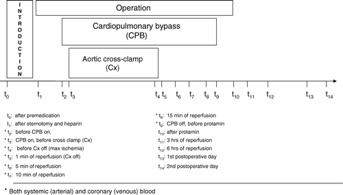
Systemic arterial blood and coronary venous blood were sampled at multiple time points as shown in and collected into precooled tubes with K3EDTA. Samples were kept on ice before centrifugation (10 min, 4°C, 3000 G) within 30 min and plasma was then immediately stored at −80°C until analysis. Urinary samples were collected by catheter at four time points; at baseline, during operation, 24 hours (day 1) and 48 hours (day 2) after end of operation. Samples were frozen and stored at −80°C until analysis.
Figure 1. Study design. The time course of operation and sampling is indicated.
Biochemical Assessments
Nonesterified 8-iso-PGF2α
Measured by radioimmunoassay without prior extraction as previously described Citation7. Detection limit was 23 pM with an intra-assay coefficient variation (CV) of 12–15%.
15-keto-dihydro-PGF2α
Measured by radioimmunoassay as previously described Citation8. Detection limit was 45 pM with an intra assay coefficient variation (CV) of 12–14%.
hsCRP
Measured on a Hitachi 917 analyzer (CRP (Latex) HS, Roche Diagnostics, Mannheim, Germany). Detection limit was 0.03 mg/l (analytical sensitive).
Troponin T
Measured by use of an Elecsys 2010 analyzer (Roche Diagnostics, Mannheim, Germany). Detection limit was 0.01 µg/l.
Albumin
Albumin concentrations (g/l) were measured by a colorometric endpoint assay on a Hitachi 917 analyzer (BCG method, Roche Diagnostics, Mannheim, Germany).
Expression of results
All plasma values of measured variables were corrected for hemodilution relative to albumin as intravascular reference. Plasma albumin was reduced to 81% of baseline levels during the initial phase of operation and fell abruptly to 57% at onset of CPB. Thereafter these albumin values were found: 60% at end of operation; 72% on day 1; and 74% on day 2.
Statistical analysis
Nonparametric tests and median values with percentiles (25% and 75%) were applied. Wilcoxon signed ranks test was performed for comparison between two dependent groups and Mann-Whitney test was used when comparing two independent groups. Spearman's correlation coefficient (rho) was applied for correlation analyses. For all analyses values below detection limit were set to the detection level at calculation. Values for p < 0.05 were considered significant. Data analyses were performed with the Statistical Package for Social Sciences (SPSS 11.5, Chicago, Illinois) or GraphPad Prism Software (GraphPad Software 4.01, Inc., San Diego, CA, USA).
Results
Duration of main critical events were: operation 142 min; CPB 63 min; and ischemia with aortic cross clamp 36 min. Median number of grafts was three. There was no hospital mortality or severe cardiopulmonary or vascular morbidity.
Oxidative stress detected by 8-iso-PGF2α
Plasma values of 8-iso-PGF2α increased, as seen in , from baseline 192 pM (t0) to 343 pM (t1) after the initial phase of the operation (p < 0.001). This was followed by a trend of further increase (NS) after onset of CPB (t3) and thereafter a gradual, but significant fall at the end of surgery (t10) and to below baseline levels on postoperative days 1 (t13) and 2 (t14). At the critical stage immediately before (t4) and 1 min after (t5) release of the aortic cross-clamp, median values were slightly (30–50 pM) higher in coronary venous blood than in arterial blood, but these differences were not significant. Urinary 8-iso-PGF2α rose significantly from baseline 300 pmol/mmol creatinine to 635 in the intraoperatively collected sample and fell to 475 and 455 on days 1 and 2, respectively.
Figure 2. Changes in plasma 8-iso- PGF2α. Blood samples were taken at various stages from a radial artery (closed symbols, t0-t14) and from the coronary sinus (open symbols, t2-t9). Median values with percentiles (25th and 75th) are shown. The results are correlated with albumin concentration.
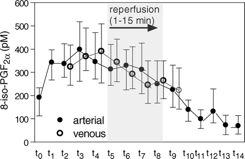
Peak plasma values of 8-iso-PGF2α correlated inversely with duration of both operation (p < 0.001) and CPB (p < 0.01), but there was no significant correlation with aortic cross clamp-time. Parallel to plasma, preoperative urine values correlated inversely with duration of the operation (p < 0.01), but not with duration of CPB or with aortic cross-clamp time. Surprisingly, analysis of ASA withdrawal prior to surgery revealed intragroup differences in 8-iso-PGF2α. Although not statistically significant, the longest withdrawal (>4 days) was associated with a 50% higher median peak value in plasma than the shortest withdrawal (466 versus 309 pM, NS). In the intra-operative urine sample, these differences were significant (697 versus 457 pmol/mmol creatinine, p = 0.025). There was a negative correlation between the dose of heparin and the rise in 8-iso-PGF2α in plasma (p = 0.035) during operation. A negative correlation was also found for heparin dose and urinary 8-iso-PGF2α both for urine sampled during surgery (p = 0.002) and on day 1 (p = 0.022).
Inflammation detected by 15-keto-dihydro-PGF2α and hsCRP
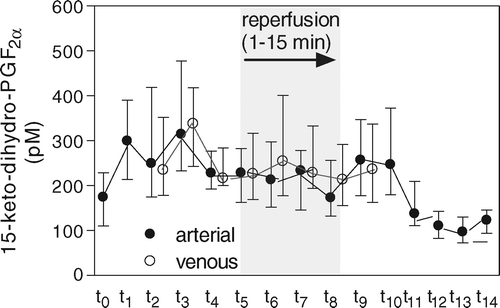
Levels of 15-keto-dihydro-PGF2α in arterial plasma presented a time-based profile partly similar to 8-iso-PGF2α (). Following sternotomy 15-keto-dihydro-PGF2α rose from baseline 174 pM (t0) to 299 pM (t1) (p < 0.001), a level that was maintained before a gradual decline (t4 −t10). In the postoperative period (t11−t14) values fell to below baseline. No differences were observed between arterial and coronary venous samples. Urinary samples showed a parallel time-based profile with elevated intraoperative values as did 8-iso-PGF2α. No significant correlation was found between 15-keto-dihydro-PGF2α and duration of operation, CPB or aortic cross-clamping. However, as with 8-iso-PGF2α, two subgroups behaved differently to withdrawal of ASA. Thus, patients with the longest withdrawal presented a higher plasma baseline (192 pM versus 107 pM, p = 0.023) and a tendency to higher median peak (t5) plasma value (247 versus 185, NS) than patients with shorter cessation. These differences were partly reflected in the urine samples by a higher median value of 15-keto-dihydro-PGF2α in samples collected during the operations than samples taken before (967 versus 679 pmol/mmol creatinine, NS).
Figure 3. Changes in plasma 15-keto-dihydro-PGF2α. Blood samples were taken at various stages from a radial artery (closed symbols, t0-t14) and from the coronary sinus (open symbols, t2-t9). Median values with percentiles (25th and 75th) are shown. The results are correlated with albumin concentration.
hsCRP was maintained at a low level (1.2–1.6 mg/l) during operation and the first post-operative hours (t12), but rose to high levels (114 mg/l) on day 1 and even higher levels (224 mg/l) on day 2 (p < 0.001). hsCRP showed a positive correlation with duration of CPB (p = 0.04), but not with operation or aortic cross clamp-time. No correlation between oxidative stress (8-iso-PGF2α) and inflammation (15-keto-dihydro-PGF2α and hsCRP) was found.
Myocardial injury detected by troponin T
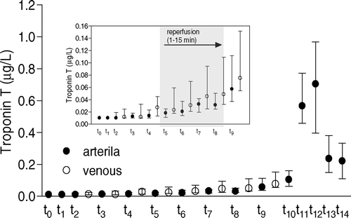
Troponin T rose slightly during the early reperfusion phase. A main peak of 0.57–0.70 µg/l was observed 3–6 hours (t11-t12) after aortic declamping (p < 0.001) to be followed by a marked fall on postoperative days 1 and 2 (p < 0.001) (). No marked differences between arterial and coronary venous blood were observed. Troponin T correlated strongly with aortic cross-clamp time (p < 0.0001) and CPB time (p = 0.006), but not with duration of the operation. No correlations were found with time off ASA, dosage of heparin or peak values of 8-iso-PGF2α and 15-keto-dihydro-PGF2α.
Figure 4. Changes in plasma troponin T. Blood samples were taken at various stages from a radial artery (closed symbols, t0-t14) and from the coronary sinus (open symbols, t2-t9). Median values with percentiles (25th and 75th) are shown. The results are correlated with albumin concentration.
Discussion
Main observations in the present study were: 1) There were no significant differences in isoprostane release between coronary venous and systemic arterial blood; 2) Isoprostane release was predominantly seen in the initial stages of surgery and 3) No correlation existed between isoprostane release and troponin T. A further observation was an apparent influence of ASA and heparin upon isoprostane release.
We could not confirm previous reports that reperfusion of the globally ischemic heart led to a detectable oxidative stress of myocardial origin in the present clinical setting Citation1, Citation10. However, it may be that such a situation was partly masked by a high systemic isoprostane level (see below). Also detection in coronary venous blood of primary ROS adducts by electron spin resonance (ESR) might have revealed a more distinct myocardial component of oxidative stress Citation10.
By frequent sampling during surgery, it was possible to follow the exact time course of oxidative stress with a rapid rise in 8-iso-PGF2α and an inflammatory process followed by a parallel rapid rise in 15-keto-dihydro-PGF2α. Our findings with both parameters elevated after initial surgery including sternotomy but before start of CPB and with little further increase thereafter were surprising and are partly conflicting with those of previous studies Citation1–3. In support of our findings, a recent study reported that surgical trauma alone, and not CPB, dominated the rise in proinflammatory variables at an early stage of CABG Citation11. Also the inverse relation between 8-iso-PGF2α (peak level) and duration of operation as seen in our study may underline the influence of the surgical trauma itself. The cytokine parameter of inflammation hsCRP did not respond during the early part of the operation, but was elevated to about 35 and 65 times higher values on postoperative days 1 and 2, respectively, thus reflecting the overall perioperative trauma to the patients. No correlation between oxidative stress (8-iso-PGF2α) and inflammation (15-keto-dihydro-PGF2α and hsCRP) was found in this study.
Importantly, and as we have previously shown Citation12 in patients with acute myocardial infarction (AMI) treated with primary percutaneous coronary intervention (PCI), there was no significant correlation between 8-iso-PGF2α and the myocardium-specific injury marker troponin T in individual patients. In contrast to 8-iso-PGF2α, troponin T showed a minor but significant rise that correlated positively with the duration of ischemia. Seen together, it seems that the oxidative stress parameter 8-iso-PGF2α reflected the sequence of additive traumas, whereas the myocardial marker troponin T showed a transient but moderate washout profile that paralleled the duration of ischemia during aortic cross-clamping. This indicates that the myocardium was protected by moderate hypothermia plus cardioplegia Citation9 or by opioid and anesthetic induced activation of beneficial preconditioning pathways Citation13.
An unexpected finding in this study was that baseline 8-iso-PGF2α (192 pM) was 3 to 5 times higher than in studies of healthy volunteers Citation7 and in previous PCI studies by our group Citation12, Citation14. The most likely reason for an elevated baseline 8-iso-PGF2α may relate to an altered balance between prooxidant and antioxidant forces prior to the operation. Surprisingly, when the time interval from preoperative withdrawal of ASA was taken into consideration, a potential explanation might be found. The arbitrary subgroup with ≤ 4 days off ASA presented lower values in 8-iso-PGF2α and in 15-keto-dihydro-PGF2α than the subgroup with > 4 days off. These results indicate that withdrawal of ASA somehow reversed or even induced a negative overshoot of some of its main properties: as a well-known cyclooxygenase inhibitor Citation15; and, as shown in animal experiments, as an inhibitor of plasma and tissue oxidases Citation16 and as an activator of endothelial nitric oxide (NO) synthase Citation17. Both latter properties strengthen the beneficial NO axis in inhibition of oxidative stress and may have been lost by discontinuation of therapy. Furthermore, the absence of the ASA metabolite salicylic acid as a potent scavenger may also have contributed to a high level of 8-iso-PGF2α Citation18. The question of whether preoperative ASA-withdrawal also conferred any cardiovascular risk Citation19 could not be answered in the present study but warrants attention.
Another important drug related observation was the negative correlation between the dosage of heparin and the levels of 8-iso-PGF2α in urine and in plasma, respectively. Heparin may have antioxidant properties in vivo, by enhancing extracellular superoxide dismutase (SOD) activities and also by iron chelation Citation20. Both properties may reduce generation of ROS and subsequently isoprostane production. Accordingly, in a placebo-controlled study of patients who had their gallbladder removed, heparin prevented an increase in plasma malondialdehyde which is another known biomarker of oxidative stress Citation21.
The above explanations for antioxidant properties of ASA and heparin stem largely from preclinical research and cannot easily be translated into a clinical reality. Also, in the present clinical study several confounding factors like a potentially nonhomogeneous population of patients and small differences in anesthetic management may have influenced the outcome of main parameters. Further the small study size (only twenty patients) and sample variation are essential limitations. Thus the intriguing possibility of both ASA and heparin having antioxidant properties in CABG has to be confirmed in new controlled studies.
Three main conclusions can be made from the present study. First, a considerable oxidative stress occurred early during conventional CABG, but was mainly induced by the surgical trauma. Second, in individual patients no correlations were found between key parameters of oxidative stress, inflammation and myocardial injury. The intriguing possibility that cardiovascular drugs in common use, such as heparin and ASA, influence the prooxidant/antioxidant balance during the operation needs confirmation in future studies.
The study was supported by grants from The Norwegian Council for Cardiovascular Disease, Trondheim University Hospital Research Fund and the SINTEF Health Research. The support by cardiologist Arve Tromsdal for echocardiografic control of the coronary sinus sampling and Stian Lydersen for statistical advice is gratefully acknowledged. We also kindly thank Eva Sejby for 8-iso-PGF2α and 15-keto-dihydro-PGF2α measurements and the staff at Department of Clinical Chemistry, Trondheim University Hospital with vitamins, troponin T, albumin and hsCRP measurements.
References
- Ferrari R, Alfieri O, Curello S, Ceconi C, Cargnoni A, Marzollo P, et al. Occurrence of oxidative stress during reperfusion of the human heart. Circulation. 1990; 81: 201–11
- Delanty N, Reilly MP, Pratico D, Lawson JA, McCarthy JF, Wood AE, et al. 8-epi PGF2 alpha generation during coronary reperfusion. A potential quantitative marker of oxidant stress in vivo. Circulation. 1997; 95: 2492–9
- Ulus AT, Aksoyek A, Ozkan M, Katircioglu SF, Basu S. Cardiopulmonary bypass as a cause of free radical-induced oxidative stress and enhanced blood-borne isoprostanes in humans. Free Radic Biol Med. 2003; 34: 911–7
- Cavalca V, Sisillo E, Veglia F, Tremoli E, Cighetti G, Salvi L, et al. Isoprostanes and oxidative stress in off-pump and on-pump coronary bypass surgery. Ann Thorac Surg. 2006; 81: 562–7
- Basu S, Helmersson J. Factors regulating isoprostane formation in vivo. Antioxid Redox Signal. 2005; 7: 221–35
- Morrow JD, Hill KE, Burk RF, Nammour TM, Badr KF, Roberts LJ 2nd. A series of prostaglandin F2-like compounds are produced in vivo in humans by a non-cyclooxygenase, free radical-catalyzed mechanism. Proc Natl Acad Sci USA. 1990;87:9383–7.
- Basu S. Radioimmunoassay of 8-iso-prostaglandin F2alpha: An index for oxidative injury via free radical catalysed lipid peroxidation. Prostaglandins Leukot Essent Fatty Acids. 1998; 58: 319–25
- Basu S. Radioimmunoassay of 15-keto-13,14-dihydro-prostaglandin F2alpha: An index for inflammation via cyclooxygenase catalysed lipid peroxidation. Prostaglandins Leukot Essent Fatty Acids. 1998; 58: 347–52
- Braimbridge MV, Chayen J, Bitensky L, Hearse DJ, Jynge P, Cankovic-Darracott S. Cold cardioplegia or continuous coronary perfusion? Report on preliminary clinical experience as assessed cytochemically. J Thorac Cardiovasc Surg. 1977; 74: 900–6
- Clermont G, Vergely C, Jazayeri S, Lahet JJ, Goudeau JJ, Lecour S, et al. Systemic free radical activation is a major event involved in myocardial oxidative stress related to cardiopulmonary bypass. Anesthesiology. 2002; 96: 80–7
- Prondzinsky R, Knupfer A, Loppnow H, Redling F, Lehmann DW, Stabenow I, et al. Surgical trauma affects the proinflammatory status after cardiac surgery to a higher degree than cardiopulmonary bypass. J Thorac Cardiovasc Surg. 2005; 129: 760–6
- Berg K, Jynge P, Bjerve K, Skarra S, Basu S, Wiseth R. Oxidative stress and inflammatory responses during and following coronary interventions for acute myocardial infarction. Free Radic Res. 2005; 39: 629–36
- Hausenloy DJ, Yellon DM. Protecting the myocardium at time of reperfusion. Meeting report of the Hatter Institute 4th International Workshop on Cardio-protection. Basic Res Cardiol. 2005; 100: 383–6
- Berg K, Wiseth R, Bjerve K, Brurok H, Gunnes S, Skarra S, et al. Oxidative stress and myocardial damage during elective percutaneous coronary interventions and coronary angiography. A comparison of blood-borne isoprostane and troponin release. Free Radic Res. 2004; 38: 517–25
- Roth GJ, Stanford N, Majerus PW. Acetylation of prostaglandin synthase by aspirin. Proc Natl Acad Sci USA. 1975; 72: 3073–6
- Wu R, Lamontagne D, de Champlain J. Antioxidative properties of acetylsalicylic acid on vascular tissues from normotensive and spontaneously hypertensive rats. Circulation. 2002; 105: 387–92
- Taubert D, Berkels R, Grosser N, Schröder H, Grünemann D, Schömig E. Aspirin induces nitric oxide release from vascular endothelium: A novel mechanism of action. Br J Pharmacol. 2004; 143: 159–65
- Powell SR, Hall D. Use of salicylate as a probe for •OH formation in isolated ischemic rat hearts. Free Radic Biol Med. 1990; 9: 133–41
- Fischer LM, Schlienger RG, Matter CM, Jick H, Meier CR. Discontinuation of nonsteroideal anti-inflammatory drug therapy and risk of acute myocardial infarction. Arch Intern Med. 2004; 164: 2472–6
- Karlsson K, Marklund SL. Heparin-induced release of extracellular superoxide dismutase to human blood plasma. Biochem J. 1987; 242: 55–9
- Dzieciuchowicz L, Checinski P, Krauss H. Heparin reduces oxidative stress in the postoperative period. Med Sci Monit 2002; 8: CR657–CR660