Abstract
Objectives. CD40 is a marker of immunological activation and is expressed in the atherosclerotic lesions. We studied whether CD40 and cholesterol synthesis pathways are associated with each other. Design. Forty-three subjects were randomly assigned to receive either simvastatin (n = 14), atorvastatin (n = 15), or placebo (n = 14) for eight weeks. Plasma samples were obtained before and at the end of the follow-up. sCD40 levels were measured in duplicate using an enzyme-linked immunosorbent assay. Cholesterol, its precursor lathosterol, the plant sterols campesterol and sitosterol as well as 27-hydroxycholesterol were quantified by gas-liquid chromatography-mass spectrometry. Results. sCD40 was inversely correlated with the lathosterol to cholesterol ratio (r = − 0.47, p = 0.002), an indicator of cholesterol synthesis rate, as well as apolipoprotein A–I (r = − 0.38, p = 0.01) in addition to being directly correlated with 27-hydroxycholesterol (r = 0.40, p = 0.008). In multivariate linear regression analysis these three predictors explained 37% of the total variability of sCD40 levels. Simvastatin or atorvastatin treatment had no significant effect on sCD40 levels. Conclusion. These results indirectly suggest that sCD40 concentrations are related to cellular cholesterol levels. This may be a novel indication for the relationship between immunological processes and cholesterol metabolism.
Hypercholesterolemia is a risk factor for coronary heart disease and may also sustain subclinical inflammation. Oxidized low density lipoprotein (LDL) particles in the artery wall stimulate immune cell adhesion to endothelial cells Citation1, and oxidized lipids may act as antigens for T cells, possibly promoting the development of atherosclerotic lesions. CD40 to CD40ligand (CD40L) interaction is needed both for the initiation and sustaining of inflammatory responses. CD40 is an important molecule in antigen presentation and B cell differentiation. The CD40 system has been shown to be upregulated in patients with moderate hypercholesterolemia Citation2, and it is involved in several stages of the formation of atherosclerotic lesions Citation3. In such lesions, high levels of CD40 and CD40L are expressed on macrophages, T cells, endothelial cells and vascular smooth muscle cells Citation4. Ligation of CD40L to the artery wall cell CD40 receptor leads to the production of adhesion molecules, proinflammatory cytokines, chemokines and metalloproteases Citation2, which promotes homing and extravasation of leukocytes at the sites of inflammation.
Hypercholesterolemia and chronic low-grade immunological activation are pivotal in the development of atherosclerosis. However, the interconnections between these two factors are not well known. The present study was designed to examine whether the CD40 system, as measured by the plasma level of soluble CD40 (sCD40), is associated with cholesterol metabolism in hypercholesterolemic patients. Lathosterol is a cholesterol precursor, and the lathosterol to cholesterol ratio served as a marker of cholesterol synthesis Citation5. 27-hydroxycholesterol is an oxygenated derivative of cholesterol and a major oxysterol in circulation Citation6; it served as a marker for cholesterol excretion pathways. The plasma lathosterol to cholesterol ratio as well as campesterol to cholesterol were selected as markers of cholesterol absorption. In addition, we studied whether short-term statin treatment affects the plasma levels of sCD40.
Materials and methods
The patients and study design have been described previously by Päivä et al. Citation7. In short, 43 subjects aged between 31 and 69 years (29 men and 14 women) were recruited from the Tampere University Hospital and primary health care centres of neighbouring municipalities. Their average serum total cholesterol concentration was 5.9±0.9 mmol/l and serum triglycerides below 4.5 mmol/l. Patients using concurrent lipid altering medication or antioxidant vitamins were excluded. The patients were instructed to adhere to their normal diet during the study. The study protocol was approved by the Ethics Committee of the Tampere University Hospital, and written informed consent was obtained from all participants. The procedures followed with the Helsinki Declaration of 1975.
This was a randomised, double-blind and placebo-controlled trial with three treatment groups: placebo, atorvastatin 40 mg/day, and simvastatin 80 mg/day. The duration of the follow-up was 8 weeks. Seven of the patients had type 2 diabetes, 24 had coronary artery disease and 16 had hypertension. Distributions of these diseases were similar between the treatment groups.
Measurement of plasma-soluble CD40 concentrations
The plasma levels of sCD40 were measured in duplicate using a commercially available enzyme-linked immunosorbent assay (ELISA) kit (MedSystem Diagnostics GmbH, Austria). The overall intra-assay coefficient of variation was 6.4% for sCD40. The detection limit of the assay was 12 pg/ml
Measurement of plasma lipid and apolipoprotein concentrations
Plasma triglycerides as well as total and high-density lipoprotein (HDL) cholesterol concentrations were analysed by a Cobas Integra 700 automatic analyser, with reagents and calibrators recommended by the manufacturer (Hoffmann-La Roche Ltd., Basel, Switzerland). LDL concentration was calculated using Friedewald's formula. Plasma apolipoprotein B (apoB) and A–I (apoA–I) concentrations were determined with fully automated immunoturbidimetric assays by the same analyzer as the lipids, using specific COBAS INTEGRA cassettes for apoB and apoA–I (Roche Diagnostics).
Plasma sterol analysis
The quantification of the cholesterol precursor lathosterol, 27-hydroxycholesterol and of the phytosterols, campesterol and sitosterol, was performed as described previously Citation7.
Statistical analyses
Associations between study variables were evaluated by Spearman's rank order correlation coefficients. Stepwise linear regression models included sCD40 as a dependent variable and all variables having the strongest correlation with sCD40 in univariate analysis as explanatory independent variables. Overall differences between the treatment groups in their responses of sCD40 over time were compared with analysis of variance for repeated measurements (RANOVA). Within the study groups, the paired t-test was used to test the significance of differences over time (baseline versus after eight weeks of treatment). Differences between tertiles were compared with the Kruskal-Wallis test. SPSS version 11.5 (Chigago, Illinois, USA) was used to carry out the calculations. A p-value less than 0.05 was considered statistically significant.
Results
The median sCD40 level for all subjects was 96 ng/ml (37–290 ng/ml). In univariate analysis, the plasma level of sCD40 correalted negatively and significantly with HDL-cholesterol (r = − 0.33, p = 0.03), apoA–I (r = − 0.38, p = 0.01), lathosterol (r = − 0.34, p = 0.03) and the lathosterol to cholesterol ratio (r = − 0.47, p = 0.002); and positively with 27-hydroxycholesterol (r = 0.40, p = 0.008, , ). Analysis of variance confirmed the above-mentioned results between sCD40 tertiles. In multivariate linear regression analysis we used 0.03 as cut-off p-value for independent variables. Independent variables were lathosterol/total cholesterol, 27-hydroxycholesterol and ApoA–I and they had no significant bivariate correlation with each other. These three variables as well as sCD40 were in normal distribution. Lathosterol/total cholesterol, 27-hydroxycholesterol and ApoA–I explained 37% of the total variability of sCD40 levels, and 27-hydroxycholesterol appeared as the most significant individual predictor (p = 0.009) (see ). Eight weeks’ treatment with simvastatin (80 mg/day) and atorvastatin (40 mg/day) had no statistically significant effect on the plasma levels of sCD40 ().
Figure 1. Correlation of soluble (sCD40) in plasma with lathosterol/total cholesterol and with 27-hydroxycholesterol.
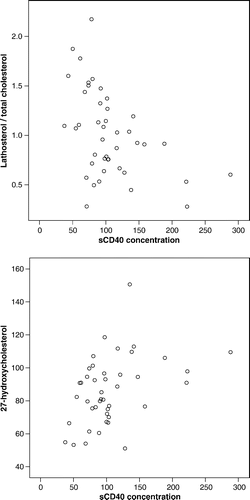
Table I. Spearman correlation analysis of soluble CD40 with lipid and cholesterol markers.
Table II. Stepwise multiple linear regression analysis with sCD40 as the dependent variable.
Table III. Plasma sCD40 (ng/ml)1 in hypercholesterolemic patients before and after eight weeks’ treatment with statin or placebo.
Discussion
We found that plasma levels of sCD40 negatively correlate with the plasma lathosterol to plasma cholesterol ratio as well as apoA–I and HDL-cholesterol; and positively with 27-hydroxycholesterol. CD40, expressed on the cell surface, is an activation marker of many cell types. We measured the soluble form of CD40, which is produced from pre-CD40 mRNA by alternative splicing to type II mRNA lacking the membrane-associated endodomain Citation8. sCD40 may also be produced by proteolytic cleavage from the cell surface CD40 Citation9. Plasma sCD40 may reflect the activation of the CD40 system: the engagement of CD40 on the surface of B cells by an anti-CD40 antibody led to enhanced sCD40 release, whereas non-activated B cells had minimal basal production of sCD40 Citation9. Moreover, cell lines having high membrane CD40 expression release large amounts of sCD40 in culture supernatants Citation10. It is suggested that enhanced signal transduction through cell surface CD40 is regulated by posttranscriptional and posttranslational production of sCD40 Citation8. Furthermore, sCD40 is a natural antagonist of CD40/CD154 interaction and may, therefore, represent negative feedback control of the CD40 system Citation9.
Cholesterol homeostasis is carefully controlled by multiple feedback mechanisms. In general, increased absorption of cholesterol and high cellular cholesterol decreases endogenous synthesis of cholesterol. Of the cholesterol metabolism markers studied, 27-hydroxysterol was the most significant predictor of sCD40 levels. 27-hydroxysterol is the most dominant oxisterol in human ateromas Citation11 where it may reflect a mechanism for eliminating excessive cholesterol and thus have a protective role Citation12. In addition, the level of 27-hyroxysterol in circulation are higher in patients with atherosclerosis than in control subjects Citation13. When combined, a decreased cholesterol synthesis rate and increased levels of 27-hydroxycholesterol may be a consequence of high levels of cellular cholesterol and, therefore, be related to sCD40. However, sCD40 had no significant correlation with total plasma cholesterol. This suggests that the cellular cholesterol synthesis rate and 27-hydroxycholesterol production are more importantly linked with the plasma levels of sCD40 than total cholesterol. However, it is of interest that patients with metabolic syndrome had higher sCD40L than patients without, supporting our observation that CD40 system and cholesterol metabolism have a connection with each other Citation14.
Statins are suggested to exert immunomodulatory and anti-inflammatory effects. With regard to the CD40 pathway, clinical trials with a moderate number of hypercholesterolemic patients have shown that several statins reduce the plasma levels of soluble CD40L Citation15–17. In addition, in human carotid arteries, atherosclerotic lesions in patients under statin therapy expressed much less CD40 compared to control patients Citation18, and short-term therapy with cerivastatin reduced CD40 on monocytes Citation19. In vitro atorvastatin inhibits the CD40 expression on vascular endothelial cells, smooth muscle cells and macrophages Citation17. However, our short-term statin therapy was not able to demonstrate similar effects, which is in line with a previous study Citation16.
As decreased cholesterol synthesis is associated with increased levels of sCD40, one may expect that statin treatment increases the levels of CD40. In our present study group simvastatin and atorvastatin treatment resulted in a significant lowering of LDL cholesterol and total cholesterol Citation7. However, sCD40 levels were similar before and after the treatment. The anti-inflammatory properties of statins may be partly due to its cholesterol-lowering effects—for example, free cholesterol accumulation in macrophages leads to the induction and secretion of proinflammatory cytokines Citation20. Statins may also enhance the immune response, since hydroxymethyl-coenzyme A reductase inhibition stimulates caspase-1 activity and Th1-cytokine release in peripheral blood mononuclear cells Citation21. In addition, an in vitro study showed that pravastatin and fluvastatin induce the production of IL-18, but the effect was not demonstrated on structural statin analog, LFA703, without an inhibitory effect on HMG-CoA reductase Citation22. Indeed, these studies suggest that decreased cholesterol synthesis may enhance immune responses, which is in line with our present findings. All in all, it seems that statins have multi-faced effects on the immune system and that these effects are also reflected on the CD40 system Citation22. In addition, the effect of statin on CD40 may depend on the duration of treatment, and different statins may have different immunological effects. Recently it was observed that simvastatin together with losartan reduced plasma sCD40L levels more than simvastatin alone Citation23.
In conclusion, a decreased cholesterol synthesis rate and increased levels of 27-hydroxycholesterol may be a consequence of high levels of cellular cholesterol, which may upregulate the CD40 system. This may be a novel indication for the relationship between immunological processes and cholesterol metabolism.
The study has received financial support from the Academy of Finland (grant no. 104821), the Medical Research Foundation of Tampere University Hospital, the Emil Aaltonen Foundation, the Paavo Nurmi Foundation, the Finnish Foundation for Cardiovascular Research, Elli and Elvi Oksanen Foundation and the Finnish Cultural Foundation. We thank Ms Marita Koli for her skilful technical assistance.
References
- Erl W, Weber PC, Weber C. Monocytic cell adhesion to endothelial cells stimulated by oxidized low density lipoprotein is mediated by distinct endothelial ligands. Atherosclerosis 1998; 136: 297–303
- Schonbeck U, Libby P. CD40 signaling and plaque instability. Circ Res 2001; 89: 1092–103
- Lutgens E, Gorelik L, Daemen MJ, de Muinck ED, Grewal IS, Koteliansky VE, et al. Requirement for CD154 in the progression of atherosclerosis. Nat Med 1999; 5: 1313–6
- Mach F, Schonbeck U, Sukhova GK, Bourcier T, Bonnefoy JY, Pober JS, et al. Functional CD40 ligand is expressed on human vascular endothelial cells, smooth muscle cells, and macrophages: Implications for CD40-CD40 ligand signaling in atherosclerosis. Proc Natl Acad Sci USA 1997; 94: 1931–6
- Simonen PP, Gylling HK, Miettinen TA. Diabetes contributes to cholesterol metabolism regardless of obesity. Diabetes Care 2002; 25: 1511–5
- Bjorkhem I, Diczfalusy U. Oxysterols: Friends, foes, or just fellow passengers?. Arterioscler Thromb Vasc Biol 2002; 22: 734–42
- Paiva H, Thelen KM, Van Coster R, Smet J, De Paepe B, Mattila KM, et al. High-dose statins and skeletal muscle metabolism in humans: A randomized, controlled trial. Clin Pharmacol Ther 2005; 78: 60–8
- Tone M, Tone Y, Fairchild PJ, Wykes M, Waldmann H. Regulation of CD40 function by its isoforms generated through alternative splicing. Proc Natl Acad Sci USA 2001; 98: 1751–6
- Contin C, Pitard V, Itai T, Nagata S, Moreau JF, Dechanet-Merville J. Membrane-anchored CD40 is processed by the tumor necrosis factor-alpha-converting enzyme. Implications for CD40 signaling. J Biol Chem 2003; 278: 32801–9
- De Paoli P, Cozzi M, Tedeschi R, Gloghini A, Cilia AM, van Kooten C, et al. High CD40 membrane expression in AIDS-related lymphoma B cell lines is associated with the CD45RA+, CD45RO+, CD95+ phenotype and high levels of its soluble form in culture supernatants. Cytometry 1997; 30: 33–8
- Garcia-Cruset S, Carpenter KL, Guardiola F, Stein BK, Mitchinson MJ. Oxysterol profiles of normal human arteries, fatty streaks and advanced lesions. Free Radic Res 2001; 35: 31–41
- Babiker A, Andersson O, Lund E, Xiu RJ, Deeb S, Reshef A, et al. Elimination of cholesterol in macrophages and endothelial cells by the sterol 27-hydroxylase mechanism. Comparison with high density lipoprotein-mediated reverse cholesterol transport. J Biol Chem 1997; 272: 26253–61
- Harik-Khan R, Holmes RP. Estimation of 26-hydroxycholesterol in serum by high-performance liquid chromatography and its measurement in patients with atherosclerosis. J Steroid Biochem 1990; 36: 351–5
- Lee WL, Lee WJ, Chen YT, Liu TJ, Liang KW, Ting CT, et al. The presence of metabolic syndrome is independently associated with elevated serum CD40 ligand and disease severity in patients with symptomatic coronary artery disease. Metabolism 2006; 55: 1029–34
- Cipollone F, Mezzetti A, Porreca E, Di Febbo C, Nutini M, Fazia M, et al. Association between enhanced soluble CD40L and prothrombotic state in hypercholesterolemia: Effects of statin therapy. Circulation 2002; 106: 399–402
- Wang TD, Chen WJ, Lin JW, Cheng CC, Chen MF, Lee YT. Efficacy of fenofibrate and simvastatin on endothelial function and inflammatory markers in patients with combined hyperlipidemia: Relations with baseline lipid profiles. Atherosclerosis 2003; 170: 315–23
- Schonbeck U, Gerdes N, Varo N, Reynolds RS, Horton DB, Bavendiek U, et al. Oxidized low-density lipoprotein augments and 3-hydroxy-3-methylglutaryl coenzyme A reductase inhibitors limit CD40 and CD40L expression in human vascular cells. Circulation 2002; 106: 2888–93
- Mulhaupt F, Matter CM, Kwak BR, Pelli G, Veillard NR, Burger F, et al. Statins (HMG-CoA reductase inhibitors) reduce CD40 expression in human vascular cells. Cardiovasc Res 2003; 59: 755–66
- Garlichs CD, John S, Schmeisser A, Eskafi S, Stumpf C, Karl M, et al. Upregulation of CD40 and CD40 ligand (CD154) in patients with moderate hypercholesterolemia. Circulation 2001; 104: 2395–400
- Li Y, Schwabe RF, DeVries-Seimon T, Yao PM, Gerbod-Giannone MC, Tall AR, et al. Free cholesterol-loaded macrophages are an abundant source of tumor necrosis factor-alpha and interleukin-6: Model of NF-kappaB- and map kinase-dependent inflammation in advanced atherosclerosis. J Biol Chem 2005; 280: 21763–72
- Montero MT, Hernandez O, Suarez Y, Matilla J, Ferruelo AJ, Martinez-Botas J, et al. Hydroxymethylglutaryl-coenzyme A reductase inhibition stimulates caspase-1 activity and Th1-cytokine release in peripheral blood mononuclear cells. Atherosclerosis 2000; 153: 303–13
- Takahashi HK, Mori S, Iwagaki H, Yoshino T, Tanaka N, Weitz-Schmidt G, et al. Differential effect of LFA703, pravastatin, and fluvastatin on production of IL-18 and expression of ICAM-1 and CD40 in human monocytes. J Leukoc Biol. 2004;77:400–7.
- Seung HH, Koh KK, Quon MJ, Lee Y, Shin EK. The effects of simvastatin, losartan, and combined therapy on soluble CD40 ligand in hypercholesterolemic, hypertensive patients. Atherosclerosis. 2006. Article in press