Abstract
Objectives. Microdialysis allows the in vivo biochemical analysis of interstitial fluids. Our aim was to reveal in vivo reliable data of the myocardium during open beating heart surgery. Design. In ten patients undergoing routine beating coronary artery bypass grafting a microdialysis catheter was inserted into the left ventricle. Measurements were performed up to 45 min after anastomosis. Data were retrospectively compared with standard on-pump procedures. Results. The myocardial lactate remained stable during anastomosis, followed by a significant decrease of lactate after revascularisation. Myocardial glucose levels showed a slight decrease, followed by a significant increase after revascularisation. Myocardial purines showed a slight increase during anastomosis, followed by a sharp decrease during reperfusion period. Conclusions. In contrast to on-pump procedures myocardial lactate and purines showed less increasing trend during the ischemic period, while myocardial glucose remained stable as a sign of preserved tissue blood flow. Myocardial microdialysis showed different values compared to the elective on-pump CABG and previous animal studies. This technique allows bedside monitoring of biochemical changes, suggesting its possible role as a clinical monitoring tool.
Coronary artery bypass grafting (CABG) using cardiopulmonary bypass is a widely used, safe, and effective procedure with a low mortality rate Citation1. However, despite of improvement in perfusion techniques, the morbidity associated with cardiopulmonary bypass is still significant Citation2. The potential benefits gained by avoiding cardiopulmonary bypass and cardioplegic arrest have led to a renewed interest in off-pump coronary artery bypass surgery Citation3.
But early detection of perioperative ischemia is also essential for immediate intervention in these patients. With the exception of septic conditions the interstitial concentrations of lactate have been shown to be closely related to variations in tissue perfusion and may thus be used as sensitive markers of myocardial ischemia Citation4. The microdialysis technique is a new and feasible technique for measuring such markers of cell injury and metabolites in the interstitial fluid of nearly every organ including the beating heart. This technique introduced to human cardiac surgery by Habicht Citation5 allows in vivo sampling and rapid biochemical analysis of interstitial fluids Citation6. Animal and isolated perfused heart experiments have also been used to monitor the different steps in the development of acute ischemic injury on a cellular level Citation7.
In this study, myocardial microdialysis was used to investigate the metabolism during and after beating heart surgery of patients with preserved ejection fraction. Our aim was to reveal in vivo reliable data of the myocardium of these patients and to depict the differences to previous on-pump studies retrospectively.
Methods
Patients
Following approval by the local ethical committee and informed, written consent, ten patients (average age 64±8) with coronary artery disease (CAD) and a left ventricular ejection fraction (LVEF) of more than 40% were included in the study. Six of the patients had a history of myocardial infarction. Cardiovascular risk factors were: hypertension: n = 10; diabetes mellitus: n = 3; former or present smoking habit: n = 4; hypercholesterolemia: n = 9; family history of cardiovascular diseases: n = 3. Patients underwent CABG with the left internal mammaria artery (LIMA) for revascularisation of the left anterior descending coronary artery (LAD). An additive venous graft or a radial artery for revascularisation of the RCA was used in six patients. Exclusion criteria included recent (≤1 month) myocardial infarction, reoperative operation, and emergency operation.
Anaesthesia
Anaesthesia was induced with etomidate (0.3–0.5 mgkg−1) and sufentanyl (0.5–1 µgkg−1) and maintained with continuous infusions of propofol (5–8 mgkg−1h−1) and sufentanyl (0.5–1 µgkg−1h−1). Muscle relaxation was achieved with pancuronium bromid (0.1 mgkg−1). All patients were mechanically ventilated in a pressure controlled mode (PEEP = 5 cmH2O, level of pressure control = 15 cmH2O, FiO2=0.5). The respiratory rate was adjusted to achieve normocapnia.
All patients were equipped with a radial arterial line, and central venous line and an automated pulmonary artery catheter for continuous determination of mixed venous oxygen saturation and semicontinuous measurements of cardiac output and right ventricular ejection fraction (Vigilance; Edwards Lifescience, Baxter, Germany, Unterschleißheim). Intraoperative fluid management was adjusted to achieve and maintain a central venous pressure between 8 to 12 mmHg and a pulmonary artery capillary occlusion pressure (PAOP) between 15 to 18 mmHg. Volume replacement was performed with Ringer's solution and gelatine polysuccinate, as appropriate. Arterial blood samples were collected during the observation period for blood gas analysis and microdialysis measurements. Nasopharyngeal and rectal temperature were continuously measured throughout observation time.
Surgical and microdialysis procedure
After a median sternotomy a longitudinal pericardotomy was performed. Before preparation of the LIMA the myocardial microdialysis catheter (CMA 70, CMA/Microdialysis AB, Sweden) was inserted almost parallel to the LAD in the apical region of the beating heart. Therefore a venous cannula was tangentially inserted through the myocardial wall. The tip of the microdialysis catheter was then inserted into the distal end of the cannula and by retracting the cannula the catheter tip was finally placed in the myocardium and sutured with 5/0 Prolene (Ethicon). The inlet of this doublelumen microdialysis catheter was connected to a battery driven infusion pump (CMA 107 pump, CMA/Microdialysis AB, Sweden) and constantly perfused with lactate-free Ringer's solution at a flow rate of 2 µl/min. The microdialysis probes used had a membrane length of 10 mm and a molecular cut off of 20 kD. Interstitial fluid was sampled via the outlet in microvials, and stored at −75°C for subsequent analysis of glucose, lactate, purines and pyruvate.
The stabilization material was the second model Octopus system (Medronic Inc, Minneapolis, MN). The coronary was opened during transient (≤1 min) occlusion of the coronary artery and a suitably sized intraluminal shunt inserted. Shunt size was estimated before opening the coronary by examination of the external appearance of the vessel; shunts were available in sizes ranging from 1.5 to 3.5 mm (in 0.25 mm increments). Shunts were inserted under direct vision, proximal end first. Coronary anastomoses were performed using 7-0 Prolene and standard techniques. Sutures for pericardial traction and Trendelenburg manipulation were used to facilitate the exposure with preservation of the hemodynamic stability. At the end of the intervention, the circulating heparin was neutralized after ACT control.
Experimental design
A possible source of error in sampling of dialysates is the circumstance that the mechanical insertion of the probe may cause bleeding, induction of inflammation and reactive changes in the tissue due to the local trauma, which potentially may influence the concentrations of the metabolites after catheter implantation. For this reason a 15 min equilibrium period was employed after probe insertion, in this time interval all metabolites showed stable values.
Microvials were exchanged 15 min after the equilibrium interval (prae 1); 15 min after shunt implantation (prae 2) and the last vial was exchanged immediately before reopening the LAD (prae 3). During reperfusion, vials were exchanged for three times in the same 15 min mode as before (post 1–3). Microdialysis catheters were removed before chest closure, inspected for blood dryness and the patient was transferred to the intensive unit.
Statistical analysis
Results are presented as means±SEM. Statistical comparison of parameters in both groups was drawn using the Mann–Whitney-U test. For baseline versus end-of-experiment values a Wilcoxon Signed Rank test was used. A p < 0.05 was considered to be statistically significant. Statistical analysis was performed with the SPSS (version 12.0) software package (SPSS Inc., Chicago, IL).
Results
The microdialysis catheters were used without any complications such as bleeding, infections, arrhythmias, etc. The insertion procedure took less than 2 min. None of the patients showed clinical or enzymatic signs of a myocardial ischemia. The course of systemic metabolism and hemodynamics showed no significant changes (). In contrast to blood levels, the results of myocardial microdialysis values showed tremendous changes during the observation period.
Table I. Cardiovascular hemodynamic values. Data are presented as median±SEM. Heart rate (HR), mean arterial blood pressure (MAP), mean pulmonary arterial pressure (PAPM), central venous pressure (CVP), cardiac index (CI), end diastolic volume (EDV), systemic vascular resistance (SVR), mixed venous saturation of oxygen (SvO2) before (prae 1/2), and after [15 min intervals, post 1–3] after anastomosis of the LAD.
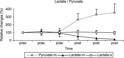
The myocardial lactate levels () remained stable before anastomosis of the LAD, there was a slight increase during the ischemic period (100% vs. 113±25%). After anastomosis, the lactate decreased tremendously to 13±6% of the baseline levels (p < 0.05). Myocardial pyruvate kept stable during ischemia, followed by a continuous increase (p < 0.05) during the reperfusion (100% vs. 350±112%) period. Plasma lactate showed no significant changes during the observation period.
Figure 1. Relative changes of the myocardial lactate and pyruvate levels before (prae 1–3) and after anastomosis of the LIMA (post 1–3; up to 45 min). During the early reperfusion a significant decrease of myocardial lactate and increase of pyruvate were observed (p < 0.05). Plasma values kept stable during the observation period.
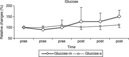
Myocardial glucose levels () showed a slight decrease during the ischemic period, followed by a significant increase (p < 0.05) after revascularisation (100% vs. 150±21%). Plasma glucose showed no significant changes during this observation period.
Figure 2. Relative changes of the myocardial glucose before (prae 1–3) and after anastomosis of the LIMA (post 1–3; up to 45 min). During the early reperfusion a significant increase of the glucose levels was found (p < 0.05). Plasma values kept stable during the observation period.
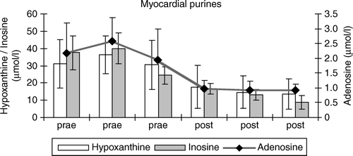
Hypoxanthine () showed a slight increase and peaked in ischemic myocardium within 20 min of LAD occlusion (31±13 vs. 36±10 µmol/l), followed by a sharp decrease during reperfusion period (p < 0.05). At the end of the observation period only 14±8 µmol/l hypoxanthine was found. Myocardial inosine and adenosine values showed a course similar to hypoxanthine levels. Adenosine had a peak of 2.6±1 µmol/l at the end of the ischemic period and a significant drop (p < 0.05) to 0.6±0.3 µmol/l after starting of reperfusion.
Figure 3. Absolute changes of the myocardial hypoxanthine, inosine and adenosine before (prae 1–3) and after anastomosis of the LIMA (post 1–3; up to 45 min). During the early reperfusion a significant decrease of the purines was found (p < 0.05).
Discussion
Off-pump myocardial revascularisation has gained increasing popularity due to its potential to avoid the damage induced by cardiopulmonary bypass, aortic cannulation, and cross-clamping. The benefit of off-pump surgery is discussed controversially. Conventional CABG with cardioplegic arrest induces global ischemia-reperfusion and myocardial injury. The procedure of off-pump myocardial revascularisation requires a period of coronary artery occlusion, which results in local ischemia. Biochemical markers have predominantly been used to identify the mechanisms mediating the inflammatory response and to compare groups of patients who had CABG with or without CPB. Diegeler and associates Citation8 found a significantly increased release of activated complement factors at different postoperative times in the CPB group. Wan and coworkers Citation9 found a significantly higher release of troponin I which correlated with the release of IL-8, and Van Dijk and associates Citation10 found a higher release of creatine kinase-MB in patients who had conventional CABG compared with OPCAB operations. On the other hand Nathoe et al. Citation11 could not find any difference in cardiac outcome after one year between those who underwent on-pump bypass surgery and those who underwent off-pump surgery. More high quality follow-up data are still needed to assess the long-term outcome Citation12.
We have recently shown that it is possible to use the microdialysis technique as an online monitoring tool during open heart surgery Citation13. No data have been available up to now, whether it is possible to perform this technique during open beating heart surgery. We assessed the myocardial tissue metabolism by using the microdialysis technique in patients undergoing off-pump CABG surgery. Our aim was to reveal in vivo reliable data of the myocardium of standard off-pump patients and to depict the differences to previous on pump studies retrospectively.
The main findings of this study are that the microdialysis technique is also useful during open beating heart surgery and reveals validated data about the redox state of the myocardial metabolism, which are different from those of CABG surgery with extracorporal circulation.
Glucose assumes a central role in the energy production in the ischemic heart, when lack of oxygen induces a shift to anaerobic metabolism with rapid stimulation of glucose uptake, glycogenolysis, and glycolytic flux Citation14. Interstitial glucose levels seem to depend on myocardial blood flow and glucose delivery to the tissue. It has been shown that during the aortic clamping myocardial blood flow and glucose delivery decline. This leads to a decreased interstitial glucose, limiting the availability of exogenous glycolytic substrate to the heart under conditions of accelerated aerobic glycolysis Citation15. In this study only a slight decrease due to implantation of the shunt was seen before anastomosis as a sign of the preserved blood flow. The trend towards the increasing of the myocardial glucose during the reperfusion period could be a sign of optimized blood flow. More substrates could be extracted by the heart than oxidized, indicating that there is a net accumulation of energy-rich substrates in the heart in that period.
Animal studies Citation16 with induced myocardial ischemia have shown a significant decrease of pyruvate, followed by a passing, sharp peak during the early reperfusion period. But in comparison to these studies myocardial pyruvate levels kept stable before anastomosis. With regard to a decrease of lactate, the continuous increase of pyruvate after revascularisation may in this study characterize the improved myocardial tissue oxygenation during reperfusion. Zemgulis and colleagues have reported about an animal model of myocardial ischemia. After CPB they observed a higher increase of the myocardial pyruvate in non-ischemic vs. ischemic areas Citation7. An explanation of this tremendous increase in pyruvate may be that the pyruvate dehydrogenase (PDH) enzyme complex was inhibited. Fink et al. proposed that systemic inflammation (e.g. extracorporal circulation) might induce inhibition of the PDH complex Citation17. So the observed peak of the pyruvate levels immediately after aortic declamping in previous on-pump studies could also be interpreted as a kind of more severe inflammatory response triggered by ischemia and reperfusion.
The quality of the left internal thoracic artery graft anastomosis with the LAD performed on the beating heart has been debated especially regarding long-term patency results. To obtain a bloodless field during anastomosis it is usually necessary to occlude the coronary target vessel for a short period of time resulting in regional myocardial ischemia. The severity of ischemia is dependent on the site of occlusion as well as coronary artery morphology and extent of collateral blood flow. Occlusion sutures have to be placed in a ‘blind fashion' and bear the potential risk of septal coronary artery branch injury. These problems may be overcome by insertion of coronary artery shunt tubes which are assumed to sustain a certain amount of coronary blood flow during anastomosis suturing without blood in the operative field eliminating the need for occluding sutures around the coronary artery Citation18. In this study initially only a slight increase of adenosine was observed, indicating the short ischemic period during the shunt implantation. It is well known that ischemia causes an increase of metabolites in purine metabolism. The outflow of purines from the myocardium showed the same pattern as the outflow of amino acids with an increase during ischemia and a further increase during the first 15 min of reperfusion. Inosine seems to be the predominant purine metabolite to be released from the ischemic myocardium even though hypoxanthine and guanosine showed the same pattern Citation19. On the other hand Bäckström et al. Citation20 only found a significant outflow of the myocardial purines in animals where myocardial infarction developed. After graft anastomosis with the LIMA a sharp significant decrease of the purines was observed as a sign of restored tissue blood flow. These data corresponded well with the myocardial glucose course and suggest that the intracoronary shunt insertion improves myocardial protection during off-pump revascularisation.
It was possible to reveal data about the metabolic changes of the myocardium during open heart surgery. Myocardial microdialysis in these patients showed different values compared to the elective on-pump CABG and previous animal studies. In contrast to on-pump procedures myocardial lactate and purines showed a smaller trend towards increasing during the ischemic period, while myocardial glucose and pyruvate kept stable during this time as a sign of preserved tissue blood flow. Myocardial glucose concentration continuously increased, indicating the success of revascularisation. This technique allows bedside monitoring of biochemical changes, suggesting its possible role as a clinical monitoring tool.
References
- Penttilä HJ, Lepojärvi MVK, Kiviluoma KT, Kaukoranta PK, Hassinen IE, Peuhkurinen KJ. Myocardial preservation during coronary surgery with and without cardiopulmonary bypass. Ann Thorac Surg. 2001; 71: 565–71
- Chang PP, Sussman MS, Conte JV, Grega MA, Schulman SP, Gerstenblith G, et al. Post-operative ventricular function, and cardiac enzymes after on-pump versus off-pump CABG surgery. Am J Cardiol. 2002; 89: 1107–10
- Angelini GD, Taylor FC, Reeves BC, Ascione R. Early and midterm outcome after off-pump and on-pump surgery in beating heart against cardioplegic arrest studies (BHACAS 1 and 2): A pooled analysis of two randomized controlled trials. Lancet 2002; 359: 1194–9
- Fink MP. Bench-to-bedside review: Cytopathic hypoxia. Crit Care. 2002; 6: 491–9
- Habicht JM, Wolff T, Langemann H, Stulz P. Intraoperative and postoperative microdialysis measurement of the human heart: Feasibility and initial results. Swiss Surg. 1998; 2: 26–30
- Ungerstedt U. Microdialysis: Principles for studies in animals and man. J Intern Med. 1991; 230: 365–73
- Zemgulis V, Ronquist G, Bjerner T, Henze A, Waldenstrom A, Thelin S, et al. Energy-related metabolites during and after induced myocardial infarction with special emphasis on the reperfusion injury after extracorporeal circulation. Acta Physiol Scand. 2001; 171: 129–43
- van Dijk D, Nierich AP, Jansen EW, Nathoe HM, Suyker WJ, Diephuis JC, et al. Early outcome after off-pump versus on-pump coronary bypass surgery. Results from a randomised study. Circulation. 2001; 104: 1761–6
- Wan S, Izzat BM, Lee TW, Wan IY, Tang TL, Yim AP. Avoiding cardiopulmonary bypass reduces cytokine response and myocardial injury. Ann Thorac Surg. 1999; 68: 52–7
- Diegeler A, Doll N, Rauch T, Haberer D, Walther T, Falk V, et al. Humoral immune response during coronary artery bypass grafting. A comparison of limited approach, “off-pump” technique and conventional cardio-pulmonary bypass. Circulation. II 2000; 102: I95–100
- Nathoe HM, van Dijk D, Jansen EW, Suyker WJ, Diephuis JC, van Boven WJ, et al. A comparison of on-pump and off-pump coronary bypass surgery in low-risk patients. N Engl J Med. 2003; 348: 394–402
- Yacoub M. Off-pump coronary bypass surgery. In search of an identity. Circulation. 2001; 104: 1743–5
- Poeling J, Rees W, Mantovani V, Klaus S, Bahlmann L, Ziaukas V, et al. Evaluation of myocardial metabolism with microdialysis during bypass surgery with cold blood- or Calafiore cardioplegia. Eur J Cardiothorac Surg. 2006; 32: 628–9
- Pietersen HG, Langenberg CJ, Geskes G, Kester A, de Lange S, Van der Vusse GJ, et al. Myocardial substrate uptake and oxidation during and after routine cardiac surgery. J Thorac Cardiovasc Surg. 1999; 118: 71–80
- Hall JL, Hernandez LA, Henderson J, Kellerman LA, Stanley WC. Decreased interstitial glucose and transmural gradient in lactate during ischemia. Basic Res Cardiol. 1994; 89: 468–86
- Metzsch C, Liao Q, Stehen S, Algotsson L. Myocardial glycerol release, arrhythmias and hemodynamic instability during regional ischemia-reperfusion in an open chest pig model. Acta Anaesthesiol Scand. 2006; 50: 99–107
- Fink MP. Bench-to-bedside review: Cytopathic hypoxia. Crit Care. 2002; 6: 491–9
- Yeatman M, Caputo M, Narayan P, Ghosh AK, Ascione R, Ryder I, et al. Intracoronary shunts reduce transient intraoperative myocardial dysfunction during off-pump coronary operations. Ann Thorac Surg. 2002; 73: 1411–7
- Wang, T, Sodhi, J, Mentzer, RM, Jr, Van Wylen, DG. Changes in interstitial adenosine during hypoxia: Relationship to oxygen supply: demand imbalance, and effects of adenosine deaminase. Cardiovasc Res. 1994;28:1320–5.
- Bäckström T, Goiny M, Lockowandt U, Liska J, Franco-Cereceda A. Cardiac outflow of amino acids and purines during myocardial ischemia and reperfusion. J Appl Physiol. 2003; 94: 1122–8