Abstract
Introduction. Cardiac syndrome X (CSX) is defined by typical chest pain, ST segment depression on ECG and normal coronary angiography. Pathology of CSX may involve microvascular dysfunction related to inflammation and abnormal pain sensitivity. Kinins are labile peptides participating in vasodilation, inflammation and pain. Their effects are mediated by two receptors: B1 and B2. The aim of the study was to assess gene expression of kinin receptors in peripheral blood mononuclear cells (PBMC) from patients with CSX. Methods. The study was carried out in 34 patients with cardiac syndrome X, 13 with unstable angina and ten healthy subjects. Total mRNA was extracted from PBMC and the number of mRNA copies was assessed by quantitive reverse transcriptase polymerase chain reaction. Results and Conclusion. The study showed 7-fold higher transcriptional activity of B1R in CSX vs. control and 3.5 higher vs. UA. B2R expression was 2.5-fold higher in CSX group vs. control and UA, while in the letter two groups it was similar. Such disturbance in kinin signaling may participate in local vasoconstriction and may reflect disturbances in kinin signaling leading to nociceptive disturbances in these patients.
Cardiac syndrome X (CSX), first described by Kemp in 1973 Citation1, is typically characterized by effort induced anginal pain with ST segment depression suggestive of myocardial ischemia and normal coronary arteries at angiography Citation2. In spite of ever-improving non-invasive techniques involved in the diagnostics of angina, about 20% of patients presenting with chest pain are still found to have normal or near normal coronary angiogram Citation3, Citation4. The mean age of cardiac syndrome X patients is 50 years old, and about 80% of this group are women Citation4, Citation5. While the prognoses of syndrome X patients as for survival and future acute cardiac events are good, the main clinical problem in these patients is pain. The pain is predominantly effort-induced, severe and long-lasting (often >30 minutes). The incidence of pain varies from patient to patient and may reach several times a day. Therefore patients with CSX require repetitive hospitalization and invasive diagnostic procedures, which is another factor that seriously affects their quality of life. According to Johnson et al. the frequency of hospitalization in cardiac syndrome X female patients was comparable to hospitalization among CAD group during 3-year follow up Citation6.
The pathogenesis of syndrome X is still unclear. Two major abnormalities are thought to play crucial role: microvascular dysfunction and abnormal pain sensitivity Citation3, Citation4. There are also other mechanisms involved in the pathology of CSX, such as prearterioli dysfunction and increased adenosine release from ischemic myocardium Citation3, Citation4, Citation7, Citation8. According to Rosen et al. it is the central nervous system that plays the key role in the abnormal pain perception in patients with CSX Citation9, whereas Lanza and Crea put forward a hypothesis of peripheral cause of pain originating around the endings of cardiac nerve fibres Citation10. The disturbances in coronary microvasculature (microvascular angina is synonymous to CSX) may be related to inflammatory process affecting endothelium, which leads to disturbed nitric oxide (NO) synthesis and increased production of vasoconstrictors (endothelin, neuropeptide Y) and activation of sympathetic system Citation3, Citation7, Citation8. The proinflammatory state in the vessels is also reflected by activation of circulating inflammatory cells Citation11.
Kinins are peptides involved in numerous physiological processes such as vasodilatation or vessel permeability, but they have also been proved to participate in the inflammatory reactions and pain. Kinins are very labile substances (half-life <1 min.) and their effect is hugely dependent on the expression rates and proportions between two receptors known as B1 and B2 Citation12. B1 receptors (B1R) are hardly expressed in healthy tissues, but their expression is markedly increased in chronic inflammation and in hypoxia Citation12, Citation13. Also B1 receptor expression is elevated in atherosclerotic vessels, as well as in peripheral blood cells from patients with atherosclerosis Citation13. B2 receptors (B2R) are expressed both under physiological and pathological circumstances. Activation of B2R leads to increased production of nitric oxide and prostanoids and is responsible for such effects as relaxation of vascular smooth muscle cells, increased permeability, but also for pain.
So far no research has been done on the role of kinin signaling in patients with the cardiac syndrome X. Since the pathology of studied disease seems to involve inflammation (also pronounced in blood mononuclears) and nociceptive disturbances, the aim of the study was to assess the gene expression of kinin receptors in peripheral blood mononuclear cells (PBMC) from patients with microvascular angina compared to patients with coronary artery disease (presenting with unstable angina) and healthy subjects.
Patients and methods
Patients
The study was carried out in 34 patients aged 39–77 (average 57 years old) with cardiac syndrome X during their symptomatic period (patients with positive exercise test admitted to the hospital due to recurrent chest pain in which coronary angiography occurred to be normal), 13 patients with unstable angina (UA) (aged 50–74; average 60) and ten sex- and age-matched healthy subjects (control group). The detailed characteristics of studies groups are shown in .
Table I. Clinical characteristics of studied groups.
In CSX group standard Bruce symptom/sign-limited treadmill exercise test was performed in according to ACC/AHA guidelines 2002. Test was found ‘positive’ for ischemia when horizontal or downsloping ST-segment depression of =0.1mV mm at 0.08s from the J point occurred in 2 contiguous leads, AND/OR if patient presented with typical chest pain consistent with an effort. Blood samples were collected within 12 h after coronary angiography confirming the diagnosis of CSX.
Patients with unstable angina (central chest pain at rest, changes in ECG suggestive to ischemia, negative tests for myocardial necrosis and coronary artery stenosis on angiography) were treated with primary coronary angioplasty. Samples from patients with UA were obtained within 6 h after coronary angiography and primary angioplasty.
In each patient the red blood cell count, white blood cell count, leukocyte differentiation, creatinine, sodium and potassium levels, glycemia and lipid parameters were measured. Also, in all participants standard 12-lead ECG and transthoracic echocardiography was performed. Patients with diabetes and left ventricular hypertrophy were excluded, as the cause of their stenocardia was assumed to be known. Moreover, patients with chronic inflammatory diseases, chronic heart failure and renal failure were excluded. Informed consent was obtained from each patient.
10 mL of blood was drawn from the basilic vein into tubes containing EDTA and peripheral blood mononuclear (PBMC) were separated by Histopaque (Sigma, USA) density gradient centrifugation.
RNA extraction
Total RNA was extracted from peripheral blood mononuclear cells (PBMCs), using the phenosol–chloroform method. All extracts were treated with DNAse I to avoid contamination by genomic DNA. The RNA extracts were qualitatively evaluated by electrophoresis in 1% agarose gel stained with ethidium bromide, and quantitated spectrophotometrically (Gene Quant II by Pharmacia).
Primers
Primers and probes for amplification of B1R and B2R were determined using the Primer Express Version 1.0 computer program (PE Applied Biosystems, USA), and their sequence homologies were checked using the GenBank database (http://www.ncbi.nlm.nih.gov/irx/genbank).
Primers sequences were following: B1R:
5′CTGCACAGAGTGCTGCCGACATT3′ (forward)
5′ACACCAGATCAGAGGCTGCCAGG 3′ (reverse)
5′CACGGTGCTAGTCCTGGTTGTGCT3′ (forward)
5′AGGTCCGCAGTGTGCCCATG 3′ (reverse)
(f) 5′TCACCCACATGTGCCCATCTACGA 3′
(r) 5′CAGCGGAACCGCTCATTGCCAATGG 3′
Real-time PCR (QRT-PCR)
In order to determine the number of mRNA copies for B1R and B2R, quantitive reverse transcriptase polymerase chain reaction (QRT-PCR) was performed using QuantiTect SYBR Green RT-PCR. Cycling conditions were as follows: RT: 50°C for 30 min; polymerase activation: 95°C for 15 min; PCR: 95°C for 15 s and 60°C for 60 s, 40 cycles; final elongation: 72°C for 10 min. The reaction mixture composition (per 10 µl of solution) was as follows: 2 x QuantiTect Sybr Green RT-PCR Master Mix-5.0 µl, QuantiTect RT Mix - 0.1 µl; Stater F(10 µM) + R (10 µM) mix - 1.0 µl, RNA - 1.0 µl and water - 2.9 µl. All experiments were performed in duplicate for each data point. QRT-PCR specificity was confirmed experimentally by PAA electrophoresis, amplimers’ melting temperature measurement using ABI PRISM7000 (SYBR Green RT-PCR Kit), and sequence analysis using ABI PRISM 377 DNA Sequencer (PE Applied Biosystems). The obtained results were recalculated to 1 µg of total RNA. Numbers of B1R and B2R mRNA copies were calculated based on the calibration curve for β-actin standards.
Statistical analysis
All values were expressed as means±standard error of mean (AVG±SE). Differences were considered to be significant at p < 0.05. In order to check the normality of the distribution, the Shapiro-Wilk test was performed. In case of a normal distribution the t-Student test was performed; otherwise the U Mann-Whitney test was used.
The study was accepted by the Ethics Committee of the Medical University of Silesia.
The investigation conforms to the principles outlined in the Declaration of Helsinki.
Results
Results are expressed as the number of mRNA copies of the assessed gene per 1 microgram of total mRNA (copies/µg).
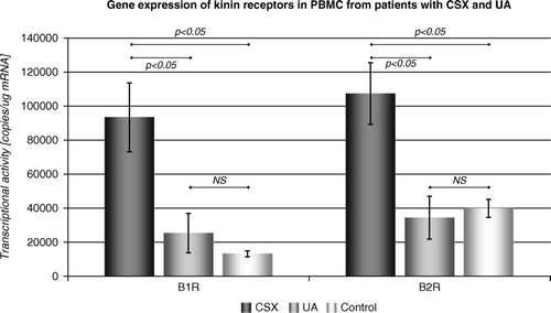
We have observed statistically higher transcriptional activity of gene encoding B1R gene in CSX group compared to the control group (93 395±20 301 copies/µg vs. 13 188±1 777 copies /µg respectively; p < 0.05) and vs. unstable angina patients (93 395±20 301 copies/µg vs. 25 365±11 553 copies /µg respectively; p < 0.05).
Likewise, we have observed elevated gene expression of B2R in CSX vs. control group (107 381±18 103 copies /µg vs. 39 908±5 309 copies /µg, respectively; p < 0.05) and CSX vs. UA (107 381±18 103 copies /µg vs. 34 431±12 598 copies /µg, respectively; p < 0.05). Moreover, transcriptional activity of studied genes did not differ between UA patients and healthy subjects. ().
Figure 1. Gene expression of kinin receptors in PBMC from patients with cardiac syndrome X (CSX) and unstable angina (UA).
We have not found any correlation between studied gene expression and any of medications () that studied patients had had administered.
There was no difference observed in expression of endogenous control gene (β-actin) among studied subgroups.
To evaluate the effect of changes in leukocyte subpopulations on expression of studies genes, lymphocyte /monocyte ratio was compared between CSX, UA and control groups showing no significant differences among subgroups (CSX vs. K: 8.32±1.52 vs. 8.52±1.46; p = NS; CSX vs. UA: 8.32±1.52 vs. 8.66±1.18; p = NS, UA vs. K: 8.66±1.18 vs. 8.52±1.46; p = NS). This allows excluding major changes in leukocyte differentiation as responsible for differences in studied gene expression.
Discussion
The role of kinin signaling in pathophysiology of cardiovascular system seems to be much wider than suspected. As stated, the final result of their action is dependent on type and proportion of their receptors in target cells. In the present study we have demonstrated that 1) transcriptional activity of gene encoding kinin B1R is more than 7-fold elevated in CSX patients vs. healthy control, and 3.5-fold higher than in UA patients, 2) gene expression of B2R is approximately 2.5-fold elevated in the studied group vs. control and vs. UA, and 3) there was no significant differences in B1R and B2R gene expression between patients with UA and healthy subjects.
The essential receptor participating in kinin signaling under physiological conditions is B2R which is expressed constitutively in most tissues Citation14, Citation15. In cardiovascular system it is predominantly expressed on the surface of endothelial cells but is also present on circulating inflammatory cells, including monocytes Citation16. Acting through nitric oxide and prostanoids, B2R takes part in vasodilation, also in coronary arteries Citation17.
In contrast to B2R, increased expression of inducible B1R is typical for chronic pathological states. B1R expression is elevated, among the others, in atherosclerosis Citation12, Citation13. Higher expression of B1R in monocytes was proved to trigger release of numerous inflammatory mediators including TNFa (Tumor Necrosis Factor a) and interleukins, such as IL-1β, IL-2 and IL-6 Citation18. The expression of kinin receptors in monocytes, however, seems to depend on the stage of activation and differentiation of the cell. Activated inflammatory cells tend to have higher expression of B1R, while B2R activity is diminished Citation19. The presence of kinin receptors on the other important inflammatory cells – lymphocytes – is rather poor and limited to T-cell subpopulation which in some states expresses mainly B1R, but this has not been widely studied vascular disease to date.
Although some authors (Lin et al. Citation20) observed only minor vascular inflammation without activation of circulating mononuclears from CSX patients, more recent studies report increased activation of mononuclears resulting in elevated superoxide generation and production of proinflammatory cytokines Citation21. Observed in our study remarkably high level of B1R gene expression, not only vs. healthy subjects but also vs. patients with unstable angina, may reflect the activation of circulating inflammatory cells in patient with CSX. The activation of circulating mononuclears, however, should be associated with diminished B2R expression, while in our experiment transcriptional activity of this gene was also elevated. There might be several explanations to this finding. Since it has been proven that B2R activation is an important mechanism in cardioprotective action of ACE inhibitors Citation22, Citation23, increased expression of this receptor may be at least partly resulting from fact that approximately 50% of CSX patients were treated with this ACE-I before (although there was no clear correlation observed in the study). On the other hand, however, even higher proportion of UA patients was on ACE-I treatment, and they did not show increased B2R expression.
Impaired nociception is another important mechanism in CSX. B1R and B2R receptors are attributed a specific role in the pain perception during inflammation which is related to the kinetics of their expressions. B2R receptors undergo a constitutive expression on non-myelinic sensory neurons and once activated by bradykinin it is being internalized. Hence, it is responsible for an acute pain reaction Citation24, Citation25. The enhanced expression of this receptor in CSX may be important factor responsible for pain hypersensitivity observed in CSX. The postulated significance of kinin receptor in abnormal nociception in CSX may suggest alternative therapeutic approach in these patients, focused on kinin signaling blockage. There is some data available to prove that B2R antagonists act as analgesics in acute conditions Citation24, whereas the antagonists of B1R receptor play a greater role in the inhibition of hypersensitivity to pain in a chronic inflammation Citation24, Citation25.
Although some mechanisms in studied disease are already well documented, the name syndrome X still remains quite adequate to what we currently know about pathophysiology and therapy of this disturbance. Despite some clear trends shown in our study (marked elevation of both B1R and B2R gene expression), the results within a group were heterogeneous (mostly due to variances in B1R expression). This diversity might result from several factors including medication (although, as stated, there was no clear correlation between drugs and studied gene expression), severity of the disease and possibly heterogeneicity of pathomechanisms underlying the disease itself. Nevertheless, kinin signaling seems to play important part in CSX. To our knowledge, this is the first study to report the role of kinin system (studied at the level of transcriptional activity of kinin receptors) in microvascular angina.
Conclusion
Patients with cardiac syndrome X present with remarkably enhanced transcriptional activity of kinin genes encoding kinin receptors in circulating mononuclears. This may reflect activation of inflammatory cells in these patients and confirm inflammatory nature of the disease, but may also suggest explanation for disturbances in pain perception in CSX.
References
- Kemp HG. Left ventricular function in patients with the anginal syndrome and normal coronary angiograms. Am J Cardiol. 1973; 32: 375–6
- Panting JR, Gatehause PD, Yang GZ, Grothues F. Abnormal subendocardial perfusion in cardiac syndrome X detected by cardiovascular magnetic resonance imaging. N Engl J Med. 2002; 346: 1948–53
- Tyminska-Sedek, K, Jakubowska-Najnigier, M, Chory z kardiologicznym zespolem X. Terapia. 2002;9, z.2/2002.
- Rogacka D, Wysocki H. Dlawica mikronaczyniowa i zaburzenia percepcji bólu jako glówne skladowe patogenetyczne kardiologicznego zespolu X. Folia Cardiol. 2003; 10: 553–61
- Kaski JC, Rosano GM, Collins P. Cardiac syndrome X: Clinical characteristics and left ventricular function long-term follow-up study. JACC. 1995; 25: 807–14
- Johnson BD, et al. Prognosis in women with myocardial ischemia in the absence of obstructive coronary disease: results from the National Institutes of Health-National Heart, Lung, and Blood Institutes-sponsored Women's Ischemia Syndrome Evaluation (WISE). Circulation. 2004; 109: 2993–9
- Kolodziej K, Kasprzak JD. Kardiologiczny zespół X. Forum Kardiołogów. 2001; 4: 1425–3674
- Vazquez-Rey E, Kaski JC. Cardiovascular syndrome X and endothelial dysfunction. Rev Esp Cardiol. 2003; 56: 181–92
- Rosen SD, Paulesu E, Wise R, Camici P. Central neural contribution to the perception of chest pain in cardiac syndrome X. Heart. 2002; 87: 513–9
- Lanza GA, Crea F. The complex link between brain and heart in cardiac syndrome X. Heart. 2002; 88: 328–30
- Leu HB, Lin CP, Lin WT, Wu TC, Lin SJ, Chen JW. Circulating mononuclear superoxide production and inflammatory markers for long-term prognosis in patients with cardiac syndrome X. Free Radic Biol Med. 2006; 40: 983–91
- McLean PG, Perretti M, Ahluwalia A. Cardiovasc Res. Kinin B(1) receptors and the cardiovascular system: Regulation of expression and function. Cardiovasc Res. 2000; 48: 194–210
- Calixto JB, Medeiros R, Fernandes ES, Ferreira J, Cabrini DA, Campos MM. Kinin B1 receptors: Key G-protein-coupled receptors and their role in inflammatory and painful processes. Br J Pharmacol. 2004; 143: 803–18
- Marceau F. Kinin B1 receptor: A review. Immunopharmacology. 1995; 30: 1–26
- Coelho MM, Oliveira CR, Pajolla GP, Calixto JB, Pela IR. Central involvement of kinin B1 and B2 receptors in the febrile response induced by endotoxin in rats. Br J Pharmacol. 1997; 121: 296–302
- Rajasekariah P, Warlow RS, Walls RS. High affinity bradykinin binding to human inflammatory cells. Biochem Mol Biol Int. 1997; 43: 279–90
- Drummond GR, Cocks TM. Endothelium-dependent relaxations mediated by inducible B1 and constitutive B2 kinin receptors in the bovine isolated coronary artery. Br J Pharmacol. 1995; 116: 2473–81
- Paegelow I, Werner H, Reissmann S. Effects of bradykinin and bradykinin analogues on spleen cells of mice. Eur J Pharmacol. 1995; 279: 211–6
- Raidoo DM, Ramsaroop R, Naidoo S, Mueller-Esterl W, Bhoola KD. Kinin receptors in human vascular tissue: Their role in atheromatous disease. Immunopharmacology. 1997; 36: 153–60
- Lin CP, Lin WT, Leu HB, Wu TC, Chen JW. Differential mononuclear cell activity and endothelial inflammation in coronary artery disease and cardiac syndrome X. Int J Cardiol. 2003; 89: 53–62
- Leu HB, Lin CP, Lin WT, Wu TC, Lin SJ, Chen JW. Circulating mononuclear superoxide production and inflammatory markers for long-term prognosis in patients with cardiac syndrome X. Free Radic Biol Med. 2006; 40: 983–91
- Yang XP, Liu YH, Mehta D, Cavasin MA, Shesely E, Xu J, Liu F, Carretero OA. Diminished cardioprotective response to inhibition of angiotensin-converting enzyme and angiotensin II type 1 receptor in B(2) kinin receptor gene knockout mice. Circ Res. 2001; 88: 1072–9
- Chen Z, Deddish PA, Minshall RD, Becker RP, Erdos EG, Tan F. Human ACE and bradykinin B2 receptors form a complex at the plasma membrane. FASEB J. 2006; 20: 2261–70
- Couture R, Harrison M, Vianna RM, Cloutier F. Kinin receptors in pain and inflammation. Eur J Pharmacol. 2001; 429: 161–76
- Moreau ME, Garbacki N, Molinaro G, Brown NJ, Marceau F, Adam A. The Kallikrein-Kinin System: Current and future pharmacological targets. J Pharmacol Sci. 2005; 99: 6–38