Venue – Stora Salen (Pathophysiology)
13.20–14.20, April 23, 2009
Imaging/diagnostics
1
A0229
The effect of alterations in pre- and after-load by using different myocardial measurement modalities of left ventricular relaxation.
Sandra Gustafsson1, Per Lindqvist1, Michael Henein1
1Hjärtcentrum, NUS
Background. Trans-mitral flow is well established measurement of left ventricular (LV) diastolic function but limited by being preload dependent. Myocardial velocities from tissue Doppler echocardiography (TDE) have been shown being a more load independent measure. Recently, speckle tracking echocardiography (STE) has emerged but not so far tested due to its load dependence. We aimed therefore to evaluate the effect of different load alterations on TDE and STE.
Methods. We examined10 young healthy subjects (mean age 23, range 21–27 years) by measuring the early diastolic (E) velocities from mitral, pulsed spectral and color TDE and STE. Velocities were measured from both the lateral and septal segments. Patients were all consequently changed into 5 different loading alterations, 1) Supine 1. 2) 45° degrees head up. 3) Standing. 4) Supine 2. 5) Elevated thigh pressure (approx. 120 mm Hg). We then compared the change in the different E velocities between phase 1–2 (preload decrease), 3–4 (preload increase) and 4–5 (afterload increase).
Results. We found a reduce in all E velocities (p < 0.05) going from supine to 45 head up position. Inversely,going from standing position to supine rest level 2, we found a significant increase in all E velocities and in both sites (p < 0.05), see figure. Thus there was no consistent difference in changes due to the septal or lateral sites. Finally, we found no changes in E velocities from mitral or myocardial velocities with increased thigh pressure.
Conclusion. Trans-mitral as well as myocardial velocities during early diastole is preload dependent in young healthy subjects. This was found using both TDE and STE. Furthermore, no consistent difference in the septal or lateral sites using changes of these myocardial velocities. Finally, by increasing afterload we found no changes in early diastolic velocities.
Imaging/diagnostics
2
A0230
Importance of apical untwist in early diastole and its relationship to filling events
Per Lindqvist1, Ulf Gustafsson1, Anders Waldenström2, Michael Henein2
1Klinisk Fysiologi, 2Kardiologi
Background. The left ventricular (LV) intracavitary flow during isovolumic relaxation (IVR) is relatively preload independent measurement of LV relaxation. It reflects pressure difference between the LV regions during a time when the mitral and aortic valves are closed. It is thus, caused by the change in cavity shape as a result of reciprocal intersegmental movements. Part of this shape change is due to the apical untwist occuring after aortic valve closure. The aim of this study was to evaluate the temporal time relationships between the IVR flow, apical untwisting and LV filling.
Methods. Fifteen healthy subjects constituted the group. Colour M-mode was used to determine IVR colour flow propagation from the base to the apex of LV. Transmitral early diastolic (E) and atrial systolic (A) flow velocities were measured using pulsed wave Doppler. Apical rotation was assessed using 2D strain. From peak R wave of the ECG we measured the time interval to the onset of IVR flow, onset of LV E wave and onset of apical untwist. We also measured the apical untwist rate during IVR time.
Results. The time interval between the R wave and the onset of the apical untwist was 369±41 ms, which coincided with the onset of IVR flow, 374±43 ms. The onset of LV E wave however, lagged by 80 ms behind these two events, 447±34 ms, p < 0.001. The delay in the onset of apical untwist correlated with its rate of untwist during IVR time (r = − 0.52, p < 0.05).
Conclusion. The sequence of normal diastolic events after aortic valve closure is apical untwisting followed by IVR flow then left ventricular filling. The time relations between those early diastolic events and apical untwist highlights the role of LV apical function in determining the pattern of its filling and as a marker of diastolic function.
Imaging/diagnostics
3
A0318
Improved Quantification of 4D Intraventricular Blood Flow in Normal and Failing Hearts
Jonatan Eriksson1, Petter Dyverfeldt1, Tino Ebbers1, Ann F Bolger2, Jan Engvall3, Carl Johan Carlhäll3
1IMH and CMIV, Linköping University, Linköping, Sweden, 2University of California San Francisco, San Francisco, CA, USA, 3Dept of Clinical Physiology, University Hospital, Linköping, Sweden
Background. The transit of blood through the beating heart is a fundamental aspect of cardiovascular function. Alterations in left ventricular (LV) flow patterns are recognized in heart failure, but quantification of the true 4D (3D + time) behavior of blood flow in dysfunctional LVs is lacking. Previously we have developed tools that allow elucidation of 4D LV blood flow organization. In an attempt to reduce user dependency and enhance robustness, we demonstrate a novel analysis approach that better integrates flow and morphological data.
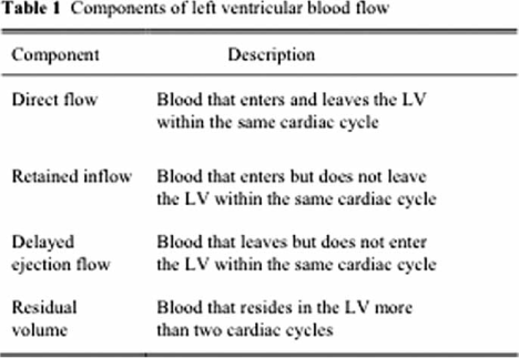
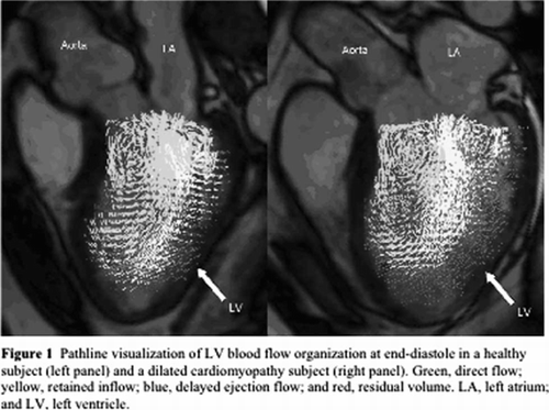
Material and methods. In six healthy subject and two dilated cardiomyopathy (DCM) patients, 4D flow data and LV long- and short-axis images were acquired using a 1.5T MRI-scanner. The LV was then segmented based on the morphological images using freely available software (http://segment.heiberg.se). The end-diastolic LV blood volume was analyzed by pathline analysis, where the trajectory taken by virtual blood particles is traced over the cardiac cycle. In-house developed software combined the LV segmentation data with the output from the pathline analysis to determine whether the traces entered and/or left the LV within the same cardiac cycle.
Results. The analysis approach presented here enabled separation and visualization of four different LV flow components (Table 1, Figure 1) in all eight data sets, and appeared less user-dependent and time-consuming than earlier techniques. By this method the volumes, as well as changes in kinetic energy during diastole and/or systole, of these flow components can be estimated. Quantitative data will be presented.
Conclusion. The present multidimensional flow analysis approach appears more user-friendly and robust than earlier techniques, and it allows better elucidation of the LV residual volume. Preliminary findings suggest that the highly organized blood flow in normal LVs is altered in DCM LVs. Such measures may be useful for improved diagnostics and management in heart failure.
Imaging/diagnostics
A0271
4
An improved method for quantification of left ventricular volumes in gated myocardial perfusion SPECT
Helen Soneson1, Fredrik Hedeer1, Martin Ugander1, Håkan Arheden1, Einar Heiberg1
1Cardiac MR-group, Clinical Physiology, Lund University Hospital
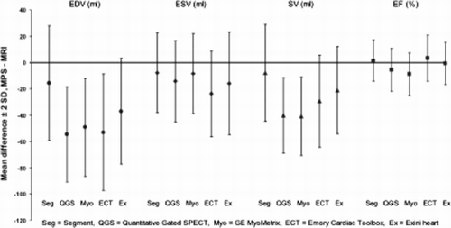
Background. Left ventricular volumes are important diagnostic and prognostic parameters for patients with coronary artery disease. Myocardial perfusion SPECT (MPS) is an established method for quantifying left ventricular volumes. The aim of this study was to develop and validate a new segmentation algorithm for the left ventricle in gated MPS and compare the results to four other commercially available software packages.
Methods. This study included 100 patients that underwent gated MPS imaging and magnetic resonance imaging (MRI). The novel, fully automatic, method was implemented in the freely available software Segment (http://segment.heiberg.se). The algorithm starts by identifying the mid-mural line of the left ventricular wall. The endo- and epicardium are then defined on the basis of the signal intensity and the requirement to preserve left ventricular mass over time. From the segmentation, left ventricular end-diastolic volume (EDV), end-systolic volume (ESV), stroke volume (SV), and ejection fraction (EF) were calculated. As a reference standard for the volumes, manually segmented left ventricles in the MR images were used for comparison. The results from Segment were then compared to results from four other commercially available software packages; Quantitative Gated SPECT, GE MyoMetrix, Emory Cardiac Toolbox and Exini heart.
Results. The results from the left ventricular volumes comparison, between MPS and MRI, are shown in the figure. Segment had the lowest bias for all four variables and the ranking is statistically significant (Fisher's Exact test, p < 0.01). However, the variability, when compared to MRI, did not differ significantly between the methods (F test).
Conclusion. Segment had significantly lower overall bias, but similar variability, compared to four other commercial available software packages for quantification of left ventricular volumes and function in gated MPS, using MRI as the reference standard.
Cardiology
5
A0354
Measurement of cardiac output with non-invasive Aesculon impedance vs. thermodilution
Petter Hedelin1, Erik Agger1, Björn Ekmehag1, Göran Rådegran1
1Avdelningen för Kardiologi, Hjärt och Lung Divisionen, Universitetssjukhuset i Lund
Background. Thermodilution (TD) is commonly used for cardiac output (CO) measurements and important in evaluating patients with pulmonary hypertension as well as severe heart failure. TD is, however, invasive, requiring right heart catheterisation. The purpose of this study was to compare the thoracic Aesculon impedance technique with TD to evaluate if Aesculon may offer a reliable non-invasive method for estimating CO.
Material and methods. CO was measured with TD, via a Swan Ganz catheter, and with Aesculon, in 33 patients, 14 females and 19 males, with a mean age±SEM of 59±2,7 years, undergoing right heart catheterisation for clinical investigation. Five measurements of CO were performed with each technique simultaneously in 33 patients at rest, and in 11 of the patients during exercise at 30 W in females and 50 W in males, and in 7 of the patients during NO-inhalation (40 ppm, 100% O2).
Results. CO correlated poorly between Aesculon and TD; at rest (R = 0,46, p < 0,001), during exercise (R = 0,35, p < 0,013) and NO-inhalation (R = 0,41, p < 0,017). CO was higher for Aesculon than TD with 0,86±0,14 l/min at rest (p < 0,001) and 2,95±0,69 l/min during exercise (p < 0,003); but similar during NO-inhalation, with a tendency (p < 0,079) only to be 0,44±0,19 l/min higher for Aesulon than TD. CO increased from rest to exercise for Aesculon and TD with 6,11±0,6 l/min (p < 0,001) and 3,91±0,36 l/min (p < 0,001), respectively; an increase that was higher (p < 0,002) for Aesculon than TD. During NO-inhalation compared to rest, CO decreased for Aesculon with 0,62±0,11 l/min (p < 0,002), but not significantly for TD with 0,21±0,12 l/min (p < 0,11).
Conclusion. Aesculon overestimates CO as compared to TD with 17% at rest and 34% during exercise. Further studies are needed to verify if Aesculon may be used to monitor relative changes of CO during pharmacological interventions in individual patients.
Venue – Sal B (New treatment)
13.20–14.20, April 23, 2009
Cardiology
6
A0317
Transcatheter Heart Valve Implantation in a Degenerated Aortic Valve Bioprothesis. First Valve-in-Valve Implantation in Sweden
Niels Erik Nielsen1, Mats Broqvist1, Éva Tamás2, Henrik Ahn2, Wolfgang Freter2, Jacek Baranowski3, Lars Wallby3, Eva Nylander3
1Kardiologiska kliniken, 2Thorax-Kärlkliniken, 3Fysiologiska kliniken
Surgical replacement of degenerated bioprosthetic valves is associated with increased perioperative risk, the patients often are elderly with considerable comorbidities. The novel technique of transcatheter heart valve implantation opens new possibilities of treating these patients.
A 75 year old man was operated June 2005 with coronary bypass and a biological valve Perimount 23 mm because of aortic stenosis. Very complicated postoperative course with reoperations because of bleeding and sternal infection. However, he recovered well. March 2008 he had a prolonged period with fever. Despite intensive investigations bacterial endocarditis was never diagnosed. August 2008 he started to get exertional dyspnoea. Echocardiography showed the biological valve to be severely stenotic with a valve area of 0.6 cm2 and a peak gradient of 92 mmHg. He had a stenosis in a vein graft, otherwise the grafts were patent. Diuretics gave temporary relief, but late 2008 he was in NYHA IIIB.
Open heart surgery was discussed, but we hesitated for more reasons: Severe complications after the first operation; liver cirrhosis with secondary hypersplenism and because of this pancytopenia with Hb about 100 g/L and platelets about 50×109/; a slightly reduce renal function; and suspicion of some reduction of his mental status, with risk of worsening after a second operation in heart-lung machine. The patient therefore was discussed for transcatheter heart valve implantation. He was informed that this was an off-label use of the device, which he accepted.
January 29 2009 a 23 mm Edwards transcatheter valve was successfully inserted by the transapical route under general anesthesia, but without cardiopulmonary bypass. Echocardiopgraphy showed a well-functioning valve with a trivial paravalvular leak. The implantation took 2 hours, the postoperative course was uncomplicated.
Transcatheter valve-in-valve implantation offers a valuable therapeutic option for patients with stenotic biological valves, especially for those at high surgical risk.
Arrhythmia
7
A0351
Totally endoscopic ablation of atrial fibrillation – preliminary results
Anders Ahlsson1, Espen Fengsrud2, Andersson Tommy2, Almroth Henrik2, Linde Peter2, Tydén Hans1, Englund Anders3
1Thoraxkliniken, Universitetssjukhuset Örebro, 2Kardiologiska kliniken Universitetssjukhuset Örebro, 3Svenskt arytmicenter Stockholm
Background. Totally endoscopic ablation (TEA) is a new method of epicardial ablation of left atrial tissue. The purpose of this study was to examine the feasibility, efficacy and safety of TEA using microwave or radiofrequency energy.
Material and Methods. TEA is performed in full anaesthesia with left single lung ventilation and CO2 insufflation in the right hemithorax. Through three working ports, the pericardium is opened, and the oblique and transverse sinus are entered. An ablation catheter is positioned on the left atrial wall and a box lesion encircling all pulmonary veins is created (video demonstration).
19 patients have undergone TEA since the start in May 2007. The indications were symptomatic AF in patients > 50 years, and patients with a BMI > 35 were excluded. The median age was 67 yrs (60–82), and 5 patients were female. The frequency of paroxysmal/persistent/permanent AF were 9/3/7, respectively, and the median duration of AF 10 years. Three patients had preoperative pacemaker implants.
Results. 9 patients were ablated using a Flex X microwave catheter (Boston Scientific, USA) and 10 patients using a Cobra Adhere XL radiofrequency catheter (ESTECH, USA). There was no hospital mortality. Major morbidity included one patient with a transient phrenical paresis postoperatively, one patient requiring rethoracoscopy due to postoperative bleeding, and in one patient a limited thoracotomy had to be performed in order to complete the ablation.
The frequency of sinus rhythm or paced rhythm at follow-up where 10/13 patients (77%) after three months and 6/10 after (60%) after 6 months. One patient died during follow-up due to cardiac arrest.
Conclusion. TEA is a feasible method of AF ablation with preliminary acceptable results. The potential clinical role of TEA has to be further evaluated in prospective, randomised trials with careful monitoring of the AF burden during follow up.
Cardiology
8
A0365
Case method assisted implementation of guidelines decreases mortality – a ten-year follow up of a randomized controlled study
Anna Kiessling1, Peter Henriksson1
1Karolinska Institutet
Aim. The aim was to determine the size of any patient survival benefit from the interactive pedagogic method case method learning (CML) to facilitate implementation of guidelines in primary care.
Material and methods. Prospective randomized controlled trial in clinical practice in the Stockholm area. New guidelines for secondary prevention in coronary artery disease (CAD) were mailed to all general practitioners (GPs) in the area and presented at a common lecture in 1995. The GPs were clustered according to their Primary Health Care Center (PHC) into two well-matched pairs and randomly allocated to active intervention with CML or usual care. GPs in the intervention group participated in recurrent CML dialogues at their PHCs during a two-year period. A locally well-known cardiologist served as facilitator. Consecutive patients (n = 255) with CAD were included. Ten-year mortality rates were obtained from the Cause of Death register and were assessed as all cause and cardiovascular mortality.
Results. The two PHC groups of patients respectively physicians were well matched and did not differ at baseline. Attendance rate at the seminars was > 82%.
19 (44%) of the included patient in the control group had deceased after ten years as compared to 10 (22%) in the intervention group (p = 0.0174; log rank test). The inclusion of the covariates age, sex, hypertension, smoking and diabetes did not change its significance. Patients treated by a specialist deceased at a rate comparable to the intervention group (23%).
Cardiovascular mortality was 32% in the control group and 16% in the intervention group (p = 0.007).
Conclusions. CML for general practitioners improved survival in patients with CAD. The hazard ratio (HR) between intervention and usual care is 0.45 (95% CI 0.20–0.95) if case method learning is used to assist implementation of evidence based care.
Cardiology
9
A0329
Long-term beneficial effects of an expanded cardiac rehabilitation after an acute myocardial infarction or coronary artery by-pass grafting: A five year follow-up of a randomized controlled study
Catrine Edström Plűss1, Ewa Billing2, Claes Held3, Peter Henriksson4, Anna Kiessling4, Monica Rydell Karlsson4, Håkan Wallen4
1Division of Cardiovascular Medicine, Danderyds Hospital, Stockholm, Sweden, 2Dept of Medical Sciences, Uppsala University, Uppsala, Sweden, 3Uppsala Clinical Research Centre and Department of Cardiology, University Hospital, Uppsala, Sweden, 4Karolinska Institutet, Dept of Clinical Sciences, Danderyd Hospital, Stockholm, Sweden
Background. Current guidelines broadly recommend comprehensive cardiac rehabilitation after an acute myocardial infarction (MI) or post coronary artery by-pass grafting (CABG). However, the evidence of the effects of cardiac rehabilitation are limited. There are few long term randomized trials comparing expanded cardiac rehabilitation and usual care.
Material and methods. A single centre prospective randomized controlled clinical trial was performed which included 224 patients with a recent MI or who were planned for CABG. Patients were randomized to either expanded cardiac rehabilitation which included a 5 days stay at a “Patient Hotel” after discharge, increased physical training, cooking sessions and, importantly, a one year stress management program, or to routine rehabilitation (“usual care”). The patients were followed clinically for one year. Five-year follow-up data were obtained from the registry of the Swedish National Board of Health and Welfare. The primary outcome is registry-based cardiac events such as cardiovascular (CV) death, myocardial infarction or readmission for CV disease.
Results. One-hundred eleven and 113 patients were randomized to expanded rehabilitation and usual care, respectively. There was a significant reduction in CV events in the expanded rehabilitation group compared to usual care when data were collected from the start point of rehabilitation (p = 0.03). The number of hospitalizations as well as the number of days of hospitalization during the 5-year follow-up were significantly lower in the patients that had received expanded rehabilitation compared to those who received usual care (p< 0.01 for both between group comparisons).
Conclusion. An expanded multifactorial cardiac rehabilitation program after an acute myocardial infarction or coronary artery by-pass grafting have beneficial long-term effects and reduces cardiovascular morbidity and hospitalizations for cardiovascular reasons.
Cardiology
10
A0319
Survival after cardiac arrest – effects of therapeutic hypothermiaCardiology
Erik Dellcrantz1, Sten Walther1,2
1Linköping University, Faculty of Health Sciences, Dept of Medicine and Health, Div of Cardiovascular Medicine, 2Swedish Intensive Care Registry
Background. Treatment of cardiac arrest has become more active during recent years. The aim of this study was to examine the use and effects of therapeutic hypothermia on short and long term survival of patients admitted due to cardiac arrest to intensive care units (ICU).
Material and methods. Admissions in the Swedish Intensive Care Registry (www.icuregswe.org) during 2003–2007 were examined. Patients with cardiac arrest were identified by the principal diagnosis at discharge from ICU. Age, gender, illness severity (APACHE II), length of stay and survival at 30 and 90 days were analyzed per treatment group (with or without therapeutic hypothermia) using t-test and Chi2 –test. Logistic regression was used to calculate odds ratios. Readmissions during the 5-year period were excluded.
Results. We identified 1675 admissions due to cardiac arrest. The proportion of patients that were treated with hypothermia increased during the study period (2003: 1%, 2004: 5%, 2005: 33%, 2006: 36% and 2007: 35%, P < 0.001). Patients with therapeutic hypothermia were more likely to be male (P = 0.001), they were younger (63 vs. 68 yrs, P < 0.001) and had higher APACHE II scores (28 vs. 26 points, P < 0.001). Survival at 30 days and 90 days after admission was greater in patients with therapeutic hypothermia (30 d: 42% vs. 30%, P < 0.001; 90 d: 39% vs. 28%, P < 0.001). The odds ratio (95% CI) for survival 90 days in the hypothermia group was 1.7 (1.3–2.1), and it was 1.6 (1.3–2.1) after adjustment for gender, age and APACHE II score.
Conclusion. Therapeutic hypothermia in patients with cardiac arrest became gradually more common during the 5-year period and it was associated with increased survival.
Venue – Sal C (Nursning Science)
13.20–14.20, April 23, 2009
Nursing science
11
A0269
Patients with unexplained chest pain-pain experience, stress, coping and health related quality of life
Margaretha Jerlock1, Catharina Welin1, Karin Kjellgren1
1Göteborgs universitet, Sahlgrenska akademin, Institutionen för vårdvetenskap och hälsa
Background. In Sweden, the number of patients discharged from hospital with a diagnosis of unexplained chest pain (UCP) has increased from 8,432 in 1987 to 17,555 in 2005.
Aim. The aim was to: identify similarities and differences in how patients with UCP and patients with ischemic heart disease (IHD) describe chest pain and determine psychosocial factors associated with UCP, coping strategies and how the chest pain experiences affect everyday life and health-related quality of life (HRQOL).
Methods. Both quantitative and qualitative methods were used. The study was carried out from December 2002 to September 2003.
Results. UCP patients perceived their condition as more painful than IHD patients and they more frequently described their chest pain as dull, sore, annoying and troublesome. UCP patients required more sensory and affective words to describe their pain.
The UCP patients explained that their pain gave rise to fear and anxiety, a feeling of uncertainty, stress and loss of strength, which to a great extent affected everyday life. The patients used cognitive coping strategies in managing stress but they had difficulty managing activities such as household chores, socialising with friends, and taking part in recreational and sexual activity. In comparison with a random population sample, patients with UCP had impaired HRQOL, they were more often worried about stress at work, perceived more stress at home, more often had sleep problems and had a sedentary lifestyle. UCP patients had experienced more negative life events and a larger proportion was immigrant. Women with UCP had higher levels of cardiovascular risk factors.
Conclusion. UCP intrudes into everyday life in a destructive manner that cannot be ignored. It is essential that ways be found to alleviate pain and to improve health and quality of life, as well as to promote physical activity and sleep.
Nursing science
12
A0299
Optimal versus Nominal programming in patients implanted with dual-chamber pacemakers. Results from the POPP study
Karin Strindlöv Carlsson1, Maria Hesselstrand1, Helene Hansson Ferreira1, Barbara Jabur Juul-Möller1, Gunilla Lilja1, Karin Wedmark1, Carl-Johan Höijer1, Eva Clausson2
1Universitetssjukhuset, Lund, 2Medtronic, Sweden
Background. Modern dual chamber (DDDR) pacemakers are shipped with a set of nominal parameters, but can be programmed in different ways. Nominal settings are different among manufacturers and are suitable, although not optimal, for most patients. Optimization aims to allow more physiologic heart rhythm and to gain battery life, but there are few guidelines regarding pacemaker programming. We wanted to study if post-operative optimization is necessary, and if it could be standardized by using a pre-decided set of parameters, optimized for AV block (AVB) and Sick Sinus Syndrome (SSS).
Material and methods. The study was performed in a prospective, randomized, single blind, cross-over design. Patients eligible for a DDDR pacemaker were asked to participate. Before pacemaker implantation, each patient was randomized to pacemaker model (pre-decided DDDR-models from Medtronic, Vitatron and St Jude Medical), and to programming sequence (nominal or optimized). Pre-decided optimized parameters were slightly different for AVB and SSS, mainly regarding stimulation rate and rate response function. Patients were seen after three and six months for data collection. Collected data included amount of ventricular pacing, battery current drain, and a symptom score. Cross-over was done after three months and the preferred programming was noted after the second period.
Results. 172 patients (105 SSS, 67 AVB) finished the study. Optimizing parameters decreased accumulated ventricular pacing by 32% (p < 0,01), and battery current drain by 17%, (p < 0,01) compared to shipped settings. Total patient symptom score improved by 43% (p < 0,01) from baseline to pacemaker implantation (shipped settings) and additionally 18% with optimized settings (p < 0,01). 67 patients (39%) preferred the optimized settings, 77 patients (45%) did not have any preference and 28 patients (16%) preferred the shipped settings.
Conclusion. Optimizing the pacemaker parameters leads to less ventricular pacing, improved patient wellbeing and better device longevity. Standardized diagnosis-based program setting seems applicable for most patients.
Nursing science
13
A0227
The use of mechanical chest compressions in the cath-lab during PCI-treatment in patients with cardiac arrest from a nursing perspective
Karl Berggren1, David Zughaft1
1Universitetssjukhuset i Lund
Introduction. Cardiac arrest (CA) in the cath-lab is not uncommon. A small number require lengthy resuscitation including manual chest compressions. Effective manual chest compressions (CC) should be performed with the cath-lab table (CLT) retracted. Performing manual CC's on the CLT during intervention is difficult. Fully extended, the CLT suffers from the trampoline effect which hampers effective CC's, and importantly, may break. Also, the CC provider gets in the way of the fluoroscopic view and is exposed to considerable amount of X-Ray. This study focuses on the algorithm and results when implementing mechanical CC's (LUCASTM) in the cath-lab during CA.
Material and methods. During 2004–2007, all patients who arrived alive in the cath-lab and required mechanical CC's were evaluated. An algorithm for the use of mechanical CCs was developed. The mechanical chest compression device LUCASTM was selected due to its excellent radiotranslucent properties and capacity to maintain circulation during CA.
Results. LUCASTM was used in 28 patients. 5 patients with myocardial rupture died. 6 of the remaining 23 were discharged from the hospital in good neurological condition. The following algorithm was developed: upon cardiac not responsive to defibrillation, begin manual CC's, apply LUCASTM and start mechanical CC's, intubate patient as soon as possible, diagnose the cause of cardiac arrest. If myocardial rupture; stop resuscitation. In the remaining patients; continue the intervention. After successful intervention, stop LUCASTM and check pressure, defibrillate if necessary. If no return of spontaneous circulation, consider left ventricular assist device or maintain LUCASTM for 30 more min before stopping.
Conclusion. Mechanical CC devices are useful in the cath-lab. It frees resources by maintaining circulation effectively. An algorithm of care during cardiac arrest and mechanical CC's are helpful when implementing mechanical CC devices in the cath-lab. Few, if any of the patients evaluated would have survived without mechanical CC's.
Cardiology
14
A0281
Rapid ambulation after coronary angiography
Johan Höglund1
1a Department of Cardiology, Linköping, University Hospital and b Department of Medicine and Care, Faculty of health sciences, Linköping University Sweden.
Background. The optimal length of patient immobilisation after coronary angiography is unknown. Short immobilisation could cause puncture site complications with modern antiplatelet therapy, while long immobilisation time increases the risk of discomfort including back problems for the patient.
Purpose. The purpose of this study was to assess the safety, as well as perceived comfort, of early mobilisation after coronary angiography in a patient population consisting of both stable angina pectoris and acute coronary syndrome (ACS).
Methods. The study was a prospective, randomised, controlled single centre trial. A total of 104 patients were randomly assigned to stay in bed either 90 min or 5h, with 60 minutes or 3 hours of femoral pressure, respectively. The primary endpoint was any incidence of vascular complication. Patients’ discomfort was measured as self-perceived grade of pain (on a visual analogue scale, VAS) in the back and/or groin.
Results. Seventy-seven percent of all patients were preloaded with clopidogrel. Forty-eight percent pre-treated with subcutaneous antithrombotics. No major vascular complications were observed. The presence of haematomas ≥5cm was 5.8% in the short immobilisation group vs. 3.8% in the control group (p = 0.816). One patient developed a pseudoaneurysm in the control group. No difference in ambulated success rate was noted. We found a significantly lower rate of perceived pain, expressed as back- and groin pain in the short immobilisation group (p = 0.001, p = 0.002 respectively), compared to the control group, at the time of mobilisation. The rate of perceived back pain remained significantly lower 4h after mobilisation, (p = 0.01).
Conclusion. Patients undergoing coronary angiography by the femoral approach and pre-treated with clopidogrel and in 48% also subcutaneous antithrombotics can safely be mobilised after 90 minutes of bed rest. Since October 2008, this procedure is accepted as the current practise at the Department of Cardiology, Linköping University Hospital.
Cardiology
15
A0327
Left ventricular untwist augments early filling.
Ulf Gustafsson1, Per Lindqvist1, Anders Waldenström1
1Heart center, Umeå University Hospital
Background. Left ventricular (LV) twist has proven to be an important factor in systolic function. Studies have indicated that the amount of twist correlates with the filling. However, no studies, as far as we know, have described how and when untwist contributes to the filling of the ventricle. We have studied the basal and apical LV untwist to examine the relationship between untwist and filling.
Material/Method. Short axis images at basal, papillary and apical levels of the LV were analysed with speckle tracking in 43 healthy subjects, 22 women, mean age 63 years. Measurements of rotation were made at 10 different time points during the cardiac cycle. The material was divided into two groups by apical untwist during the interval from mitral valve opening (MVO) to peak E velocity of more or less than 2.5 degrees.
Results. The group with more apical untwist in the first part of the early filling phase had significantly higher peak E velocities, 0.68m/s vs 0.58m/s (p = 0.015). The group with more early untwist also had borderline significant longer time to peak apical rotation, 14ms after aortic valve closure (AVC) vs 9ms before AVC (p = 0.052). There were no differences in age, heart rate, blood pressure or peak basal and apical rotation between the groups. Peak E velocity correlated with global and apical untwist during the interval from MVO to peak E (R = 0.512 p < 0.000 and R = 0.456 p = 0.002 respectively), and with global untwist during the interval from AVC to mid isovolumic relaxation period (IVR) (R = − 0.301 p = 0.05).
Conclusion. Untwist during the early filling period augments LV filling, demonstrated by increased E-velocities. However, untwisting during the IVR is correlated to lower E-velocities, which might suggest a negative effect on early filling. Therefore, untwisting during the isovolumic relaxation period could possibly be a waste of energy.
Venue – Sal Katalin (Mechanisms)
13.20–14.20, April 23, 2009
Arrythmia
16
A0294
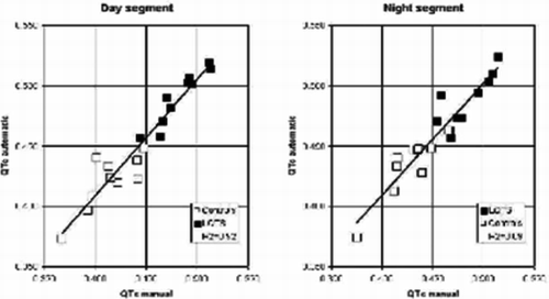
A new automatic QT-analysis of Holter recordings performs equal to manual analysis in children with the long QT syndrome
Annika Winbo1, Annika Rydberg1, Ola Gustavsson2, Marcus Karlsson2, Urban Wiklund2
1Department of Pediatrics, Umeå University Hospital, 2Department of Biomedical Engineering, Umeå University Hospital
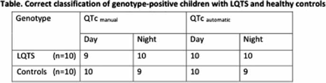
Background. Ambulatory 24-hour electrocardiographic recordings (Holter) provide information useful for diagnosis and risk stratification in children with the long QT syndrome (LQTS). Manual measurements are time-consuming and systems for automatic QT-analysis are often suboptimal in performance.This pilot study evaluates a new automatic system for QT-analysis in 2-lead Holter recordings developed at the Department of Biomedical Engineering at Umeå University Hospital.
Method. QT-intervals were measured manually and automatically in 2-lead (V2 and V5) Holter recordings from 10 patients with genotype-positive LQTS (age 11.2±4.8 years) and 10 healthy controls (age 11.3±5.0 years). Manual measurements were performed by a QT expert. The algorithm for Q-onset detection used a length-transform approach combined with a tangent-based method to derive the Q-onset point. An area-based algorithm calculated T-end. Manual and automatic measurements were done on all normal heartbeats in two 5-minute segments (day- and nighttime, respectively) chosen with respect to optimal signal quality. QT-intervals were corrected for heart rate using Bazetts's formula (QTc). The averages of QTc during 5 minutes were used for classification of LQTS (positive if QTc ≥ 450 ms).
Results. The average relative difference between manual and automatic measurements of QTc was 20 ms in day- and nighttime segments.
The agreement between genotype, manual and automatic measurements was excellent, with Cohen's kappa between 0.8–1.0 in all comparisons.
The automatic analysis correctly classified all daytime recordings (10 normal, 10 LQTS) and 19/20 of the nighttime recordings. Both the manual and the automatic analysis classified one healthy control as having LQTS in the nighttime recording.
Conclusion. In this pilot study, a new system for analysis of QTc in 2-lead Holter recordings was evaluated in genotype-positive LQTS children and healthy controls. The system's performance was equal to manual assessment, both in absolute measurements and in correct classification of LQTS.
Cardiology
17
A0296
Volume and intensity of objectively measured physical activity related to clustering of risk factors for cardiovascular disease in younger children
Tina Tanha1, Magnus Dencker1, Ola Thorsson1, Per Wollmer1, Magnus Dencker2, Magnus K. Karlsson2, Christian Linden2, Lars B. Andersen3
1Dept of Clinical Sciences, Unit of Clinical Physiology and Nuclear Medicine, Malmö University Hospital, Malmö, Sweden, 2Dept of Clinical Sciences, Clinical and Molecular Osteoporosis Research Unit, Malmö University Hospital, Malmö, Sweden, 3Institute of Sport Science and Clinical Biomechanics, University of Southern Denmark, Odense, Denmark
Aim. This study evaluates if accelerometer measured physical activity is related to clustering of risk factors for cardiovascular disease (CVD) in children aged 8 to 11 years.
Methods. Two hundred twenty-three children aged 7.9–11.1 years (boys n = 123, girls n = 100) were included. Abdominal fat mass (AFM) and total body fat mass (TBF) were quantified by Dual-Energy X-Ray Absorptiometry. TBF was also calculated as percentage of body mass (BF%) and body fat distribution (AFM/TBF). Maximal oxygen uptake (VO2PEAK) was measured during maximal exercise test. Daily physical activity was assessed by accelerometers for four days and daily accumulation of minutes of moderate-to-vigorous physical activity (MVPA) and general physical activity (GPA) were calculated per day. The cut-off point for MVPA was set at >3500 counts/minutes. Resting heart rate (HR) and blood pressure (SBP, DBP, and mean artery pressure (MAP)) were measured. Z-scores (value for the individual-mean value for group)/SD were calculated. Sum of z-scores for BF%, AFM, AFM/TBF, SBP, DBP, MAP, HR, and -VO2PEAK were calculated in boys and girls, separately, and used as indices of clustered risk.
Results. Pearson correlation between GPA and MVPA versus indices of clustered risk were for boys (−0.34 P < 0.05 and −0.31, P < 0.05) and for girls (−0.20, P < 0.05 and −0.28, P < 0.05). Boys and girls were divided according to quartiles of GPA and MVPA.
One-way ANOVA analysis indicated significant differences in sum of z-scores between quartiles of MVPA in boys (P = 0.004) and in girls (P = 0.009), whereas significant differences could only be observed for GPA in boys (P = 0.002), and no significant differences in girls (P = 0.16).
Conclusion. Low amount of moderate-to-vigorous physical activity per day was related to clustering of risk factors for CVD in this cohort of children aged 8 to 11 years. This was also observed for low amount of general physical activity in boys, but not in girls
Cardiology
18
A0338
Sympathetic nerve activity in Takotsubo Cardiomyopathy-“the Broken Heart Syndrome”
Yrsa Bergman Sverrisdóttir 1, Tomas Schultz2, Göran Matejka2, Per Albertsson2, Elmir Omerovic2
1Institute of Neuroscience and Physiology, Dept. of Clinical Neurophysiology, 2Sahlgrenska University Hospital/Department of Cardiology
Background. Takotsubo cardiomyopathy has been recognized as a novel syndrome of acute heart failure affecting mostly elderly women exposed to severe emotional stress. Increased sympathetic neuronal outflow is a hallmark of congestive heart failure (CHF), but whether this is the case in takotsubo is not known. The objective of this study was to directly evaluate sympathetic nerve traffic in patients with takotsubo.
Methods. Sympathetic nerve activity to the muscle vascular bed (MSNA) was recorded in 6 Patients (5F/1M) with takotsubo. The nerve recordings were compared with 6 (5F/1M) patients with mild-moderate CHF, on the basis of coronary heart disease (CAD) and idiopathic dilated cardiomyopathy (IDC) and 6 healthy controls matched for age, gender and BMI. MSNA was expressed as burst frequency (BF), burst incidence (BI) and burst amplitude distribution (relative median burst amplitude (RMBA%)), a sensitive indicator of sympathetic intensity.
Results. Sympathetic outflow expressed as BF and BI was significantly lower in patients with TSC as compared to patients with mild-moderate CHF (30±15 vs 69±14 BF and 51±22 vs 85±14 BI%, p < 0.005, respectively) but did not deviate from matched controls (BF: 41±8.5 and BI: 65±8.8) (p = 0.1).
Burst amplitude distribution was equal in takotsubo patients and healthy controls but was increased in patients with mild-moderate CHF (RMBA%: 37±7 vs 38±7 and 45±12, respectively).
Heart rate was significantly lower in takotsubo as compared to mild-moderate CHF (57±12 vs 81±11; p < 0.005) but did not deviate from healthy controls (57±12 vs 64±12; ns).
Conclusions. Sympathetic nerve activity whether expressed as BF, BI or burst amplitude distribution did not deviate between takotsubo patients and healthy controls. These results are contrary to that seen in CHF, a condition associated with sympathetic activation. In conclusion; our preliminary results indicate that takotsubo cardiomyopathy is not a condition associated with sympathetic activation.
Other
19
A0355
Metabolic issues in psychiatric patients
Eva Lundberg1, Annelie Nordin1, Karl-Fredrik Norrback1
1Inst Klin Vetenskap, Psykiatri, Umeå Universitet
Patients within the schizophrenic and manic-depressive spectra are at ultra-high-risk for the development of cardiovascular disorders
Background. Individuals with severe mental disorders, such as schizophrenia, have on average a 20% shorter life span (1), with coronary heart disease being the leading cause of death (2), related to the increased prevalence of the metabolic syndrome in schizophrennia and in manic-depressive disorders (3–5).
The aim of this naturalistic cross-sectional study was to investigate the prevalence of cardiovascular risk factors in a representative cohort of out-patients with a diagnosis within the schizophrenic- and manic-depressive spectra.
Material and methods. 421 patients with schizophrenia, 253 with manic-depressive illness and 273 controls were examined for metabolic aberrtions using a semi-structured interview, anthropometric measurements and an extended biochemical screening.
Results. We found a prevalence of the metabolic syndrome of 42.7% in the schizophrenic- and 27.9% in the manic-depressive group, compared with 15.8% in the control group. Compared to controls, pre-diabetic and diabetic conditions were 2–3 times higher in the diseased population.
Conclusion. Reports concerning diabetes, hyperglycemia and dyslipidemia in patients treated with antipsychotic medication, particularly of the second-generation type show an association between metabolic complications and treatment (6–9). The impact on mortality and morbidity is substantial and requires increased attention. Guidelines emphasizing screening and monitoring have been published (e.g. www.psykiatri.se; Adolfsson and Nordin 2008).
References
- Harris EC, Barraclough B. Br J Psychiatry. 1998; 173:11–53.
- Hennekens CH et al. Am Heart J. 2005; 150:1115–21.
- McEvoy JP et al. Schizophr Res. 2005; 80:19–32.
- Hägg S et al. Int Clin Psychopharmacol. 2006; 21(2):93–8.
- Fiedorowicz JG et al. Ann Clin Psychiatry. 2008; 20(3):131–7.
- ADA. Diabetes Care. 2004;27:596–601.
- Casey DE et al. J Clin Psychiatry. 2004; 65:4–18.
- Newcomer JW. CNS Drugs. 2005; 19:1–93.
- Gianfrancesco FD et al. J Clin Psychiatry. 2002; 63:920–30.
Cardiology
20
A0363
NT-proBNP in senior athletes detects severe cardiovascular disease
Anders Sahlén1, Aigars Rubulis1, Marcus Ståhlberg1, Thomas Fux1, Thomas P Gustafsson2, Tony Marklund3, Frieder Braunschweig4
1Karolinska Institutet, Hjärtkliniken, Karolinska Universitetssjukhuset, Solna, Sweden, 2Karolinska Institutet, Division of Clinical Chemistry, Danderyd Hospital, Stockholm, Sweden, 3Roche Diagnostics Scandinavia, Bromma, Sweden, 4Karolinska Institutet, Hjärtkliniken, Karolinska Universitetssjukhuset, Solna, Sweden
Background. Though sudden death occurs rarely in athletes, senior participants run the highest risk. The best strategy for pre-participation screening (PPS) has not been fully established. Cardiac biomarkers are predictive of death in other settings but their role in PPS is not known.
Material and methods. We assessed 185 participants [132 (71%) male] aged 55 or above (mean 62 ys) at a 30 km cross-country race, after carefully excluding anyone with a known cardiovascular disorder using a written questionnaire. The following biomarkers were analysed before the race: N-terminal pro-Brain Natriuretic Peptide (NT-proBNP; normal <194 ng/L); high-sensitivity C-reactive protein (CRP). Those with abnormal levels of NT-proBNP were subsequently invited to undergo non-invasive cardiac work-up.
Results. Levels of NT-proBNP were 53 (median; [range: 8–2250]) ng/L and levels of CRP were 0.4 (0.2–7.4) mg/L. Abnormal NT-proBNP was found in a subgroup of 15 subjects (8.1%; 302 [198–2250 ng/L]). Cardiovascular disease was found in 4 subjects (27% of subgroup, 2.2% of whole sample; Table 1), of which 1 sadly died of sudden heart death while training, a few months after participation (post-mortem findings in Table 1). The remaining 3 were disqualified from intense sports for fear of death and/or disease progression. Minor cardiac disorders were found in 6 (40%). There were 3 (20%) false positives.
Conclusion. In senior, self-reportedly healthy endurance athletes, severe cardiovascular disease may be more common than previously believed. NT-proBNP identifies a subset of athletes with elevated cardiovascular risk. Our data provide a rationale for larger studies evaluating the role of NT-proBNP in pre-participation screening.

Imaging/diagnostics
21
A0272
Contractile Dysfunction after Acute Myocardial Infarction; Velocity Encoded Strain vs Wall Thickening
Joey Ubachs1, Einar Heiberg1, Martin Ugander1, Henrik Engblom1, Håkan Arheden1, David Erlinge2, Matthias Götberg2, Göran Olivecrona2, Stefan Jovinge2
1Cardiac MR Group, Department of Clinical Physiology, Lund University Hospital, Lund, Sweden, 2Department of Cardiology, Lund University Hospital, Lund, Sweden
Background. One of the most important trademarks of ischemic heart disease is the extent and degree of regional myocardial dysfunction, which are important factors in determining the long-term prognosis. In the chronic stage, myocardial strain has shown to be superior to wall thickening for detecting dysfunctional myocardium after coronary occlusion. In the acute stage, this remains to be studied.
Purpose. To assess myocardial strain and myocardial wall thickening in the acute phase after coronary occlusion.
Methods. Twenty-eight patients (mean age 61, range 36–83; 26 males) presenting with first-time acute ST-elevated myocardial infarction were included in the study and treated with primary percutaneous coronary intervention (PCI) resulting in TIMI grade 3 flow.
Prior to PCI, 99mTc tetrofosmin was administered intravenously and myocardial perfusion SPECT was performed after primary PCI for determination of myocardium at risk in 22 patients. The remaining 6 patients received MRI with T2-STIR at day for determination of myocardium at risk. Fifteen patients received cardiac MRI for assessment of wall thickening at day 1 and 13 patients received MRI with velocity encoded strain at day 1.
Results. Both methods showed that they can be used to appoint the occluded vessel after acute coronary occlusion. However, when comparing both modalities to SPECT/ T2-STIR, no significant differences were found for wall thickening (R2 = 0.40, mean difference −9.6±18.6) and myocardial strain (R2 = 0.10, mean difference −14.4±13.9). The wide range of strain between patients and the long-axis motion within wall thickening, make both modalities limited for quantitative analysis. Also, the amount of dysfunctional adjacent and remote myocardium was a major limitation for quantitative analysis.
Conclusion. MRI wall thickening and velocity encoded strain can appoint the correct culprit vessel after acute myocardial infarction. However, no accurate quantification of the myocardium at risk can be performed due to involvement of adjacent and remote myocardium.
Poster presentations
Arrhythmia
22
A0234
Secondary prevention with Implantable Cardioverter Defibrillator (ICD) – a retrospective study of how the guidelines are implemented
Rasmus Borgquist1, Jesper Alex-Petersen2
1Dept. of Cardiology, Lund University Hospital, Lund, Sweden, 2Lund University Medical Faculty, Lund, Sweden
Background. Although the incidence of cardiac arrest in northern European countries is similar, significantly less ICDs are implanted in Sweden compared to some of the other countries in this region. There are also studies showing that gender and ethnicity might affect the probability to receive ICD-therapy.
Materials and methods. All patients in the Malmö-area who were diagnosed with cardiac arrest, or sustained ventricular tachycardia between 1999 and 2008 were included. Comparisons were made between patient who received ICD therapy and those who did not. Predictors for receiving ICD treatment were investigated.
Results. Of 395 patients included in the study 272 could be considered for ICD-therapy according to guidelines. 78 received an ICD, 182 had contraindications and hence did not receive an ICD. Twelve had no contraindications, but did not receive an ICD. Six of these twelve were still alive by the study end. No correlation with gender or ethnicity regarding probability of receiving ICD was found. High age and preserved left ventricular ejection fraction decreased the likelihood of receiving ICD therapy.
A common reason for not implanting an ICD was that the arrhythmia was judged to be caused by an acute myocardial infarction (n= 93). However, a significant minority of these patients (n = 35) had no prior chest pain, and no ECG changes conclusive for myocardial infarction.
Conclusions. In accordance with guidelines, age, co-morbidity and left ventricular function predicted who received ICD therapy, whereas gender and ethnicity did not have an influence.
A few patients did not receive ICDs despite clear-cut indications, and a significant minority were judged to have myocardial ischemia as contraindication, without substantial evidence to support this diagnosis. Appropriate attention to, and management of, these patients in the future may save lives and bring ICD implant rates in Sweden up to similar levels as in neighbouring countries.
Arrhythmia
23
A0301
The Y111C-KCNQ1 founder mutation is a substantial cause of LQT1 in Sweden
Annika Winbo1, Annika Rydberg1, Eva-Lena Stattin2, Ulla-Britt Diamant3, Steen Jensen3
1Department of Clincal Sciences, Division of Pediatrics, Umeå University Hospital, 2Department of Medical Biosciences, Medical and Clinical Genetics, Umeå University Hospital, 3Department of Public Health and Clinical Medicine, Division of Medicine, Umeå University Hospital
Background. The long QT syndrome (LQTS) is a known cause of sudden death in young individuals. In Scandinavia, LQT founder mutations explain 73% of LQTS in Finland, while in Norway the mutational spectrum seems diverse without founder mutations. This study investigates the occurrence of the Y111C-KCNQ1 mutation in the Swedish population, where the LQTS mutational spectrum is previously unknown.
Material and methods. Index cases were recruited from clinical practice and national referrals. Sequencing of the KCNQ1 gene was done in index cases with clinically suspected diagnosis of LQTS. Cascade screening with direct mutation analysis was performed in family members. Genealogical investigation in LQTS-families was done using local parish registers, records of census and genealogical databases
Results. A total of 123 Y111C-KCNQ1 mutation-carriers have, so far, been identified in 28 index families. Presently, Y111C constitutes a third of all identified LQTS mutations in index cases analyzed at the Department of Clinical Genetics, Umeå University Hospital, Sweden.
A common founder for 9 of the families, a woman who married twice, born in 1694 in northern Sweden has been identified. Seven other index families share a common ancestor born in the early 19th century. Ancestors of 26 index cases have been found to originate from the same geographic region, a river valley in northern Sweden.
In 2009, analysis of microsatellite markers will provide formal evidence as to whether the Y111C mutation is the first Swedish LQT founder mutation.
Conclusion. The Y111C mutation, the first Swedish LQT founder mutation by genealogical and geographical evidence, is a substantial cause of LQTS in Sweden. Genetic analysis will conclusively reveal whether the occurrence of the Y111C mutation in the Swedish population is “hot spot” or a founder effect.
Arrhythmia
24
A0307
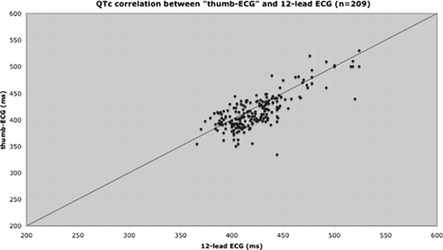
Assessment of QTc intervals using 1-lead handheld ECG
Jacob Broms1, Mårten Rosenqvist1, Börje Darpö2
1Kardiologkliniken Södersjukhuset, Stockholm, 2Medivir AB
Background. QT prolongation on the ECG is a result of prolonged myocardial repolarization and can be acquired (often drug induced) or congenital. QT prolongation has been linked to ventricular tachyarrhythmias, specifically torsade de pointes, which may lead to sudden death.
The heart rate corrected QT interval (QTc) is often monitored in patients who are started on Class III and Ia antiarrhythmics to allow the identification of subjects with pronounced QT prolongation. There is a need for simple devices to facilitate this monitoring process and to allow for ambulatory out-of-hospital use.
The objective of this study was to compare QTc intervals generated from standard, 12-lead ECGs with those generated from a hand-held 1-lead ‘thumb-ECG’ (Zenicor-EKG®). The thumb-ECG measures standard limb lead I between left and right thumb.
Material and methods. Patients in sinus rhythm were recruited at the cardiology department, Södersjukhuset and in the emergency room, Danderyds sjukhus.
First phase. ECGs were registered in pairs (one 12-lead ECG and one thumb-ECG) on a single occasion from 45 patients. Second phase: ECGs were registered in pairs from 20 patients on four different occasions with a minimum of four days interval. Totally 240 ECG-pairs were registered.
Results. A QTc-value from the thumb-ECG was possible to calculate in 87% of the registrations corresponding to 88% of the patients. When QTc could not be calculated it was mainly due to invisible T-wave in the 1-lead ECG.
72% of the thumb-ECGs QTc-values were within 30ms from the 12-lead ECG registration.
90% of the QTc-values correlated within +/ − 50ms.
Conclusion. QTc registration with 1-lead thumb-ECG correlates well with standard 12-lead ECG and could be a valuable tool for long term out-of-hospital monitoring of QTc prolongation.
Key Words. QT intervals, QT prolongation, thumb-ECG
Arrhythmia
25
A0316
Cryoballoon pulmonary vein isolation
Fariborz Tabrizi1, Göran Kennebäck1, Jonas Schwieler1, Hamid Bastani1, Frieder Braunschweig1, Bita Sadigh1, Nikola Drca1, Anna Grahn1, Christer Wredlert1, Mats Jensen-Urstad1
1Dept of Cardiology, Karolinska University Hospital
Background. Linear pulmonary vein isolation (PVI) with radiofrequency energy is widely used for catheter ablation in symptomatic patients with pharmacologic refractory paroxysmal atrial fibrillation (AF). A novel technology is cryothermal energy applied via a double lumen balloon catheter (ArcticFront, CryoCath).
Material and methods. We tested this technique in 75 consecutive patients with paroxysmal AF (median age 57; range 31–75; 21 women) who had failed anti-arrhythmic therapy. We used a 23 or 28 mm balloon depending on pulmonary vein diameter. The end-point was pulmonary disconnection. PV conduction was verified before and after ablation by means of circular mapping-catheter in accordance with established criteria. If necessary touch-up ablation was performed using an 8 mm Freezor Max catheter. The movement of the diaphragm was repeatedly verified using fluoroscopy when ablating the right-sided PVs.
Results. 273/304 (90%) of targeted veins were successfully isolated solely with the balloon. In 28 veins, the isolation was completed using the Freezor Max catheter. In 3 veins isolation failed. Procedure and fluoroscopy time were 204±59 and 46±19 minutes. Mean freeze time per vein was 15±8 minutes. Reversible phrenic nerve palsy was seen in 10 patients (one moderately symptomatic during two months). Four patients underwent 2 cryoballoon procedures. A substantial number of veins not isolated with the balloon were due to phrenic nerve palsy. After a median follow-up of 13 months (range 4–28), 68% were free of symptomatic AF and an additional 17% were significantly improved. 40% were still on AA at the time of evaluation.
Conclusion. Cryoballoon isolation of the pulmonary veins is feasible. In the majority of patients PVI can be achieved with a limited single balloon approach. Reversible nerve palsy was a limiting factor in 13% of treated patients. In this series touch-up was necessary in 22% of patients. Long-term outcome remains to be evaluated.
Arrhythmia
26
A0322
Quality of life is improved in patients with atrial fibrillation after pulmonary vein isolation
Carina Carnlöf1, Per Insulander1, Mats Jensen-Urstad1
1Dept of Cardilogy Karolinska University Hospital
Background. Atrial fibrillation (AF) is the most common arrhythmia and many AF patients experience a significantly impaired health-related quality of life (HRQOL). AF is also associated with a high risk of stroke and death. Many pharmacological treatments for AF are ineffective and may have adverse effects. New methods, such as pulmonary vein isolation (PVI) have been developed to treat AF. The aim of this study was to investigate HQQOL in severely symptomatic AF patients before and after PVI.
Material and methods. Forty patients with severely symptomatic AF were included. 36 completed the study with self-reported HRQOL questionnaires (SF-36) before and after PVI. A standardized control group was used.
Results. HRQOL before PVI was significantly lower in all domains except for bodily pain compared to the control group. All subscales of the SF-36 improved significantly after PVI except for bodily pain, which remained unaltered.
Conclusion. Health-related quality of life is improved in severely symptomatic AF patients after pulmonary vein isolation.
Arrhythmia
27
A0328
Arrhythmia-specific protocol U22 in supraventricular tachycardia: Improvement in well-being after catheter ablation
Milos Kesek1, Titti Tollefsen1, Niklas Höglund1, Folke Rönn1, Steen M Jensen1
1Hjärtcentrum, Norrlands Universitetssjukhus
Purpose. Main indication for ablation of supraventricular tachycardia is symptomatic relief. An evaluation of the treatment by general measures of quality of life, like SF-36, is however hampered by the fact that these scales do not measure the specific symptoms. U22 (Umea 22 Arrhythmia Questions) quantifies multiple symptom aspects associated with arrhythmic spells. Discrete 0–10 scales measure the influence of arrhythmia on well-being, intensity of discomfort during a spell, the type of dominant symptom and a time aspect that summarizes the duration and frequency of spells.
Methods. Symptoms were measured with U22 and SF-36 on hospital admission and 6 month later in patients with accessory pathway (AP) and atrioventricular nodal re-entry tachycardia (AVNRT) scheduled for catheter ablation. The diagnosis was established during the subsequent ablation. Catheterisation reports were reviewed by a blinded, experienced operator. Patients with a primarily successful ablation were included. Data are presented as mean±SD. Paired t-test is used for comparison.
Results. Fifty-eight patients (27 men and 31 women), ablated with primary success during 2006–2008 for AP (n = 23, age 43.5±18.5) and AVNRT (n = 35, age 56.2±13.3), completed the 4 forms (U22 and SF-36 at baseline and at follow-up, 204±37 days after ablation).
The score for well-being (0–10, 10 being best) increased from 6.0±2.6 to 7.9±1.9 (p < 0.0005). The score for arrhythmia as cause for impairment in well-being (0–10, 10 being highest) decreased from 7.5±2.8 to 2.0±3.1 (p < 0.0005). The time-aspect score for arrhythmia (0–10) decreased from 4.7±1.5 to 1.4±1.8 (p < 0.0005). The two SF-36 summary measures PCS and MCS increased from 46.9 (9.4) to 48.4 (10.7) and from 44.9 (12.5) to 49.1 (9.9) (p = 0.04 and 0.002).
Conclusion. The U22 protocol detected a prominent increase in measures of arrhythmia-related well-being after successful ablation of AP and AVNRT. In comparison, the improvement observed in SF-36 was relatively small.
Arrhythmia
28
A0360
Stepwise ablation of persistent and permanent atrial fibrillation
Mats Jensen-Urstad1, Fariborz Tabrizi1, Jonas Schwieler1, Göran Kennebäck1, Hamid Bastani1, Frieder Braunschweig1, Nikola Drca1, Bita Sadigh1, Jari Tapanainen1, Per Insulander1
1Dept of Cardiology, Karolinska University Hospital
Background. Pulmonary vein isolation (PVI) with radiofrequency energy is widely used in symptomatic patients with pharmacologic refractory paroxysmal atrial fibrillation (AF). Promising results have been presented in patients with persistent/permanent AF using a stepwise approach and to PVI adding ablation of regions with fractionated electrograms (CFAE), linear lesions in the roof, mitral isthmus, and coronary sinus. A common finding was AF converting to atrial tachycardias which could be further targeted.
Materials and methods. Since January 2008 we have treated 40 (24 men, 6 women) patients with persistent (5) or long-lasting persistent/permanent (35) AF (persistent AF for at least 3 months before ablation) using this approach. Fourteen had undergone a previous PVI procedure. Eight patients with permanent AF had EF ≤30. All ablations were performed during AF, except in 5 patients where initial PVI was done during SR but AF was later induced.
Results. In addition to PVI, ablation of CFAEs was done in 34 patients; a roof line in 33; ablation along CS from lower LA and inside CS in 17; and a mitral isthmus line in 9. The cavotricuspid isthmus was ablated in all patients. Seventeen patients converted to SR during the procedure; in the others successful cardioversion was done. Regularization to atrial tachycardia, which was targeted, occurred in 19 patients. Two patients underwent 2 procedures. Procedure time: 270±65 minutes. Fluoroscopy time: 69±26 minutes. Radiofrequency time: 102±31 minutes. No serious complications occurred. Thirty patients have a follow-up > 3 months (mean 6.2±2.6 months). 50% are still in SR, 19% are improved, and 31% failed. Several of the unsuccessful patients are scheduled for a second procedure.
Conclusion. Ablation of persistent and permanent AF, also in patients with heart failure, using a stepwise approach is feasible with a high acute success rate. Short term results are promising.
Arrhythmia
29
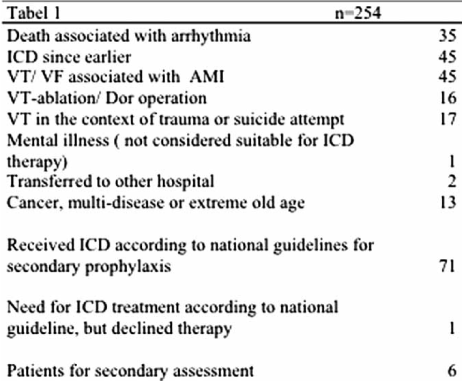
A0361
ICD therapy after cardiac arrest
Carina Carnlöf1, Katarina Ringdahl1, Fredrik Gadler1
1Dept of Cardiology, Karolinska University Hospital
Background. At Karolinska University hospital annually about 200 patients are hospitalized after cardiac arrest or life-threatening ventricle tachycardia. According to national guidelines a patient who has survived ventricular fibrillation or ventricle tachycardia with cardiac arrest should be treated with an implantable defibrillator.
Material/methods. The aim of this study was to identify whether the patients admitted to Karolinska during 2007 after cardiac arrest were offered and received appropriate treatment according to national guidelines for secondary prophylaxis regarding ICD therapy.
All files of patients admitted to Karolinska during 2007 with the diagnosis of either cardiac arrest or sustained ventricular tachycardia where identified. The files were reviewed as regards to treatment and outcome.
Results. In total 254 patient files were reviewed with the following results (see Table 1).
Conclusion. The most common cause for a ptient not receiving ICD therapy was that the ventricular arrhythmia was considered secondary to an ischemic event. If a life-threatening ventricular tachycardia is primary or secondary to an ischemic event can sometimes be difficult to determine. The moderate enzyme leakage caused by a primary arrhythmia can be misinterpreted as secondary to an ischemic event. Of the 254 patients with a diagnosis of severe ventricle tachycardia72 patients were treated with ICD therapy according to the national guidelines. Only six patients should be assessed again.
Arrhythmia
30
A0371
TIR-results from the National quality registry for anticoagulation treatment in Sweden
Mårten Rosenqvist1, Peter J Svensson2, Anders Själander3, Viveka Frykman4, Lars Wallentin5
1Södersjukhuset, Stockholm, 2Koagulationscentrum Malmö, 3Medicinkliniken Sundsvall, 4Hjärtkliniken Danderyds sjukhus, 5UCR Uppsala
The Swedish national anticoagulation registry Auricula started in 2006. It is growing rapidly, and has now over 16.000 patients treated with warfarin and more that 350.000 INR values are registred. The most common and also fastest growing treatment indication is atrial fibrillation. Over 40 primary health care centers and anticoagulation clinics from all over the country are now participating in the registry. More centers are joining continuosly. Of all patients in Sweden treated with warfarin, approximately 10% are now registred in Auricula. 30% of the patients are over 80 years of age. The quality of the warfarin treatment in Sweden is generally very high, with a mean time in range (TIR) of 69% (fig 1) range between 62–85% for 2008. High INRs,>8 is registered for 0.01% and > 5 is 0.61% of the INR samples. The TIR is consistently high even in older age groups. The warfarin dose is showing a striking age dependent decrease with age, almost linear from the age of forty (45mg/w) up to the age of ninety (22mg/w).
From this national wide registry we conclude that in general the quality of oral anticoagulant therapy in Sweden is very good and comparable with randomized controlled clinical trials with warfarin. More than 30% of the patients are more than 80 years of age. Even these elderly patients show a comparable and high TIR.
We conclude that these good results are highly dependent of the well organized anticoagulation treatment in Sweden.
Cardiology
31
A0252
Long term stability of heart rate variability in chronic stable angina pectoris, and the impact of an acute myocardial infarction
Inge Björkander1, Thomas Kahan1, Lennart Forslund2, Mats Ericson3, Nina Rehnqvist4, Paul Hjemdahl5
1Karolinska Institutet, Department of Clinical Scineces, Danderyd Hospital, Stockholm, Sweden, 2Medical Products Agency, Uppsala, Sweden, 3Stockholm University College of Physical Education and Sports, Stockholm, Sweden, 4Swedish Council on Technology Assessment in Health Care (SBU), Stockholm, Sweden, 5Department of Medicine, Karolinska University Hospital (Solna), Stockholm, Sweden
Background. Heart rate variability (HRV) reflects the balance between cardiac parasympathetic and sympathetic autonomic influences. Reduced HRV has adverse prognostic implications. The time course for changes in HRV over prolonged periods of time, and the influence of an acute coronary event on HRV are not well established.
Materials and methods. HRV was assessed in patients with chronic stable angina pectoris, who were followed for three years within the Angina prognosis study in Stockholm (APSIS). Patients who suffered an acute myocardial infarction after the study were re-examined after this event. We assessed HRV by the simple geometric method differential index, and traditional time and frequency domain measurements of HRV.
Results. The differential index was essentially unchanged during the study (i.e., the ratio month 36/month 1 was 1.00±0.06, n = 261). Also most other time and frequency indices of HRV (SDNN, r-MSSD, SDNNIDX, total power, and VLF, LF, HF, respectively; n = 63) remained largely unchanged; pNN50 and LF/HF were, however, less reproducible. In 21 patients with a subsequent acute myocardial infarction SDNN, SDNNIDX, total power, LF, and LF/HF were reduced following the event, whereas differential index, pNN50, and HF remained unchanged.
Conclusion. Differential index and other indices of HRV are stable and reproducible in patients with chronic stable angina pectoris. We have previously shown that the simple differential index method provided equally good or better prognostic information regarding cardiovascular death in stable angina pectoris than conventional, more laborious HRV methods in the time or frequency domain. The present results show that high frequency HRV (reflecting cardiac parasympathetic activity) and the differential index changed little following an acute coronary event, and may be suitable for predictions of the future risk of sudden death even in the presence of a recent acute coronary event.
Cardiology
32
A0257
Left ventricular asynchrony and raised filling pressure predict limited exercise performance assessed by 6 minute walk test
Michael Henein1, Per Lindqvist1, Gani Bajraktari2, Shpend Elezi2, Venera Berisha2, Nehat Rexhepaj2
1Hjärtcentrum, Norrlands Universitetssjukhus, Umeå, 2Service of Cardiology, Internal Medicine Clinic, University Clinical Centre of Kosova, Prishtina, Kosovo
Background and Aim. Six minute walking (6-MWT) may serve as a reproducible test for assessing exercise capacity in heart failure patients and can be clinically predicted. We aimed in this study to identify any additional ventricular function predictors of 6-MWT in patients with left ventricular (LV) ejection fraction (EF) <45%.
Methods. This study included 77 consecutive patients (60±12 years, and 33.3% were female) with stable heart failure who underwent 6-MWT and Doppler echocardiographic examination in the same day. LV end-diastolic and end-systolic dimensions, shortening fraction (SF), EF, myocardial velocities were measured. E:A ratio, t-IVT, and Tei index were also calculated. Patients were divided into two groups based on the 6-MWT distance (limited performance ≤300m and good performance >300m).
Results. Of all LV functional measurements, E’ wave (r = 0.61, p < 0.001), E/E’ ratio (r = − 0.49, p < 0.001), t-IVT (r = − 0.44, p < 0.001), Tei index (r = − 0.43, p < 0.001) and NYHA class (r = − 0.53, p < 0.001) had the highest correlation with the 6-MWT. Patients with limited 6-MWT performance had lower SF (p = 0.02) and EF (p = 0.017), longer t-IVT (p= 0.001), higher Tei index (p = 0.002) and higher E/E’ ratio (p < 0.001) compared with good performance patients. In multivariate analysis, only E/E’ ratio [0.8 (0.66–0.96), p = 0.017], and t-IVT [0.77 (0.62–0.95), p = 0.018] independently predicted poor 6-MWT performance (<300m).
Conclusions. In heart failure patients, the higher the filling pressures and the more asynchronous the left ventricle, the poorer is the patient's exercise capacity. These findings highlight specific LV functional disturbances that should be targeted for better optimization of medical and/or electrical therapy
Cardiology
33
A0265
The combined effect of low-grade albuminuria and a reduced glomerular filtration rate for the prediction of cardiovascular disease
Elisabet Nerpin1, Björn Zethelius1, Johan Ärnlöv1, Elisabet Nerpin2, Johan Ärnlöv2, Erik Ingelsson3, Ulf Risérus4, Samar Basu 4, Johan Sundström5, Anders Larsson5
1Department of Public Health and Caring Sciences/Geriatrics, Uppsala University, Uppsala, Sweden, 2Department of Health and Social Sciences, Högskolan Dalarna, Falun, Sweden, 3Depatrment of Medical Epidemiology and Biostatistics, Karolinska Institutet, Stockholm, Sweden, 4Section of Clinical Nutrition, Uppsala University, Uppsala, Sweden, 5Department of Medical Sciences, Uppsala University, Uppsala, Sweden
Background. The combined impact of reduced glomerular filtration rate (GFR) and microalbuminuria on the risk for cardiovascular disease is scarcely studied. Thus, we aimed to identify optimal cut-offs for albuminuria and GFR for the prediction of cardiovascular mortality in a community-based cohort of elderly men and to investigate whether the combined addition of these kidney markers adds independent prognostic information.
Material and methods. In a sub-sample, without cardiovascular disease at baseline, of the community-based Uppsala Longitudinal Study of Adult Men (ULSAM, n = 649, mean age 71 years, median follow-up 12.9 years; 86 cardiovascular deaths during follow-up), GFR (cystatin C-based) and urinary albumin excretion rate (UAER, overnight urine collection) were calculated.
Results. The following cut-off points were identified in order to achieve optimal model discrimination based on the integrated discriminative improvement: UAER 6.25 µg/min and GFR 45 ml/min/1.73m2. In Cox-proportional hazard models adjusted for established risk factors (age, systolic blood pressure, antihypertensive treatment, total cholesterol, HDL cholesterol, lipid lowering treatment, diabetes, smoking, body-mass-index and previous cardiovascular disease), participants with low-grade albuminuria only (>6.25 µg/min, HR 1.75, 95% CI 1.05–2.89), participants with reduced GFR only (<45 ml/min/1.73m2, HR 2.56, 95% CI 1.05–6.28) and participants with both low-grade albuminuria and reduced GFR (HR 5.91, 95% CI 2.87–12.18) were at higher risk for cardiovascular mortality compared to participants with normoalbuminuria and normal GFR.
Conclusion. Albuminuria and GFR predicted cardiovascular mortality in elderly men independently of each other and of established risk factors. Men with both low-grade albuminuria and reduced GFR were at particularly increased risk. The optimal cut-off point for albuminuria for the prediction of cardiovascular mortality was well below the current diagnostic threshold for microalbuminuria (>20 µg/min), while the optimal cut-off for GFR was similar to the diagnostic threshold for renal failure in the elderly (<50 ml/min/1.73m2).
Cardiology
34
A0266
Low incidence of cholesterol measurments in primary care patients at high cardiovascular risk
Anders Rane1, Åke Ohlson-Önerud2, Marie-Louise Ovesjö1
1Karolinska Universitetssjukhuset Huddinge. Avdelningen klinisk farmakologi, 2Pfizer AB
Low incidence of cholesterol measurements in primary care patients at high cardiovascular risk
Background. Systematic prevention and disease management are crucial in large patient groups such as patients with cardiovascular disease or diabetes mellitus. General Practioners play a key role in treating risk factors such as dyslipidemia. We have studied the extent of statin treatment and annual cholesterol measurements in primary healthcare.
Material and methods. We retrieved data from medical records (Swedestar®) of twelve urban primary healthcare centres serving a population of 125 267 citizens in Stockholm County. The study period was 2004–2007. The following patients were selected: previous myocardial infarction, diabetes mellitus or previous stroke/TIA. Patients with more than one of the diagnosis were included in all relevant groups. Any measurement of plasma cholesterol within 90 days before or 360 days after the first statin prescription was identified.
Results. The mean age was 71.3 yrs (62% male) in the MI group, 64.6 yrs (54.6% male) in the DM group, and 70.7 yrs (49.7% male) in the stroke group. In the MI group, 25.2% also had diabetes mellitus. In the MI group 69.2% were prescribed a statin, in the DM group 47.4% and in the stroke group 46.5%. Cholesterol measurement within 90 days before the first statin prescription was performed in 30.7% in the MI group; in 48.4% in DM group and in 41.0% in the stroke group. Within 360 days after the first statin prescription, 50.2% in the MI group, 55.5% in the DM group and 45.5% in the stroke group had a cholesterol measurement.
Conclusions. Many patients in either category were not treated with statins. Treatment appears often to be initiated without plasma cholesterol measurement before or after starting the statin treatment. Only about half of the patients were followed-up.
Cardiology
35
A0277
Serum Cathepsin S is independently associated with cytokine mediated inflammation in a community- based sample of elderly men.
Elisabeth Jobs1, Elisabet Nerpin1, Magnus Jobs1, Johan Ärnlöv1, Ulf Risérus2, Samar Basu2, Erik Ingelsson3, Elisabet Nerpin4, Johan Ärnlöv4, Magnus Jobs5, Anders Larsson6
1Department of Health and Social sciences, Högskolan Dalarna, Falun, Sweden, 2Department of Public Health and Caring sciences/ section of Clinical Nutrition, Uppsala University, Uppsala, Sweden, 3Department of Medical Epidemiology and Biostatistics, Karolinska Institutet, Stockholm, Sweden, 4Department of Public Health and Caring sciences/Geriatrics, Uppsala University, Uppsala, Sweden, 5Department of Medical Sciences/clinical virology, Uppsala University, Uppsala, Sweden, 6Department of Medical Sciences, Uppsala University, Uppsala, Sweden
Background. Previous experimental and clinical studies suggest that Cathepsin S, a lysosomal protease, is activated in obesity and in the development of atherosclerosis, and that the action of cathepsin S, to some degree is, mediated via increased inflammation. However, data on the relationship between circulating cathepsin S and markers of inflammation in the community are scarce.
Material and methods. Serum levels of Cathepsin S, and two different circulating markers of cytokine-mediated inflammation (high sensitivity C- reactive protein [CRP] and interleukin 6 [IL-6]) were analysed at a re-examination of a community-based cohort of elderly men (Uppsala Longitudinal Study of Adult Men, ULSAM; n = 1070) when the participants were 70 years. Moreover, CRP and IL-6 was measured at a reinvestigation when the participants were 77 years.
Results. In cross-sectional analyses, higher serum levels of Cathepsin S was significantly associated with higher serum CRP (-coefficient for 1 standard deviation (SD) increase 0.14, 95% CI (0.07–0.20), p < 0.001) and higher serum IL-6 (-coefficient for 1-SD increase 0.07, 95% CI (0.01–0.13), p < 0.03) after adjustment for age, lifestyle factors (body mass index, physical activity, smoking) and cardiovascular risk factors (hypertension, dyslipidemia, previous cardiovascular disease and diabetes). Moreover, the multivariable longitudinal associations between Cathepsin-S at age 70 and CRP and IL-6 at age 77 were similar (CRP: -coefficient for 1-SD increase 0.11, 95% CI (0.03–0.19), p < 0.005; IL-6: -coefficient for 1-SD increase 0.09, 95% CI (0.01–0.20), p < 0.03)
Conclusions. Higher serum levels of Cathepsin- S were independently associated with higher CRP and IL-6 in a community-based sample of elderly men. Our data provides support for the notion that Cathepsin S involved in inflammatory processes, possibly leading to atherosclerosis. Further studies are warranted to investigate the role of cathepsin S in the development of cardiovascular disease.
Cardiology
36
A0279
Predictors of direct admittance to a Coronary Care Unit among ambulance transported patients with suspected acute coronary syndromes.
Annica Ravn-Fischer1, Per Johanson1, Stefan Kihlgren1, Johan Herlitz1
1Sahlgrenska Universitetssjukhuset
Background. Independent predictors for direct admission to a Coronary Care Unit (CCU) among patients transported by ambulance due to suspected acute coronary syndrome (ACS) were investigated in a university hospital setting over a four year period.
Patients and Methods. All patients in the community of Göteborg who were transported by ambulance between 2004 and 2007 having symptoms raising suspicion of ACS and from whom therefore an ECG was sent directly by telemedicine to the CCU (n = 3244) were included in this survey. Decision about direct admission to the CCU were made of either a physician or a nurse at the CCU based on ECG patterns and clinical signs.
Results. An ECG was sent to the CCU in 3244 patients and 30% of them (n = 976) were directly admitted either to the CCU or to the Cath.lab. Strong independent predictors for direct admission were ECG signs of; a) ST-elevation (OR 43.3; 95% CI 32.3–58.0), b) Left bundle branch block (OR 3.1; 2.5–5.4). c) ST-depression (OR 2.0; 1.4–2.9). Other predictors positive for direct admission to the CCU were: Arrhythmia (OR 3.0; 1.6–5.5), Cold sweat (OR 2.2; 1.6–2.9), Ongoing chest pain (OR 2.1; 1.5–2.9) and Syncope (OR 2.1; 1.2–3.9). Predictors negative for direct admission were: Increasing age (OR 0.987; 0.979–0.996) and Female gender (OR 0.62; 0.48–0.81).
Conclusion. Different ECG patterns and ongoing symptoms were positive predictors for direct admission to the CCU and having ST-elevation on ECG was by far the most dominant predictor. Increasing age and female gender however were negative predictors which reduce the chance for direct admission to the CCU.
Cardiology
37
A0284
An evaluation of secondary prevention in coronary artery disease at a university hospital
Johan Herlitz1, Lillemor Stensdotter1, Mona From Attebring1
1Med Inst, Avd för Molekylär och klinisk medicin, Sahlgrenska universitetssjukhuset, Göteborg
Background. Traditional risk factors for coronary artery disease (CAD) can be favourably modified after discovery of the disease. Our aim was to evaluate the secondary prevention after hospitalisation of CAD at a university hospital.
Methods. Patients were invited between 6 and 10 months after hospitalisation due to CAD for a further screening including a 24 hour blood pressure recording, analysis of blood lipids and an oral glucose tolerance test.
Results. In all 250 patients have taken part in the survey. Mean age was 66±9 years (range 31–85 years) and 27% were females. Fourteen% had suffered from stable CAD and 86% unstable CAD. The proportion of patients who at follow up were on aspirin, betablockers, lipid lowering drugs and ACE-inhibitors were 93%, 88%, 94% and 44% respectively. The proportion of patients with elevated low density lipoprotein (LDL ≥2.5 mMol/l) was 39%, elevated mean blood pressure during 24 hours (≥140/90 mmHg on day or ≥130/80 mmHg at night in non diabetics and ≥130/80 mmHg on day or ≥120/70 mmHg at night in diabetics) was 41%. Thirteen% were smokers and 9% were snuffers. Mean heart rate during day time of > 80 beats per minute was seen in 13%.
Among non-diabetics 61% had impaired glucose tolerance.
Among all patients 72% had any of the following: present smoking, elevation of LDL, elevation of mean blood pressure or elevation of mean heart rate.
Conclusion. There is room for considerable improvement in the secondary prevention of coronary artery disease even at a university hospital. A large proportion of patients have elevation of cholesterol levels despite the fact that nearly all were on treatment with lipid lowering drugs.
Cardiology
38
A0304
High prevalence of persistent lipid abnormalities in high-risk patients treated with statins in Sweden-Results from the Dyslipidemia International Study
Gunilla Journath1,2, Anders Broijersen2, Pia Lundman3
1Department of Medicine, Clinical Epidemiology Unit, Karolinska Institutet, 2Merck Sharp & Dohme (Sweden) AB, 3Department of Clinical Sciences, Division of Cardiovascular Medicine, Karolinska Institutet, Danderyd Hospital
Objective. To assess the prevalence of lipid abnormalities in patients treated with statins.
Methods. Analysis included 958 Swedish patients from Dyslipidemia International Study (DYSIS), a large cross-sectional study carried out in 12 countries in Europe and Canada. Since June 2008, consecutive statin-treated patients 45 years or older (mean age 68 (61–75) years old) with available lipid values were recruited by primary care physicians, cardiologists, endocrinologists and internists. A clinical examination and patients’ latest lipid values on statin (≥3 month duration) were recorded.
Results. Of the 958 patients 59% were male, 12% current smokers, 28% had a family history of premature cardiovascular disease (CVD), 76% hypertension and 43% diabetes mellitus (DM). Coronary heart disease was present in 38% of patients, cerebrovascular disease in 13% and peripheral arterial disease in 6%. One third of the study population (33%) was obese (BMI ≥30 kg/m) and 67% had metabolic syndrome (IDF definition). Table 1 shows achievement of lipid targets.
Conclusion. A majority of statin-treated patients in Sweden did not reach current LDL cholesterol targets and/or had abnormal levels of HDL-cholesterol and triglycerides. These persistent lipid abnormalities will continue to contribute to CV risk in statin-treated patients and the results demonstrated the gap between guideline recommendations and clinical practice.
Cardiology
39
A0326
Left ventricular remodelling in HCM distorts radial and longitudinal function.
Ulf Gustafsson1, Stellan Mörner1, Anders Waldenström1
1Heart center, Umeå University Hospital
Background. Patients with hypertrophic cardiomyopathy (HCM) are known to have reduced longitudinal function and increased radial LV function. However, the relationship and the timings of these and the rotational function have not been displayed. We investigated these three functional dimensions independently and the timings to peak amplitudes and onset of relaxation.
Material/Method. From standard echocardiographic images we measured the lateral AV-plane displacement and radial movement of the posterolateral wall by M-mode, and the apical rotation by speckle tracking. The study population consisted of 16 patients with HCM and 16 controls (mean age 52 and 56 years, 4 and 9 women respectively).
Results. Patients with HCM had significantly lower lateral AV-plane displacement, 11.2mm vs 13.6mm (p = 0.018), longer time to peak longitudinal displacement, 70ms vs 8ms after aortic valve closure (AVC) (p = 0.003) and later onset of longitudinal relaxation, 115ms vs 100ms (p = 0.019). They also had significantly higher fractional shortening, 42.2% vs 35.9% (p = 0.002) and radial displacement, 12.4mm vs 9.4mm (p = 0.006). No difference in amplitudes or timings of apical rotation was seen. Time to onset relaxation was 5ms vs 11 ms after AVC radial and 9ms vs 10ms after AVC circumferential for HCM patients and controls respectively.
Conclusion. While peak longitudinal displacement was delayed in patients with HCM, radial and circumferential peak motions were still well timed to end of systole. Besides reducing the longitudinal function and increasing the radial function, HCM seems to only affect the timing of onset relaxation in the longitudinal dimension.
Cardiology
40
A0330
Bacterial endocarditis among abusers of alcohol and i.v.-drugs. A retrospective study.
Niels Erik Nielsen1, Hanna Amberntsson1
1Kardiologiska kliniken
Background. The incidence of bacterial endocarditis is 6/100.000/year. Intravenous drug abuse increases the risk up till 60 times, and studies have shown that i.v-drug abusers suffer from a higher risk of complications both throughout treatment and after, and also have a higher risk of relapse. We investigated our patient population to see if the above was true for our region, and in case evaluate if this should affect our future treatment of these patients.
Material & Methods. The study was a retrospective cohort study performed at the University Hospital of Linköping. Hospital records were collected from a total of 60 cases of bacterial endocarditis during 1986–2007, 30 abusers (both abuse of alcohol and intravenous drugs) and 30 patients without known abuse. Several factors concerning the episodes of endocarditis were studied.
Results. See table
“Complications during follow-up” mailet till Martina Åkerlund, Malmö Kongressbyrå enligt abstract instruktioner.
Conclusion. Abusers, and especially abusers of intravenous drugs, arrive at the hospital in a worse condition and suffer from a higher frequency of complications including death both in the short and the long term. However, the difference in complication frequency is not of such nature that this study can recommend a different treatment for drug abusers than for non-abusers.
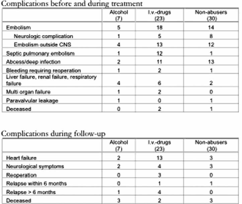
Cardiology
41
A0339
Takotsubo cardiomyopathy in Western Sweden
Elmir Omerovic1, Tomas Schultz1, Truls Råmunddal1, Per Albertsson1, Göran Matejka1
1Department of Cardiology, Sahlgrenska University Hospital
Introduction. Takotsubo cardiomyopathy has been recognized as a novel syndrome of acute heart failure affecting mostly elderly women exposed to severe emotional stress. In this study we present clinical characteristics of the large takotsubo cohort from Västra Götaland.
Methods. Between January 2005 and January 2009, all consecutive patients diagnosed with takotsubo at Sahlgrenska University Hospital were registered for prospective follow-up. All patients were admitted to the emergency department due to acute chest pain, signs of acute heart failure or cardiac arrest.
Results. We have registered 82 takotsubo patients; 10% males and 90% females, age 19–84. Thirty-day mortality rate was 9.7%. Four patients (2 males and 2 females) had significant coronary artery narrowing on coronary angiogram. UCG demonstrated in the majority of patients large akinetic area involving 2/3 of the apical portion of left ventricle with decreased ejection fraction. Interestingly, some patients have shown other patterns of reversible regional akinesia such as mid-wall only or inferior-wall only. In one male patient takotsubo was induced by habitual angina pectoris. Six pts developed thrombo-embolic complications (lung, brain and kidney) and three patients have developed cardiac arrest. All surviving patients recovered to normal cardiac function. Only 5% of patients were non-Swedish. Three patients had history of recidivism. In two patients with cardiogenic shock treatment with inotropic agents and intra-aortic balloon contra-pulsation acutely worsened hemodynamic status due to increased intracavitar gradient in left ventricle.
Conclusion. Takotsubo cardiomyopathy is a novel syndrome that affects both men and women of different age. Elderly Swedish women are mostly affected. The syndrome is associated with significant mortality and thrombo-embolic complications. Future studies are needed to elucidate the epidemiology, pathophysiology and long-term prognosis in this patient category.
Cardiology
42
A0352
Fibrinolytic therapy and bleeding complications – risk predictors from RIKS-HIA
Lisa Wernroth1, Ulf Stenestrand2
1Uppsala Clinical Research Center, 2Universitetssjukhus i Linköping
Background. Fibrinolytic treatment for ST-elevation myocardial infarction (STEMI) is associated with increased bleeding risk but is still widely used world-wide, mainly because of limited access to primary PCI. The aim of this study was to analyse contemporary fibrinolytic treatment patterns, in-hospital bleeding risk and prognosis during 2001 to 2005 in unselected Swedish patients.
Methods. The RIKS-HIA registry covers almost all Swedish patients treated for acute coronary syndromes. Major in-hospital bleeding was defined as lethal or intracranial bleedings or bleedings requiring surgery or blood transfusion. Survival status of the 14732 patients was obtained from the National Cause of Death Register.
Results. The number of patients receiving fibrinolysis as reperfusion therapy decreased from 4357 in 2001 to 1437 patients in 2005. Major in-hospital bleedings increased from 1.3% (including 0.8% lethal or intracranial) in 2001 to 3.9% (1.5%) in 2005, p < 0.001. History of serious bleeding, higher age, female gender, clopidogrel treatment prior to admission, pre-hospital administration of fibrinolysis, and fibrin-specific fibrinolytics were identified as predictors for bleeding. Major in-hospital bleeding was the strongest predictor of adverse prognosis with more than three-fold increase in 1-year mortality.
Conclusion. During 2001 to 2005 the use of fibrinolytic treatment markedly decreased while the incidence of major bleedings was more than doubled, the latter might in part be explained by increasing use concomitant anti-platelet therapy, pre-hospital treatment, fibrin-specific fibrinolytics and rescue PCI. Future close monitoring of bleeding complications is warranted, especially when considering the increased use of various combinations of antithrombotic drugs in conjunction with fibrinolysis and the great impact of bleedings on long-term mortality.
Cardiology
43
A0364
Echocardiographic Findings in Senior Endurance Athletes with Exercise-Induced Biomarker Release
Anders Sahlén1, Kambiz Shahgaldi1, Reidar Winter1, Marcus Ståhlberg1, Cecilia Linde1, Frieder Braunschweig1
1Karolinska Institutet, Hjärtkliniken, Karolinska Universitetssjukhuset, Solna, Sweden
Background. Strenuous exercise leads to elevation of N-terminal pro-Brain Natriuretic Peptide (NT-proBNP), a sensitive marker of cardiac dysfunction. Such release is reportedly proportionate to the levels at baseline. We sought to identify the structural determinants of baseline NT-proBNP in endurance athletes.
Materials and methods. 69 subjects (64±5 years; 83% male) underwent blood tests before and after a 30-km foot race [NT-proBNP, troponin T (TnT) and haemoglobin (Hb)]. Echocardiography was performed after a prolonged rest and included left ventricular mass index (LVMI), left ventricular ejection fraction (LVEF), left atrial area (LAA) planimetered in the apical 4-chamber view and indexed for body surface area. The ratio between transmitral E-wave and early diastolic relaxation (Em; colour-coded tissue Doppler imaging) was recorded.
Results. NT-proBNP at baseline (62 ng/L; IQR 31–134) and post race [224 (126–452)] correlated strongly (r = 0.90, p < 0.01). TnT post-race was 0.02 (0.01–0.05) µg/L. The LVMI was 104±29 g/m2, LVEF was 57±8%, LAA was 11±2 cm2/m2 and E/Em was 8±3. NT-proBNP correlated with: age (r = 0.42, p < 0.001), LVMI (r = 0.40, p = 0.001), LAA (r = 0.38, p = 0.002; Fig 1) and inversely with Hb (r = − 0.47, p < 0.001). Post-race TnT correlated with: LAA (r = 0.39, p = 0.001) and LVMI (r = 0.28, p = 0.02). E/Em did not correlate with either biomarker.
Conclusion. NT-proBNP release during strenuous exercise is largely determined by the levels present at baseline. Both biomarkers correlate with LV mass and atrial size. This may suggest that there is an association with filling pressures that escapes detection by E/Em due to limited sensitivity of this method in healthy subjects. Volume expansion leading to dilutional pseudo-anaemia is common in athletes and may explain the inverse association between NT-proBNP and Hb.
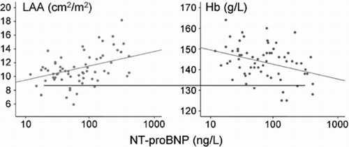
Cardiology/coronary
44
A0276
Can lipid/apolipoprotein factors account for residual risk in patients attaining current LDL-C targets?
Anders G. Olsson1, Christina Lindahl2, Rana Fayyad3, Sonal Bhatia3, Ingar Holme4
1Hälsouniversitetet, Linköping, 2Pfizer AB, Sverige, 3Pfizer Inc. New York, 4Oslo University Hospital, Oslo
Objectives. In this study, we assess if levels of apolipoprotein B (apoB), the ratio of apoB to apoA1 (apoB/apoA1) and non-HDL cholesterol (non-HDL-C) could account for the residual risk in patients who attain LDL-C goals.
Methods. The IDEAL study allocated 8888 patients to receive simvastatin (SIM) 20–40 mg/d or atorvastatin (ATO) 80 mg/d. In this subanalysis of subjects achieving LDL-C goals of 2.5 or 2.0 mmol/L, the ability of lipoprotein components to predict any CV event were assessed for each variable by comparing 1st (Q1) vs 4th (Q4) quartiles and assessing the effect of a 1 standard deviation (SD) increase. Adjustments were made for age, sex, smoking, hypertension, diabetes and previous statin treatment.
Results. 12 and 38% of SIM patients and 51 and 81% of ATO patients reached the 2.0 and 2.5 mmol/L goals, respectively. For any CV event the hazard ratios (HR) of Q4 vs Q1 for apoB, apoB/apoA1 and non-HDL-C were 1.23 (95CI:0.96,1.58,p = 0.11), 1.53 (1.19,1.98,p = 0.001) and 1.46 (1.14,1.88,p = 0.003), respectively. HR of 1 SD increase of the same variables were 1.19 (1.09,1.29,p = 0.001), 1.22 (1.12,1.33,p = 0.0001) and 1.18 (1.09,1.27,p = 0.0001), respectively. Similar results were obtained for the 2.5 mmol/L goal and for men and women.
Conclusions. In CHD patients who reach the 2.0 mmol/L LDL-C goal, there are still significant correlations to CV events for apoB, apoB/apoA1 and non-HDL-C. Our findings indicate that these variables are responsible for a residual risk of CV disease and suggest that patients could benefit by further reducing apoB, apoB/apoA1 and non-HDL-C.
Cardiology/coronary
45
A0283
Physical activity associated with less hospitalization and higher quality of life six years after an acute coronary syndrome.
Sara Ekman1, Henrik von Sydow2, Daniel Thunborg3, Mikael Dellborg1, Per Johansson1
1Dept of Cardiology, Sahlgrenska University Hospital, 2Dept of Internal Medicine, Kungälv hospital, 3Med Faculty, Linköping University
Background. Strong evidence supports physical activity to be effective as a treatment for coronary heart disease. Performed regularly it is known to reduce several riskfactors as well as improving quality of life. The main objective with this study was to assess the level of physical activity and its relation with known riskfactors and cardiovascular events as well as quality of life in patients with coronary heart disease.
Materials and methods. 177 consecutive patients that underwent coronary angiography at a university hospital due to an acute coronary syndrome in 2001 were investigated six years later. Factors as level of physical activity, weight and quality of life were assessed by questionnaire and information from electronic charts was collected from a parallel study.
Results. 45,3% of the patients were physically active, i.e. reported a physical activity analogous with a 30 min swift walk at least three times a week. The group of physically active patients included fewer overweighted (p = 0,011) and reported higher quality of life (p < 0,001), less limited physical capacity (p < 0,001) and worry about a cardiovascular event (p = 0,011) and was to a lesser degree hospitalized due to angina or myocardial infarction (p = 0,03).
Conclusion. The main findings of this study are that physical activity is associated with a lesser degree of hospitalization for angina or myocardial infarction and a higher quality of life. Physically active patients reported a less restricted physical capacity and were neither as overweighted, nor as concerned about cardiac events as those with a more sedentary life
Cardiology/coronary
46
A0291
Long-Term Effects of Spinal Cord Stimulation in Refractory Angina Pectoris-3-year Results from the European Angina Registry Link Registry
Paulin Andréll1, Wei Yu2, Lars Gillberg3, Kenneth Pehrsson4, Agneta Ståhle5, Clas Mannheimer1
1Multidisciplinärt smärtcentrum, medicinkliniken, Sahlgrenska Universitetssjukhuset/Östra, Göteborg, 2Capio, St Görans sjukhus, Stockholm, 3Anestesikliniken, Centralsjukhuset, Kristiandstad, 4Kardiologiska kliniken, Karolinska Universitetssjukhus/Solna, Stockholm, 5Institutionen för Neurobiologi, Vårdvetenskap och Samhälle, sektionen för sjukgymnastik, Karolinska Institutet, Stockholm
Background. Refractory angina pectoris is defined as severe angina due to coronary artery disease resistant to optimal pharmacological therapy and/or revascularization. The aim of the study was to assess the long-term effect of spinal cord stimulation (SCS) on angina symptoms and quality of life in patients with refractory angina.
Methods. The European registry for refractory angina is a prospective, three-year follow-up study. During 2003–2005, all patients with refractory angina referred for SCS treatment at 10 European centers were consecutively included. The patients were followed-up up to three years after implantation regarding angina symptoms and quality of life. The quality of life was assessed using generic (Short Form 36, SF-36) and disease specific (Seattle Angina Questionnaire, SAQ) quality of life questionnaires.
Results. Initially, 235 patients were included in the study. After screening, in order to determine suitability for SCS implantation, 122 patients were implanted and followed-up. The implanted patients reported improved CCS class from 3.3 to 2.7 (p < 0.0001), fewer angina attacks (p < 0.0001) and reduced short-acting nitrate consumption (p < 0.0001). Before implantation the quality of life was severely impaired both with regard to SAQ and SF-36. However, after implantation the quality of life was significantly improved in all dimensions of the SAQ and in all dimensions of the SF-36 except for the dimension “general health” after 3-year follow-up. During the 3-year follow-up period 26 patients died (21.3%).
Conclusions. Patients with refractory angina pectoris have a high mortality rate and the patients suffer from severe angina pectoris which limits daily life and impairs quality of life. SCS treatment improves anginal symptoms and increases quality of life in patients with refractory angina pectoris. The effects of SCS seem to be persistent after 3-year of follow-up. SCS might be a suitable alternative for pain relief in this patient population with severe coronary artery disease.
Cardiology/coronary
47
A0308
Matrix metalloproteinase-9 and cortisol reactivity in patients with myocardial infarction.
Aleksander Szymanowski1, Johnny Nijm1, Lennart Nilsson1, Lena Jonasson1, Margareta Kristenson2
1Department of Medical and Health Sciences, Division of Cardiology, Linköping University, Sweden, 2Department of Medical and Health Sciences, Division of Social Medicine and Public Health Science, Linköping University, Sweden
Background. Increased circulating levels of matrix metalloproteinase (MMP)-9 may indicate an increased risk of coronary plaque rupture. We have recently shown that a dysregulated cortisol pattern is associated with systemic inflammatory activity in coronary disease. In this study, we investigated whether also circulating MMP-9 levels were related to a disturbed hypothalamic-pituitary adrenal axis in patients with myocardial infarction (MI).
Material and methods. Thirty patients with a first-time MI were assessed at day 1–3 and after 3 months. At 3 months, all subjects were also subjected to a psychological stress test. Serum samples were assayed for total MMP-9, active MMP-9, tissue inhibitor of MMP (TIMP)-1, C-reactive protein (CRP) and interleukin-6 (IL-6). Free cortisol was measured in 24-h urine and in repeated saliva samples.
Results. At day 1-3, active MMP-9, MMP-9/TIMP-1 ratio, IL-6 and CRP were all significantly increased in patients compared to healthy controls. At 3 months, active MMP-9 (86 + / − 25 vs 75 + / − 33 ng/ml, p < 0.05) and MMP-9/TIMP-1 ratio (2.3 + / − 1.3 vs 1.4 + / − 0.7, p < 0.01) were still higher in patients while IL-6 and CRP were “normalized”. The 24-hour urinary cortisol and salivary evening cortisol were always higher in patients and independently correlated with MMP-9 and TIMP-1 at 3 months. During the stress test, the increase in cortisol was significantly larger in controls compared to patients. Active MMP-9 and MMP-9/TIMP-1 ratio were both significantly reduced in controls 24 hours after the stress tests (−15% and −19%, respectively) while they remained unchanged in patients.
Conclusion. Increased levels of active MMP-9 and MMP-9/TIMP-1 ratio were associated with a dysregulated cortisol pattern in MI patients. The data also raise the intriguing question that MMP-9 is differently regulated in patients during stressful conditions.
Cardiology/coronary
48
A0310
T cell activation and apoptosis in coronary artery disease
Aleksander Szymanowski1, Lena Jonasson1, Karin Backteman2, Jan Ernerudh2
1Department of Medical and Health Sciences, Division of Cardiology, Linköping University, 2Department of Clinical and Experimental Medicine, Division of Immunology, Linköping university
Background. An inflammatory response, including T cell activation, has been reported in patients with coronary artery disease (CAD). Lymphocyte apoptosis is a key event in the resolution of cellular immune activation. Here we examined T cell activation and apoptosis in varying clinical manifestations of CAD.
Material and methods. Blood was collected from patients with chronic stable angina (SA, n = 35) and acute coronary syndrome (ACS, n = 18) before (day 0) and 3 months after revascularization. Twenty-two healthy subjects served as controls. CD4+ and CD8+ cells were assessed by flow cytometry for activation markers (CD69, HLA-DR, CD56, loss of CD28) and apoptosis.
Results. CD4+ cells were increased in CAD patients at all time points compared to controls though mostly pronounced in the ACS group. CD8+ cell fractions did not differ between any groups. Neither did T cell activation differ from controls in either SA or ACS patients at any time point with one exception; the CD4 + CD28− population was significantly expanded in both patient groups at all time points. T cell apoptosis showed opposite patterns in SA vs ACS patients. Day 0, it was increased in SA patients and decreased in ACS patients compared to controls. At 3 months, CD4+ apoptosis was normalized in both patient groups whereas CD8+ apoptosis increased further in SA patients, 3.6 (1.1–5.6)%, remaining significantly reduced in ACS patients, 0.9 (0.5–1.7)%, vs controls, 1.3 (0.6–4.0)%.
Conclusion. There was evidence for an immune activation in the peripheral blood of patients with CAD. However, the T cell activation profile did not differ between different clinical manifestations of CAD providing no explanation for marked differences in T cell apoptosis between SA and ACS patients. We hypothesize that increased T cell apoptosis in SA patients reflects a compensatory mechanism during a chronic inflammatory process and that this protection may have failed in ACS patients.
Cardiology/coronary
49
A0311
The influence of streamlined teamwork in TAVI from a nursing perspective
Åsa Fåhraeus1, David Zughaft1
1Universitetssjukhuset i Lund, HIA/Hjärtangio
Background. Transcatheter Aortic Valve Intervention (TAVI) is an interventional treatment option for aortic stenosis. TAVI requires a multi-disciplinary team involving about 16 people. In order to achieve a safe and streamlined teamwork a well functioning crew is desirably. While the physicians must focus on their specific task, the nurse staff has a prominent coordinating role in TAVI.
Aim. To retrospectively investigate the influence of streamlined teamwork correlated to a feeling of coherence and job satisfaction in a TAVI team.
Method and materials. The first TAVI in Sweden was performed in Lund, in January 2008. Totally 20 patients who underwent TAVI in Lund 2008 were evaluated and the outcome was analyzed. The TAVI team consists of about 7 physicians of different specialities, 2 technicians and 7 nurses. To evaluate the multidisciplinary teamwork the Safety Attitude Questionnaire (SAQ) – OR version was distributed to 10 nurses in the TAVI team. The control group included 10 nurses working in a regular Operating Room (OR). Specific SAQ terms investigated were Teamwork Climate, Safety Climate and Job Satisfaction calculated with the Likert Scale.
Results. No serious technical or medical complications were observed or documented in the (OR) or in the Intensive Care Unit. The response rate of the SAQ was 95%. In the TAVI team 66% of the responders were scored positive due to SAQ terms. In the control group the rate was 20% due to lack of experienced coherence.
Conclusion. The nurses of the TAVI team responded more positive than the control group according to the SAQ. The result of the study is that the TAVI team experienced a better functioning teamwork climate, safety environment and satisfaction with their work. There are reasons to believe that safe streamlined teamwork might improve the outcome for the TAVI treatment.
Cardiology/coronary
50
A0337
In Stent Restenosis – Benign or Dangerous? Clinical Presentation of Coronary Restenosis in Sweden
Elmir Omerovic1, Truls Råmunddal1, Lars Grip1, Göran Matejka1, Per Albertsson1, Jan Boren2
1Sahlgrenska Universitetssjukhuset/Kardioilogen, 2Wallenberglaboratoriet vid Sahlgrenska Akdemin
Background. Restenosis after percutaneous coronary intervention (PCI) is thought to be a benign event clinically manifested as stable exertional angina. The aim of this prospective multicenter registry study was to investigate to which extent patients (pts) with restenosis present as acute coronary syndromes in Sweden.
Methods. We searched Swedish Coronary Angiography and Angioplasty Registry (SCAAR) data. All registered cases of PCI for restenosis (in-stent, after balloon angioplasty) occurring between 1995 and 2005 in Sweden were scrutinized. Both multivessel and single vessel interventions were included. Restenosis presentation was classified into four categories: 1) stable angina, 2) unstable angina/non-STEMI, 3) STEMI and 4) other reasons. Restenosis episodes were based on symptoms and were defined clinically rather than angiographically as routine angiographic screening was not performed.
Results. We identified 4660 cases of restenosis in 1962 patients. Coronary restenosis presented in 40.4% of cases as stable angina, in 45.5% as unstable angina/non-STEMI, in 10.1% as STEMI and in 4.0% as “other reasons". Cardiogenic shock was reported in 48 patients. Restenosis after balloon angioplasty had lower incidence of STEMI v. in-stent restenosis (6.9% v. 13.8%). Women had higher incidence of unstable angina/non-STEMI (52.3%) but lower incidence of STEMI (9.6%) compared to men (43.6% and 12.2% respectively). Mortality rate was 1.7% at 30 days, 3.2% at 6 months and 4.6% at one year in patients with restenosis
Conclusion. Majority of patients with coronary restenosis present either as acute myocardial infarction or as unstable angina requiring hospitalization and new interventions. Women may have higher risk to develop acute coronary syndrome due to restenosis. Prevention of restenosis may be an important target for improvement of “hard” clinical outcomes in pts undergoing coronary revascularization.
Cardiology/coronary
51
A0357
Paternal history of coronary heart disease, as opposed to maternal, predicts coronary heart disease in 60-year-olds
Karin Leander1, Max Vikström1, Mats Halldin1, Mai-Lis Hellénius2, Ulf de Faire3
1Institute of Environmental Medicine. Div of Cardiovascular Epidemiology, Karolinska Institutet, Stockholm, 2Dep of Medicine, Clinical Epidemiology Unit, Karolinska University Hospital, Solna, 3Institute of Environmental Medicine. Div of Cardiovascular Epidemiology, Karolinska Institutet, Stockholm, and Dep of Cardiology, Karolinska University Hospital, Solna
Background. We explored the relation between paternal and maternal history of coronary heart disease (CHD), respectively, and risk of CHD in a population-based cohort of 60-year-old men and women from Stockholm.
Materials and methods. A cohort of 3740 individuals (participation rate 78%), free from earlier signs and diagnoses of CHD, was prospectively followed from baseline 1.7.1997–30.6.1998 until 31.12.2005 – on average 7.4 years. Incident cases of CHD were identified using data from the national cause of death registry and the inhospital registry. Exposure data was collected through questionnaires and physical examinations including blood sampling. Hazard Ratios (HR) with 95% confidence intervals (CI) for CHD were calculated both unadjusted and adjusted for risk factors (hypertension, hypercholesterolemia and type II diabetes) using Cox proportional hazard model.
Results. Paternal history of CHD, defined as a CHD diagnosis in the father before the age of 66, was present in 21% of individuals with incident CHD (n = 166) and in 12% among those without CHD during follow-up (n = 3523). The adjusted HR of CHD was 2.20 (95% CI 1.40–3.45) for men reporting paternal history of CHD compared with men with no paternal history. The corresponding HR for women was 1.58 (0.79–3.18). Maternal history of CHD was not associated with increased risk of CHD in either men or women.
Conclusion. Our study shows that risk of CHD in 60-year-olds is more clearly influenced by paternal factors than maternal.
Cardiology/coronary
52
A0367
CABG still provides long-term survival benefit over medical treatment
Helena Rexius1, Rolf Ekroth1, Gunnar Brandrup-Wognsen1, Micael Nilsson1, Anders Jeppsson1, Ingvar Karlberg2, Anders Odén3
1Department of Cardiothoracic Surgery, Sahlgrenska University Hospital, 2Department of Public Health and Community Medicine, Göteborg University, 3Dep of Mathematical Sciences, Chalmers University of Technology
Background. The survival benefit of CABG over medical treatment in high-risk patients that was demonstrated more than twenty years ago has been questioned. Long waiting lists for CABG are detrimental but offer the possibility to compare the risk of death before and after surgery. We hypothesized that the risk of death is still lower after surgery than preoperatively.
Methods. The death hazard functions were calculated by the use of Poisson regression before and after surgery in a cohort of 10,657 consecutive CABG patients. The risk functions were related to the patients’ background risk profile and the preoperative risk were compared with the risk one and five years after inclusion on the waiting list, respectively.
Results. In the one-year perspective, CABG reduced the overall risk of death in 55.6% of the patients (average absolute risk reduction 2.5%, relative risk reduction 45%, number needed to treat (NNT) 39.7). In the five-years perspective, CABG reduced the overall risk for death in 67% of the patients (average absolute risk reduction 9.6%, relative risk reduction 46%, NNT 10.4). The relative risk reduction was highest in high-risk patients with preoperative unstable angina (mean relative risk reduction at five years 75%) and/or impaired cardiac function (mean risk reduction 65%). In patients with low risk (single or multi vessel disease without unstable angina, impaired cardiac function or proximal LAD stenosis) there was no gain in survival.
Conclusions. CABG still provides long-term survival benefit over medical treatment in the majority of patients selected for CABG. The gain is largest in high-risk patients. Our data reinforce guidelines that advocate initial medical treatment in low-risk patients and CABG in high-risk patients.
Experimental/physiology
53
A0246
Comprehensive physiological exercise echocardiography: A pilot study.
Per Lindqvist1, Stellan Mörner1, Karin Holmström1, Maria Backlund1, Anders Waldenström1, Michael Henein1
1Hjärtcentrum, Norrlands Universitetssjukhus,Umeå
Background. Exercise related symptoms, chest discomfort or breathlessness are very common in cardiovascular medicine. While spirometry is used to assess lung function exercise echocardiography is the counterpart for cardiac function.
Objective. We aimed to assess the feasibility of combining exercise echocardiography and cardiopulmonary exercise testing in providing comprehensive assessment of cardiac and respiratory function.
Methods. We studied 10 normal volunteers at rest and during incremental increase in exercise workload by 10 watt every other minute until they reached exhaustion. The subjects exercised on a supine bicycle having had a Doppler echocardiographic assessment of cardiac physiology and natriuretic peptides (BNP) at rest and at suboptimal and peak exercise. They also had cardiopulmonary exercise assessment throughout the exercise duration.
Results. Measurements of cardiac function were taken at heart rate of 108 bpm, although all subjects exercised till exhaustion, in order to secure good quality measurements. Peak exercise workload was 95±15 Watt and peak MVO2 was 20.3±4.2 ml/min/kg. Left ventricular stroke volume increased from 73±13 to 84±14 ml (p < 0.01) and BNP from 18.5±11.3 to 32.9±21.0 pg/ml (p < 0.01). Blood pressure rose from 140±10 to 176±19 mmHg, p < 0.001. No volunteer developed Doppler signs of raised left atrial pressure, symptoms or ECG abnormalities.
Conclusion. This small pilot study suggests that comprehensive cardiopulmonary exercise approach is feasible and measurements are fairly consistent. Although it does not provide detailed evaluation of pulmonary function, analysis of cardiac data should guide towards the likely explanation of patient's symptoms.
Experimental/physiology
54
A0273
The enlargement of ventricles and atria seen in athlete's heart is balanced between the left and right side
Henrik Mosén1, Katarina Steding1, Håkan Arheden1
1Hjärt MR gruppen, avd. för klinisk fysiologi, Lunds Universitetssjukhus
Objective. The aim of this study was to investigate if long tem endurance training can cause a balanced enlargement between the left and right atrium and the left and right ventricle.
Background. It is well known that long term endurance training induces morphological changes of the left ventricle. Few studies have investigated the effects of training on the volumes of the right and left atria and the right ventricle. Different pathologies can cause an enlargement of the chambers of the heart, e.g. dilated cardiomyopathy. We hypothesized that an enlargement of a healthy heart caused by long term endurance training should be well balanced between the left and right side.
Methods. Eighteen elite triathletes (6 female) and 27 healthy normal subjects (12 female) underwent cardiac magnetic resonance imaging (CMR). Left and right end-diastolic volume (LVEDV, RVEDV) and left and right atrial volumes (LA, RA) were calculated from short-axis images using planimetry. Mann-Whitney non-parametric test was used to compare atrial and ventricular volumes between groups. Linear regression was performed to assess the relationship between LVEDV and RVEDV and between LA and RA.
Results. Male triathletes had significantly larger LA and RA volumes (p < 0.0001, p < 0.01) and LV and RV EDV (p < 0.0001, p < 0.001) when compared to male normals. Female triathletes had significantly larger LV and RV EDV (p = 0.015, p < 0.01). There were no significant differences in atrial volumes between female triathletes and female normal subjects (LA p = 0.247 and RA p = 0.147). Linear regression showed a strong correlation between LVEDV and RVEDV (R2 = 0.83, p < 0.0001) (Fig.1A). The correlation between LA and RA was somewhat weaker, yet significant (R2 = 0.56, p < 0.0001) (Fig 1B).
Conclusion. This study has shown that long term endurance training is associated with a balanced enlargement of the left and right atrial and ventricular dimensions in both males and females.
Experimental/physiology
55
A0288
Growth factor stimulation of hyaluronan synthesis in cultured cardiomyocytes and fibroblasts
Urban Hellman1,2, Anna-Maja Åberg1, Nina Gennebäck1, Stellan Mörner1, Anna Engström-Laurent1, Anders Waldenström1, Li-Ping Ma3,4, Michael Fu4, Linus Malm5, Göran Larsson5
1Department of Public Health and Clinical Medicine/Medicine, Umeå University, Umeå, Sweden, 2Department of Medical Biosciences/Medical and Clinical Genetics, Umeå University, Umeå, Sweden, 3Department of cardiology/Changhai Hospital,The Second Military Medical University/Shanghai, China, 4Department of Molecular and Clinical Medicine, Wallenberg Laboratory, Institute of Medicine, Sahlgrenska Academy, Gothenburg University, Gothenburg, Sweden, 5Department of Medical Biochemistry and Biophysics, Umeå University, Umeå, Sweden
Background. Hyaluronan (HA) is a glycosaminoglycan located in the interstitial space and is essential in both structural and cell regulatory functions in any connective tissue in health and disease. We have previously shown that cardiac tissue use two different HA synthases in the hypertrophic process and that HA synthesis is up-regulated in experimental myocardial infarction. It is unknown which cells are involved in the process and whether the different HA synthases are activated in different cells and are responsible for different sizes of HA.
Material and methods. Cardiomyocytes (HL-1) and fibroblasts (NIH 3T3) were cultivated in claycomb and DMEM media resp. for 24 H in presence of the following growth factors (GF), PDGF-BB, TGFbeta2, and FGF2. HA concentration was determined by an ELISA kit. HA size was determined using Dynamic Light Scattering (DLS).
Results. Cardiomyocytes only produce HA when stimulated by PDGF-BB at high concentrations. Fibroblasts are stimulated by all three GFs. No HA synthesis was seen in control groups where no GF was added except when a mixture of cardiomyocytes and fibroblasts (80/20) were co-cultivated. FGF2 induced high mw HA synthesis in fibroblasts and PDGF-BB induced HA synthesis of lower mw in cardiomyocytes.
Conclusion. We show for the first time that cardiomyocytes can produce HA. Fibroblasts are stimulated to HA synthesis by all three GFs and there is a dose dependent response where the highest dose used induced less HA. This phenomenon is in accordance with earlier works. Interestingly the co-cultivation of cardiomyocytes and fibroblasts induced HA synthesis without addition of GF's indicating that one cell type is cross talking to another to produce HA.
Experimental/physiology
56
A0300
Volume Tracking-a Novel Visualization and Quantification Method for Blood Flow From Phase Contrast MRI
Johannes Töger1, Håkan Arheden1, Einar Heiberg1, Gustaf Söderlind2
1Department of Clinical Physiology, Lund University, Lund, Sweden, 2Numerical Analysis, Department of Mathematics, Lund University, Lund, Sweden
Background. The dynamics of intracardiac blood flow are not yet completely understood. Three-dimensional phase contrast MRI may provide new insights. However, cardiac blood flow is highly complex, and visualizations must be designed with care to display data in an intuitive and informative way. Additionally, quantitative measures are of great interest when following patients over time. Therefore, the purpose of this study was to develop and test the feasibility of Volume Tracking, a novel method for visualization and quantification of blood flow.
Materials and methods. Four healthy volunteers underwent Phase Contrast Magnetic Resonance Imaging (PC-MRI). To follow an arbitrary blood volume in the heart, a novel mathematical representation of the tracking problem was formulated and solved using state-of-the art numerical methods. Visualization was performed using the software Ensight (CEI, USA).
Results. The proposed method is feasible. Arbitrary volumes can be interactively selected and followed over time (Figure 1). Additionally, parameters such as kinetic energy in the followed volume can be quantified (Figure 2).
Conclusion. Volume Tracking, a new method for visualization and quantification of cardiac blood flow was developed and tested. A major advantage over existing methods is the ability to quantify parameters such as kinetic energy and momentum.
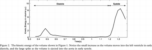
Experimental/physiology
57
A0305
Cardiac gene regulation in patients with Hypertrophic Cardiomyopathy
Nina Gennebäck1, Gerhard Wikström2, Urdan Hellman1, Stellan Mörner1, Danièle Charlemagne3, Anders Waldenström1
1Department of Public Health and Clinical Medicine/Medicine, Umea university, Umea, Sweden, 2Department of Medical Sciences, Cardiology, Uppsala university, Uppsala, Sweden, 3INSERM U 942, Paris, France.
Material and methods. Endomyocardial biopsies were taken from consecutive patients with HCM and immediately put in RNAlater. Control biopsies were treated likewise. mRNA was purified and analysed using whole-genome expression and real-time PCR. The raw data was analysed with bioinformatic software.
From the micro array raw data, 18 189 genes, 300 genes were selected according to significance level of up- or down- regulation. Those 300 genes were entered into the software and networks as well as a biomarker profile were developed.
Results. The biomarker profile had significant p-values for cardiovascular diseases such as; congenital heart defects (0,00001), ischemia (0,00009) and heart arrest (0,00005). The network analysis found two networks, consisting of 23 and 4 genes respectively. Both networks include transcription factors, structural proteins and regulatory proteins. In the larger network early growth response 1 (EGR1) is a key component, involved in the inhibition and the activation of several other network components.
Conclusion. We show results from the first gene array analysis in myocardial biopsies from patients with HCM. The bioinformatics software verifies a significant correlation to cardiac diseases and feasibility of endomyocardial biopsies for such analysis, showing the great power of this technique for clinical investigation of such patients.
Experimental/physiology
58
A0324
Interventricular interaction mediated through septal movement; A quantitative magnetic resonance imaging study in healthy subjects and patients with pulmonary regurgitation
Sigurdur Sverrir Stephensen1,2, Einar Heiberg1, Håkan Arheden1, Marcus Carlsson1
1Department of Clinical Physiology, Lund University Hospital, 2Department of Pediatric Cardiology, Lund University Hospital
Introduction. Interventricular interaction is mediated in part through septal movement. If the epicardial border of the septum moves towards the left ventricle (LV), stroke volume (SV) is added to the LV. The aim was to quantify the contribution of septal motion to the SV in healthy volunteers and patients with moderate to severe pulmonary regurgitation (PR) due to repaired tetralogy of Fallot (TOF).
Methods. 12 healthy volunteers (40.3±14.8 years, 6 females) and 9 patients with PR due to repaired TOF (11.8±1.7 years, 7 females) were included. Steady state free precession cine MRI (TR/TE/ =2.8/1.4ms/60°, 8 mm slice thickness, 30 time phases) in the short axis view covering the heart was acquired. LV and RV endocardial and epicardial contours were delineated and the percentage LVSV resulting from septal motion was defined from the 3-dimensional contours in a specially adapted segmentation program (Segment v1.8, http://segment.heiberg.se). Septal motion towards the LV was denoted a positive percentage contribution of the LVSV and septal motion towards the RV as negative.
Results. The contribution to the LVSV by septal motion was 7.9±3.5% in healthy volunteers. In contrast, the septal motion in TOF patients with moderate to severe PR was towards the RV, thereby contributing to the RVSV. This was reflected in a negative contribution to LVSV, −12.8±6.2% (p < 0.001 vs normals).
Discussion. Interventricular septal movement contributes significantly to the LVSV in healthy individuals. The septal motion is reversed and contributes significantly to the RVSV in TOF patients with RV volume load due to PR.
Experimental/physiology
59
A0340
Cardiac output is not affected by passive leg raising in supine healthy humans
Andreas Otto1, Marcus Carlsson1, Håkan Arheden1, Martin Ugander1
1Dep of Clinical Physiology, Lund University Hospital
Background. Passive leg raising in the supine position can be used to increase mean arterial blood pressure, but its affects on cardiac output are debated. Also, a simple noninvasive method to change cardiac output for physiological studies of cardiac function by MRI would be desirable.
Aim. To assess the effects of passive leg raising in the supine position on cardiac output.
Methods. Six healthy individuals aged 23–35 years, one female, were studied. Stroke volume, heart rate and cardiac output were measured in the ascending aorta by free breathing velocity encoded MRI (Philips 1.5T). Measurements were undertaken at baseline as the mean of three consecutive measurements, and every other minute for 20 minutes following a 45 degree passive leg raise. Data were normalized to baseline and are illustrated as the mean +/− SEM.
Results. Passive leg raising resulted in an unchanged stroke volume (p = 0.25), unchanged heart rate (p = 0.14) and unchanged cardiac output (p = 0.60) after 20 minutes. There was no change over the 20 minutes. See figure.
Conclusions. Passive leg raising in the supine position does not affect cardiac output in healthy individuals.
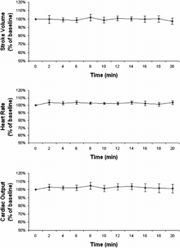
Experimental/physiology
60
A0356
A newly identified genetic marker, rs599839, modulates the risk of myocardial infarction through an interaction with serum lipids levels in the Stockholm Heart Epidemiology Program.
Bruna Gigante1, Max Vikström1, Karin Leander1, Shu Ye2, Ulf de Faire3
1Division of Cardiovascular Epidemiology, IMM, Karolinska Institutet, Stockholm Sweden, 2Centre of Clinical Pharmacology, William Harvey Research Institute, Barts and the London School of Medicine and Dentistry, London, UK, 3Institute of Environmental Medicine, Division of Cardiovascular Epidemiology, Karolinska Institutet, Dept of Cardiology, Karolinska University Hospital, Stockholm, Sweden
Objective. An intergenic anonymous genetic marker rs599839A/G has been recently found to modulate serum lipids levels and the risk of myocardial infarction (MI). The aim of the present study was to investigate if rs599839A/G influences the risk of MI through the regulation and the interaction with serum lipids and biomarkers in the Stockholm Heart Epidemiology Program (SHEEP).
Methods and Results. Serum lipids were stratified according to rs599839 genotype and differences (expressed as median and interquartile range) between cases and controls in men (n = 852/1054) and in women (n = 361/507) were assessed by Kruskall Wallis test. In male cases, presence of the GG genotype was associated with reduced total-cholesterol (GG: 5.60 (4.80–6.98) vs AA 6.10 (5.50–6.90), p = 0.0003), LDL-cholesterol (GG: 3.64 (3.30–4.91) vs AA 4.22 (3.67–4.80), p = 0.001) and ApoB serum levels (GG 1.32 (1.22–1.71): AA: 1.61 (1.38–1.85), p = 2*10–6) as compared to the AA genotype. Genotype and allele frequencies at rs599839 did not differ among MI cases and controls. We then tested through a logistic regression analysis if genetic variants at rs599839 locus modulate the risk of MI through an interaction with total-, LDL-cholesterol and ApoB serum levels. In the presence of high (>75th percentile) ApoB serum levels the GG + GA genotype was associated with a significant reduction in MI risk in men (beta-coefficient±SEM: −0.34±0.11, p = 0.002).
Conclusions. Our study confirms and extends previous data on the relationship between rs599839 locus and serum lipids levels. It also suggests that rs599839 modulates the risk of MI in men through an interaction with circulating lipids levels.
Imaging/diagnostics
61
A0231
The relationship between conventional right heart pressure measurements and those estimated by Doppler echocardiography
Per Lindqvist1, Michael Henein1, Sharzad Shirinpor2, Gerhard Wikström2
1Hjärtcentrum, NUS, Umeå, 2Thoraxcentrum, Akademiska sjh. Uppsala
Background. Doppler echocardiographic estimates of right heart pressures are routinely used for studying cardiac and pulmonary patients. Reports however, highlighted important limitations that may affect the clinical value of such measurements.
Methods. We studied 207 consecutive patients who underwent routine right heart catheterization (RHC), who also had Doppler echocardiographic (DE) examination within a week of the RHC. From the invasive data we measured peak pulmonary artery systolic pressure (PASP) and mean right atrial (RA) pressure. From the DE studies we measured peak trans-tricuspid retrograde pressure drop, as a representative of RV systolic pressure, and mean right atrial pressure was estimated using the inferior vena cava lumen diameter and its lumen variation due to normal respiration (adding 5,10, 15 and 20 mmHg respectively)
Results. RHC measured PASP was 15–103 (mean 48±22 mmHg) and RAP was 3–30 (11±5 mmHg). DE estimated PASP was 16–115 (49±22 mmHg) and RAP 5–20 (8±4 mmHg). RHC PASP correlated well with peak retrograde tricuspid regurgitation pressure drop, (r = 0.87, p < 0.001), as well as the Doppler estimated PASP (r = 0.89, p < 0.001), irrespectively of different RHC RAP.
Conclusion. Peak pulmonary artery systolic pressure correlates with estimated values obtained from continuous wave Doppler recordings of tricuspid regurgitation pressure drop. The subjectively estimated right atrial pressure from vena caval index do not improve the relationship.
Imaging/diagnostics
62
A0232
Echocardiographically estimated right atrial pressure correlates poorly with catheter based values
Per Lindqvist1, Michael Henein1, Sharzad Shirinpor2, Gerhard Wikström2
1Hjärtcentrum, NUS, Umeå, 2Thoraxcentrum, Akademiska sjh, Uppsala
Background. Echocardiography is an invaluable investigation for the diagnosis and follow-up of patients with pulmonary hypertension. This is based on the summation of retrograde trans-tricuspid pressure drop and the estimated right atrial pressure (RAP).
Methods. We used Doppler echocardiography (DE) to study 207 patients within 7 days of a routine right heart catheterization (RHC) and pressure measurements. From RHC we measured pulmonary artery systolic pressure (PASP) and RAP. RHC RAP were categorised to normal (3–7 mm Hg), modestly increased ( 8–11 mm Hg) and severely increased (>11 mm Hg). Right atrial pressure from DE was estimated using the inferior vena cava lumen diameter and its lumen variation due to normal respiration (adding 5, 10, 15 and 20 mmHg respectively).
Results. Echo estimated RA pressure correlated poorly with actual pressure measurements, r = 0.50, p < 0.001. After adding the retrograde trans-tricuspid pressure drop to the DE estimate of RA pressures similar correlations were found in the three categories of RHC measured RAP, r2 = 0.76–0.79.
Conclusion. These findings demonstrate significant inaccuracies in the daily practice of echocardiography in estimating RA pressure. More accurate parameters need to be identified that would preserve the clinical reliability of echocardiography as an essential non-invasive cardiac pressure estimation investigation.
Imaging/diagnostics
63
A0248
Reduced left atrial systolic twist function: An unrecognised disturbance in patients with paroxysmal atrial arrhythmia and raised atrial pressure
Mark Henein1, Per Lindqvist1, Michael Henein1
1Hjärtcentrum, Norrlands Universitetssjukhus, Umeå
Background. Patients with proxismal atrial arrhythmia may be clinically normal but may demonstrate signs of raised left atrial (LA) pressure on Doppler echocardiography.
Objective. We aimed to study in detail LA pump function in a group of patients with non-sustained atrial arrhythmia and Doppler evidence for raised LA pressure.
Methods. We studied 15 such patients, age 70±7 years (10 male) using Doppler echocardiography and compared them with 16 normal controls, age 58±13 year (10 male). Raised LA pressure markers were taken from left ventricular (LV) spectral Doppler filling pattern (Dominant E wave, with short deceleration time <140 ms and E/A >1.5). LA diameters and area were measured from a frozen frame of the 4 chamber view. LV systolic untwist was measured using speckle tracking technology. LA systolic twist function was taken as the maximum degree of mitral annular twist occurring during LA systole (after the P wave of the ECG). No patient had LV ejection fraction <50% or significant valve disease.
Results. In addition to the inclusion criteria for raised LA pressure, patients had significantly higher E/E’ 9.4±5.3 vs 6.1±1.8 (p > 0.01). LA diameters and area were not different from normal (NS). LA systolic twist was reduced in patients with respect to controls 1.4±0.5 vs 3.0±0.6 degree (p < 0.0001). LA strain at the lateral and septal walls, however, was not different from controls (NS). LV degree of untwist was not different between the two groups, 5.6±2.3 vs 6.8±1.2 (NS).
Conclusion. In patients with normal LV systolic function and raised filling pressures LA systolic twist function is significantly impaired, even in the presence of preserved intrinsic atrial myocardial function. This finding suggests the need for optimal LA pressure offloading therapy as an attempt to recover its twist function and possibly alleviate a substrate for recurrent arrhythmia.
Imaging/diagnostics
64
A0249
Effect of raised left atrial pressure on its regional and segmental chamber function: the role of speckle tracking
Mark Henein1, Per Lindqvist1, Stellan Mörner1, Michael Henein1
1Hjärtcentrum, Norrlands Universitetssjukhus, Umeå
Background. Left atrial (LA) myocardial fibres are predominantly longitudinal, originating at its rear and inserting at the mitral annulus circumference. With the stationary origin of these fibres LA sytolic function is exhibited by the movement of its basal region.
Objective. We aimed to assess the effect of raised LA pressure on its segmental and regional function.
Methods. We studied 15 patient, age 70±7 year (10 male) with normal left ventricular (LV) size and ejection fraction >50%, but who had raised LA pressure (dominant E wave, with short deceleration time, <140 ms, and E/A >1.5) on LV Doppler filling. LA segmental and regional functions were studied by speckle tracking technique, from which strain and velocity measurements were made at the annular, mid-cavity and rear levels of the lateral and septal walls. No patient had impaired LV or valve disease. Patient's results were compared with 16 normal controls, age 58±13 year (10 male).
Results. LA diameters and area were not different between patients and controls (NS). LA lateral and septal velocities incrementally increased towards the annulus 1.6±1.3 vs 2.7±1.4 vs 5.0±1.9 and 1.9±1.2 vs 4.1±1.3 vs 6.1±1.3 (p > 0.001 for all) with respect to its rear. In patients, segmental velocities followed the same pattern, but demonstrating reduced velocities only at the septal mid-cavity segment 2.9±1.0 (p < 0.01) with respect to controls. LA strain followed the normal velocity pattern at the lateral wall (p < 0.02) but at both lateral and septal walls in patients (p < 0.05).
Conclusions. As is the case in the LV, LA segmental velocity analysis highlights the annular region as the main contributor to overall chamber systolic function, thus emphasising the spring-like function of the base of the heart. Although segmental strain varied in normals it was more uniform when LA pressure was raised, thus reflecting the effect of increased wall stress on all segments.
Imaging/diagnostics
65
A0250
Atrial interaction in patients with ventricular outflow tract obstruction
Mark Henein1, Michael Y Henein1, YY Lam2
1Hjärtcentrum, Norrlands Universitetssjukhus, Umeå, 2Prince of Wales University, Hong Kong
Background. Mechanisms behind left and right ventricular interaction are fairly established and are mainly based on pressure difference between chambers. Atrial interaction is by far less understood.
Aim. The objective of this study was to assess the nature of atrial interaction in patients with long standing ventricular outflow tact obstruction.
Methods. We studied 82 patient with ventricular outflow tract obstruction, 41with aortic stenosis (AS group), age 35±12 year, 17 female and 41 with pulmonary stenosis (PS group), age37±10 year, 22 female and compared them with 27 normal controls, age 29±6.7 year, 14 were females. All individuals were studied by Doppler echocardiography and non had dilated ventricle or atrial fibrillation.
Results. The two patient groups had similar degree of outflow tract obstruction, AS gradient 67±18 mmHg and PS gradient of 62±14 mmHg. Left and right ventricular ejection fraction were 71±8% and 69±6% & 54±12% and 54±13% in the two groups, respectively compared to 68±6 and 53±13% in normals. LV and RV myocardial early diastolic velocities were reduced in both groups compared with controls (p < 0.01). In contrast, right atrial myocardial velocities were raised in AS patients as were left atrial velocities in PS patients. LV late diastolic filling stroke distance correlated with PS gradient, r= 0.62 (p < 0.001) as did RV stroke distance with AS gradient, r = 0.62 (p < 0.001).
Conclusions. Ventricular outflow tract obstruction results in myocardial hypertrophy and reduced early diastolic lengthening velocities. The raised contra-lateral atrial systolic velocity and the association of its stroke distance with the severity of outflow tract obstruction represent a clear evidence for atrial interaction through the shared outer myocardial fibres. These findings may explain the basis of atrial arrhythmia known in these conditions.
Imaging/diagnostics
66
A0254
Effect of valve replacement surgery for aortic stenosis on left atrial function
Mark Henein1, Per Lindqvist1, Anders Holmgren1, Anders Waldenström1, Michael Henein1
1Hjärtcentrum, Norrlands Universitetssjukhus, Umeå
Background. Atrial arrhythmia is a known complication to long standing aortic stenosis. It may also be a post-operative complication, particularly in the early peri-operative period.
Objective. We aimed at studying possible effect of valve replacement surgery for aortic stenosis on left atrial (LA) function using speckle tracking velocities and strain rate.
Methods. We studied 20 patients, age 60±12 years (13 male) with severe aortic stenosis before and 6 months after valve replacement surgery using Doppler echocardiography. Left atrial function was assessed using speckle tracking technique based velocities and strain measurements. All patients had normal ejection fraction of >50%. No patient had arrhythmia or other valve disease.
Results. All patients had normal LA size and dimensions and non developed post-operative arrhythmia. LA systolic velocities at the lateral and septal wall did not change after surgery 7.9±3.2 vs 8.6±3.2 cm/s and 6.4±1.5 vs 6.1±1.8 cm/s (NS), respectively. LA strain rate followed the same pattern 1.4±0.8 vs 1.1±0.6 and 1.5±0.9 vs 1.3±0.6 (NS) at the lateral and septal wall, respectively.
Conclusion. In patients with maintained left ventricular systolic function and normal left atrial size and function aortic valve replacement for aortic stenosis does not have a direct effect on normal left atrial function. These findings may support early valve surgery before patients develop left ventricular dysfunction and raised left atrial pressure.
Imaging/diagnostics
67
A0258
Myocardial determinants of right ventricular ejection in pulmonary hypertension: 2D strain rate and spectral Doppler study
Michael Henein1, Avin Calcutteea1, Per Lindqvist1, Wei Li2, Margaret Hodson2
1Hjärtcentrum, Norrlands Universitetssjukhus, Umeå, 2Royal Brompton Hospital, London, UK
Background. Symptoms of pulmonary hypertension (PHT) are explained by a decrease in cardiac output caused by an afterload-induced right ventricular (RV) failure.
Objective. We aimed to assess RV regional systolic function and its relationship ejection time relations in pulmonary hypertension patients.
Methods. We studied 20 normal individuals (age 59 + 15 years, 7 male) and 37 primary PHT patients (age 66 + 12 years, 14 male) using Doppler echocardiography and strain technology. Offline analysis was performed using 2D strain software. The time from the onset of Q-wave (ECG) to peak ejection, and to peak systolic strain rate at basal and RVOT levels was measured.
Results. In normals, Q-wave to peak RV ejection was 228±22 ms (T1) and Q- peak systolic strain rate of basal RV free wall, mid segment and RVOT was 231±36 (T2), 237±46 (T3) and 234±38 (T4) ms, respectively (T2 vs T3, p = NS; T2 vs T4, p = NS; T3 vs T4, p= NS). T1 correlated with T4 (r = 0.7, p = 0.04) but not with T2 (p = 0.15) or T3 (p= 0.1). In PHT patients, Q-peak ejection was 197±41 ms (T5) and Q- peak systolic strain rate of basal RV free wall, mid segment and RVOT was 200±40 (T6), 197±48 (T7) and 216±58 (T8) ms, respectively (T6 vs T7, p = NS; T6 vs T8, p = NS; T7 vs T8, p = NS). T5 correlated with T7 (r = 0.6, p = 0.05) but not with T6 (p = 0.07) or T8 (p = 0.08).
Conclusion. Outflow tract function determines RV ejection time relations. In PHT and outflow tract dilatation, this close relationship is retrogradely transferred to mid RV cavity region. Thus, pacing outflow tract in such patients may optimise RV ejection performance.
Imaging/diagnostics
68
A0263
Assessment of biatrial remodeling in patients with atrial fibrillation: An echocardiographic insight on its impact on right and left ventricular mechanical burden
Paula Lipponen Anderson1, Anna Beckman1, Samir Saha1, Camilla Johansson1
1Sundsvalls sjukhus, avd för klin fysiologi
Background. Atrial fibrillation (AFIB) is one of the very common arrythmias encountered in routine clinical practice. However, the exact mechanism is still unclear though a reentry circuit originating from atrial tissue is considered to be the holy grail of the disease. Whatever be the location of the trigger, perpetuation of fibrillation requires biatrial remodelling(RA, LA). However, in a routine clinical laboratory more emphasis is placed on the LA and its mechanical consequence on the left ventricle (LV). We sought to study whether both atria are remodelled in patients with AFIB and whether both LV and RV are also involved mechanically.
Materials and methods. 99 non consecutive human subjects (31 Controls in sinus rhytm (SINUS) and 68 patients with AFIB admitted in the Sundsvall Regional hospital underwent routine echocardiography on a Philips IE33/HD11 apparatus. Echocardiographic measurements included estimation of cardiac dimensions, LVEF, LV filling pressure (by E/E'ratio). Pulmonary arterial systolic pressure (PASP) was obtained using standard methods. Atrial breadths and lengths (2D) were measured from apical 4-C projection at end systole and were indexed to body surface area. LV systolic function was assessed by AV plane excursion and LVEF% while RV mechanical function was measured by TAPSE. Unpaired t test was performed to compare the data between the two groups (vide Table).
Results. Presented in the table below. Data are mean±SD. I = index
Conclusion. The data indicate that atrial fibrillation is associated with biatrial remodelling. This leads to mechanical disturbances of the RV and LV. While PASP is mildly but non significantly higher in the AFIB group, the LV filling pressure remained normal in both the groups. The finding may support the newer concept of pathophysiology and LV, RV mechanical consequences of this intractable illness.
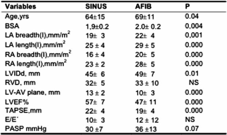
Imaging/diagnostics
69
A0274
Characterization of the cardiogenic impedance waveform measured with chronic pacemaker leads in heart failure patients
Fredrik Gadler1, Karlheinz Seidl2, Georg Noelker3, Wolfgang Kranig4, Johan Brandt5, Nils Holmström6, Johannes Sperzel7
1Karolinska University Hospital, Stockholm, Sweden, 2Klinikum der Stadt, Herzzentrum Ludwigshafen am Rhein, Germany, 3Klinikum Coburg, Coburg, Germany, 4Hertzzentrum, Bad Rothenfelde, Germany, 5Lund University Hospital, Sweden, 6St Jude Medical AB, Sweden, 7Kerckhoff Klinik, Bad Nauheim, Germany
Continuous intracardiac impedance signals can be used to monitor cardiac contractions. These cardiogenic impedance (CI) signals can be acquired using a research tool available in the Promote™ CRT-D (St. Jude Medical). The objective of this study was to characterize the CI signals during different hemodynamics.
Cardiac resynchronization systems were implanted in 27 heart failure patients with LBBB (NYHA class II–IV). Impedance data were collected at two occasions, 1–4 weeks apart, 20 weeks after the implantation and acquired in different body positions. Atrial triggered biventricular pacing with AV- and VV-delays recommended by QuickOpt™ timing optimization was used.
CI was measured between several electrode configurations using quadrapolar impedance (current injection and voltage sensing between separate electrodes) or bipolar (current and voltage sharing electrodes). The data were analyzed with respect to amplitude and morphology changes. Characteristic impedance templates of the heart cycles at different conditions were used to compare the waveforms.
The peak-to-peak amplitude from RA ring to LV ring bipolar CI signal was smaller for non-ischemic (17±10 ohm) than for ischemic patients (28±17 ohm) (p = 0.02). The systolic slope of the same signal was also smaller for the non-ischemic (−130±85 ohm/s) than for the ischemic patients (−265±191 ohm/s) (p = 0.01). Patients with hypertrophic cardiomyopathy had higher RV tip to ring bipolar slope (685±1060 ohm/s) than non-hypertrophic (260±184 ohm/s) patients (p = 0.03). Lead location in the coronary vein influenced the signal amplitude and morphology for RV to LV quadrapolar and RA ring to LV ring bipolar CI signals.
CI signals provided mechanical information of time intervals, amplitude and systolic slope related to the heart contractions. Ischemic patients displayed greater RA to LV impedance and systolic impedance slopes than non-ischemic. Patients with hypertrophy tended to have greater RV tip to ring systolic impedance slopes. The four CI vectors were stable in time but differed substantially across patients.
Imaging/diagnostics
70
A0275
Do intracardiac impedance parameters reveal mechanical asynchrony in heart failure patients?
Fredrik Gadler1, Karlheinz Seidl2, Georg Noelker3, Wolfgang Kranig4, Johan Brandt5, Nils Holmström6, Johannes Sperzel7
1Karolinska University Hospital, Stockholm, Sweden, 2Klinikum der Stadt, Herzzentrum Ludwigshafen am Rhein, Germany, 3Klinikum Coburg, Coburg, Germany, 4Hertzzentrum, Bad Rothenfelde, Germany, 5Lund University Hospital, Sweden, 6St Jude Medical AB, Sweden, 7Kerckhoff Klinik, Bad Nauheim, Germany
Continuous intracardiac impedance signals can be used to monitor cardiac contraction. These cardiogenic impedance (CI) signals can be acquired using a research tool available in the Promote™ CRT-D (St. Jude Medical). The study objective was to evaluate changes of CI-parameters that automatically are calculated from the CI-signal.
Methods. CRT systems were implanted in 27 HF patients with LBBB (NYHA class II–IV). CI-data were collected at two occasions, 1–4 weeks apart, 20 weeks after implantation. Data were acquired in supine position at atrial-triggered biventricular pacing (BiV) and RV-only pacing (RVP) with QuickOpt™ settings. Impedance was measured between several electrode configurations using quadrapolar (current injection and voltage sensing between separate electrodes) or bipolar (current and voltage sharing electrodes) impedance. Two CI-parameters were studied; “fractionation” (FR), which is the normalized curve length between two R-waves, and “diastolic dispersion” (DD), which is the waveform elevation during diastole. The changes of the CI-parameters were compared to echocardiographic references at BiV and RV.
Results. When BiV was compared to RVP, hemodynamics improved significantly, as indicated by the echocardiographic variables and CI parameters below. Echo: Systolic LV Diameter 2.2%; Diastolic LV Diameter 2.0%; Systolic LV Volume 2.0%; Diastolic LV Volume 2.2%; Aortic minus Pulmonary Pre-ejection time 78%; LV dP/dt (max) 44%; Mitral Regurgitant Area 17%; Time from QRS to peak systolic movement of posterior wall 12%; and Time from QRS peak to E-wave 4.1%. CI parameters: RA to RV Quadrapolar DD 14%; RV tip to ring Bipolar FR 9% and DD 15%; RA ring to LV ring Bipolar FR 5% and DD 11%. Other configurations did not change significantly.
Conclusion. The change of hemodynamics between synchronized BiV- and RV-only pacing was significant for both echo variables and CI parameters. The data suggest that CI parameters may be used for interventricular timing optimization or pacing site optimization.
Imaging/diagnostics
71
A0314
Feasibility to Assess Pulmonary Blood Density using Magnetic Resonance Imaging
Mikael Kanski1, Håkan Arheden1, Martin Ugander1, Roger Hesselstrand2
1Avdelningen för Klinisk Fysiologi, 2Avdelningen för Reumatologi
Background. Systemic sclerosis (SSc) patients have an increased risk of developing pulmonary arterial hypertension (PAH). The assessment of PAH lacks reliable non-invasive measures. Therefore, the aim of this study was to use MRI to measure the pulmonary blood volume (PBV), the PBV variation (PBVV), and the pulmonary blood density (PBD) in early-stage SSc and to make a preliminary comparison to healthy individuals.
Material and methods. Thirty-nine SSc patients (25 women, mean 58 years) and 14 healthy individuals (three women, mean 26 years) underwent cardiac MRI. PBV was calculated as the product of cardiac output determined by velocity encoded MRI, and the pulmonary transit time (PTT) determined as the time for a 2 ml contrast bolus to pass from the pulmonary trunk to the left atrium. The pulmonary volume (PV) was determined by planimetry using transverse MR images covering the lungs. The PBD was defined as PBV/PV. Also, the blood flow in the pulmonary artery and the pulmonary veins was measured using velocity encoded MRI. The PBVV was calculated by integration of the difference in arterial and venous flow over the cardiac cycle. PBV and PBD was measured in four healthy individuals (all men, mean 31 years). In 10 healthy individuals (three women, mean 24 years), the PBVV was assessed.
Results. Stroke volume and PBV in 36 SSc patients and four healthy subjects were respectively (mean±SD) 77±20ml vs 98±12ml, 467±111ml vs 640±194ml. PBD was 17±5% vs 20±5%. PBVV/SV in 32 SSc patients and 10 healthy individuals was 40±8% vs 49±21%.
Conclusions. This study shows the feasibility to assess the pulmonary blood density using MRI. Preliminary data shows no difference between newly diagnosed SSc patients and healthy individuals considering PBD, nor PBVV. The PBD may be a prognostic measure of PAH. Further studies are needed to assess the usefulness of the PBD.
Imaging/diagnostics
72
A0321
The potential additive value from combining contrast echo and tissue Doppler during dobutamine stress echohcardiography
Georgios Vavilis1, Kambiz Shahgaldi1, Aristomenis Manouras1, Jacek Nowak1, Reidar Winter1, Lars-Åke Brodin2
1Karolinska Unversity Hospital, Huddinge, 2School of Health and Technology, KTH syd
Background. Contrast echo (CE) and tissue Doppler (TD) derived strain and strain rate analysis are both well validated for dobutamine stress echocardiography (DSE). CE has better signal-to-noise ratio in the apical region, while on the other hand TD is more feasible in the mid-basal regions due to angular problems and the relative lack of apical movement.
The aim of this study was to reveal a possible regional superiority for CE in detection of apical ischemia and a corresponding better accuracy for TD in detecting ischemia in the mid-basal segments.
Material and methods. 120 patients with coronary angiogram and DSE including both full contrast and tissue Doppler data were identified from clinical DSE studies. 16 patients were fitted in to a model with three groups with distinguishable apical ischemia (6 pts), mid-basal ischemia (4 pts) or no ischemia (6 pts), according to the coronary angiogram and a total of 288 segments were analysed using CE and TD.
A segment was considered ischemic when there was a detectable wall motion defect during stress using CE analysis or a post/peak systolic strain ratio >35% ( ps/ max) or presence of detectable post systolic strain rate (PSSR)
Results. CE was superior in comparison to TD for detection of apical ischemia while the opposite was true for mid-basal segments.
Apical sensitivity, specificity and accuracy was 69–89–80%, 64–42–53% and 53–31–44% for CE, PSSR and ps/ max respectively. For mid-basal segments the corresponding sensitivity, specificity and accuracy was 58–100–90%, 92–91–91%, and 80–91–88%.
Conclusions. CE and TD seems to have overlapping regional accuracy i.e. CE seems to more accurate in the apical region while TD is more accurate in mid-basal segments. Thus, it could be useful to combine these techniques during DSE. Larger clinical studies are needed to confirm these results before implementing a combined approach in clinical routine.
Imaging/diagnostics
73
A0325
Are measurements of systolic myocardial velocities and displacement with colour- and spectral TD compatible?
Evangelia Nyktari1, Arben Shala2, Reidar Winter2, Jacek Nowak2, Kambiz Shahgaldi3, Reidar Winter3, Kambiz Shahgaldi4, Lars- Åke Brodin4
1Cardiology Department, Heraklion University Hospital, Stavrakia, Crete, Greece, 2Department of Clinical Physiology, Karolinska University Hospital in Huddinge, 3Department of Cardiology, Karolinska University Hospital in Huddinge, 4School for Technology and Health, Royal Institute of Technology, Flemingsberg, Stockholm, Sweden
Background. Tissue Doppler echocardiography is today an established non-invasive method of quantification in cardiac ultrasound. Tissue Doppler in pulsed mode (pulsed/spectral TD) and colour TD are the two TVE modalities today available. We sought to further investigate the relation between these two modalities based on the distinct methodological background of each method.
Methods. In this study, left ventricular longitudinal systolic velocities and displacement during the ejection phase was quantified in 24 healthy individuals patients (4 women and 20 men, 34±12 years) using colour TD with high frame rate (270±28Hz) on unfiltered data as well as spectral TD. Measurements were performed at the basal septal and lateral wall. Spectral calculations were performed according to a calibration method recently proposed where the outer and inner spectral borders of the velocity signal were time integrated and maximal minimal and mean tissue velocities and displacement were obtained.
Results. Colour- and spectral TD measurements using the mean velocities were highly concordant. The mean difference between the two methods for velocity estimation was near to zero at both sites of mitral annulus: −0.1±0.54 cm/sec for the septal wall and −0.09±0.97 cm/sec for the lateral wall). Additionally, even when measuring displacement the two modalities showed good agreement: −0.22±0.74mm for the septal and 0.27±1.31mm for the lateral wall. Furthermore, the limits of agreement between the two modalities were smaller than the limits of interobserver variability for each modality and particularly for spectral TD.
Conclusion. When considering the physical principles of both colour- and spectral TD the two methods show good agreement. This finding in conjunction with the higher interobserver variability of spectral TD measurements as documented in this and in previous studies suggests that the use of colour TD when performing TVE may be preferable.
Imaging/diagnostics
74
A0353
Feasibility and accuracy of 3-dimensional echocardiography for measurements of the right ventricular volumes and function
Ellen Ostenfeld1, Marcus Carlsson2, Johan Holm3
1Kardiologiska Kliniken, Universitetssjukhuset MAS, 2Hjärt-MR gruppen, Avdelning för Klinisk Fysiologi, Universitetssjukhuset i Lund, 3Kardiologiska Kliniken, Universitetssjukhuset i Lund
Background. The size and function of the right ventricle (RV) are intricate to assess in an exact manner, even though they have great diagnostic, therapeutical and prognostic value. The complex irregular structure of the RV is non-suitable for simple algorithms for calculating volumes and function. A large panel of different 1- and 2-dimensional echocardiographic (2DE) modalities has instead been used as estimata. We examined the accuracy and feasibility of 3-dimensional echocardiography (3DE) for the RV volumes and function compared to magnetic resonanse imaging (MRI).
Methods. 29 patients referred for 2DE on clinical indication were examined with 3DE and MRI. The x4-transducer was used for 3DE (Sonos 7500, Philips, USA). The 3DE data were interpreted with Tomtec 4D-RV Analysis 1.0 © (Unterschlessheim, Germany). A 1,5 T MRI scanner (Philips Intera, Netherlands) was used and MRI-images were analysed using Segment 1.699d. The quality of the 3DE images were graded as non-feasible, poor, fair or good, depending on endocardial border detection.
Results. With 3DE, 5 patients were non-feasible for examination, 10 had poor acoustic window, 11 were fairly visualized and only 3 had good acoustic window. The correlation between 3DE and MRI was fair for end-diastolic (EDV) (r = 0.70; P < 0.05) and end-systolic volumes (ESV) (r = 0.80; P < 0.05) and moderate for ejection fraction (EF) (r = 0.66; P < 0.05). When excluding patients with poor acoustic window, the accuracy (EDV and ESV computing r = 0.83 and 0.95, respectively; P < 0.05) and EF (r = 0.78; P < 0.05) was better.
Conclusion. 3DE is moderately to fairly accurate for RV volume and functional measurements. The accuracy increases from fair to good when excluding those with poor acoustic window. This exclusion on the other hand decreases the feasibility of the 3DE technique for assessing the RV.
Nursing science
75
A0261
Content in nurses’ counselling of hypertensive patients after consultation training
Eva Drevenhorn1, Ann Bengtson2, Karin I Kjellgren2
1Region Skåne, primärvård, 2Sahlgrenska akademin, Göteborgs universitet
Background. Nurses in hypertension care have an important role in minimising risk factors for cardiovascular diseases, but the care can be improved. The aim of this study was to explore the content of nurses’ consultations with hypertensive patients before and after consultation training.
Material and methods. Nine-teen nurses from a randomised study of nurse-led hypertension clinics at health centres received three days residential training in patient-centred counselling and cardiovascular prevention. To assess the result two consultations with hypertensive patients in clinical practice before and after the training were audio-recorded. Content analysis was used for the analysis.
Results. Diet and exercise were the most frequent topics in the consultations both before and after the training. Discussions about alcohol and patient's responsibility for treatment increased after the training (Figure). Time spent for talk on various issues, other health problems, history and appointment scheduling decreased in the consultations after the training.
Conclusion. After the consultation training the nurses succeeded in emphasizing important issues for risk factor control to a greater extent.
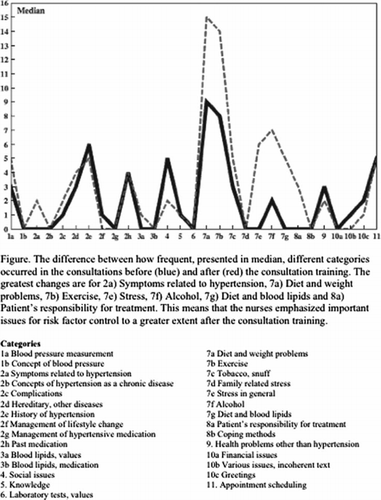
Nursing science
76
A0286
Quality from patients perspective – a patient questionnaire at Hälsoenheten
Vesna Stefan1, Anna-Karin Bryder1
1Hälsoenheten, Heart and Lung Center, Lund University Hospital
Background. The Hälsoenheten is a secondary prevention outpatient unit at Heart and Lung Center, Lund University Hospital. Patients referred to the unit have been treated with PCI (percutaneous coronary intervention) or CABG (coronary bypass operation) and/or have had a coronary infarction. During 2006 there were 460 new patients and the follow- up time for each patient was one year.
The staff is organized in teams consisting of nurses, doctors, secretaries, physiotherapists, psychologists, and social welfare officers.
The purpose of this study was to evaluate if the unit had met the treatment goals from the patients’ perspective and a process perspective.
Material and methods. Hälsoenheten had during the year of 2006 approximately 3000 patient visits and 2000 patient phone calls. During the years of 2004 and 2006 patients were asked to fill in a questionnaire using a follow-up evaluation software named KUPP ( KUPP is a questionnaire which measures quality from the patients perspective). In total 234 patients answered the questionnaire using a computer platform at the unit directly following the consultation.
Results
Patients
found that nurses and doctors were able to form a correct opinion of their needs.
found that the support they received from the staff helped them to understand the meaning of lifestyle changes, and how they could help themselves to change it.
found that they were treated with respect and that they had great opportunities to influence their care.
were pleased in how easy they could reach the nurse responsible for their care.
Conclusion. The results from a questionnaire (KUPP) to the patients supported that the treatment goals were reached at our department.
Nursing science
77
A0287
Travel to work as daily physical activity. An exploratory study
Background. The travel to/from work is potentially the most important opportunity for everyday physical activity for people with sedentary occupations. In Sweden on average 29% of all travel to/from work is by foot or bicycle. The potential is probable much higher.
Method. To test this conjuncture, a questionnaire about travel habits was directed to a particular category of people who can see the ill-effects of obesity and heart-diseases where lack of physical may have contributed.
A sample of 61 respondents were asked to record all her/his travel to/from work during four days.
Results. A basic division of the responders was between those living in town, that is the built up area of Linköping municipality (73%) and those living out of town (23%). In the latter group, 60% of the work trips were made by car and 40% by public transport and consequently no trip by foot or bicycle. In the former larger group the choice of travel mode is given in the table.
Plats För Tabell
It was also found that in the subcategory of work trips involving also leaving/fetching children at nurseries, the bicycle share was still 57%. Women are somewhat keener bicyclists than men.
Conclusion. The difference made by living within walking/bicycling distance from your workplace is striking. If Swedes in general could be as health- conscious as health professionals a great contributor to public health would be obtained.

Other
78
A0244
Cardiovascular disease in Hodgkin lymphoma long term survivors
Anne Andersson1, Beatrice Malmer1, Ulf Näslund2, Björn Tavelin3, Gunilla Enblad4, Anita Gustavsson5
1Institutionen för strålningsvetenskaper, Onkologi, Norrlands Universitetssjukhus, 2Institutionen för kardiologi, Hjärtcentrum, Norrlands Universitetssjukhus, 3Onkologiskt Centrum, Umeå, 4Institutionen för onkologi, radiologi och immunologi, Sektionen för onkologi, Uppsala Universitet, 5Institutionen för onkologi, Lunds Universitetssjukhus
Background. Previous studies have shown increased cardiovascular mortality as late side effects after treatment of Hodgkins lymphoma (HL), mainly due to the treatment given. This study investigated the risk for being admitted to hospital for cardiovascular disease (CVD) in HL survivors in Sweden, and stratified the cohort on family history of first degree relatives (FDR) with CVD, to identify subgroups that could benefit for surveillance.
Material and methods. HL patients diagnosed between 1965 and 1995 (n = 6.946) and their first-degree relatives (N =17 858) were identified through the Swedish Cancer Registry and the Swedish Multi Generation Registry. For the HL and FDR cohort, in-patient care for CVD was registered through the Hospital Discharge Registry. Standard incidence ratio (SIR) of developing CVD for the HL cohort was calculated and the cohort was stratified on positive or negative family history of CVD.
Results. A markedly increased risk for in-patient care of CVD was observed in HL patients with HL diagnosed at age 40 years or younger and with more than 10 years follow-up. The risk was especially increased in the cohort with more than 20 years follow up. The risk for coronary artery disease was SIR 3.06 (95% CI 2.41–3.83) for the whole HL cohort after 20 years of follow up and for the HL kohort with a family history of coronary artery disease SIR 5.53 (95% CI 3.89–7.62).
Conclusions. The present study shows a markedly increased risk for CVD in HL survivors overall. Family history of CVD gives an even greater risk. The Swedish Hodgkin lymphoma intervention and prevention study (SHIP) is ongoing with the aim of preventing late side effects after treatment of HL where co-morbid factors and family history is built into the risk model. The concept of the study will be presented.
Other
79
A0256
Is Gender Always Reported in Blood Pressure Research?
Anna Hofsten1, Anna Hofsten2, Helander Ingrid3
1Högskolan i Gävle, 2Inst för Folkhälso- och Vårdvetenskap- Allmänmedicin – Uppsala universitet, 3Medicinska biblioteket – Uppsala universitet
Background. Blood pressure research is an extensive, expensive and profitable activity. There are physiological, anatomical and pathological differences between men and women and it is therefore of importance to see how gender is reported in the scientific abstracts.
Methods. The PubMed database was searched for journal articles indexed with the MeSH terms “blood pressure” and not “female” or “male” with publication date 1980–2007 (I) and 2000–2007 (II). Only journal articles with abstracts, in English and on humans were accepted, reviews were not included. To find the genus neutral words which had been used the latest 80 abstracts without “female” or “male” were read (III).
Results.
I – Articles published 1980–2007
The literature search gave us 63 322 articles, 9 823 (16%) of these were not registered as female or male.
II – Articles published 2000–2007
The literature search gave us 22 374 articles, 2 741 (12%) of these were not registered as female or male.
III – Gender neutral words used instead of male or female
In the latest published abstracts (071001–071231) the words used instead of male or female were:
patients (22)
subjects (7)
volunteers/participants/individuals (8)
and respondents, family members, children, neonates, Indians and divers (1 each).
Some articles were wrongfully classified by the National Library of Medicine.
Conclusion. A large proportion of articles in blood pressure research does not describe gender, but a small tendency of becoming more precise can be seen over time.
It is very important to be correct and informative in a short abstract. The National Library of Medicine has the possibility to fill in a code for male and/or female to every journal article, but this must be described by the authors. We have an old non informative medical tradition of using gender neutral descriptions, as patients or subjects; this must come to an end.
Other
80
A0292
Heart patients self-experienced health before and after physical rehabilitation
Katarina Ferm1, Henri von Eggers Patron1
1HLD Usil
Title. Heart patients self-experienced health before and after physical rehabilitation
Background. Coronary disease is one of our big widespread diseases. Every year about 10 000 new individuals is estimated to become ill. Physical training within heart-rehabilitation lowers mortality rates. The aim of this study was to measure how the patients estimated their physical and mental health before and after physical rehabilitation.
Methods and Materials. Patients under 70 years, with the diagnosis myocardial infarction and/or who underwent CABG-operation were offered physical rehabilitation in a group, lead by a physiotherapist.
The rehabilitation took part twice a week for six weeks. During the training-sessions patients underwent regular training such as cycling, weight-lifting/aerobics and relaxing but also alternative training such as Nordic walking, water-aerobics and they also were invited to try a class at the nearest trainingcenter. Before the first, and after the last session, the patients estimated their physical and mental health in two different QoL-questionnaires, EQ5D and SF-36.
Patients with difficulties in understanding the Swedish language was excluded from the study.
Results. 70 patients that participated in the physical rehabilitation answered the questionnaires. The results showed that the patients self-estimated health increased after six weeks of training. In the SF-36 questionnaire the heart patients estimated their health equal to or better than the normal population in seven of eight parameters. At VAS in EQ5D the estimations rose from initial 72,3 to 83,7. In the other five dimensions that EQ5D measured it was an improvement in four. The fifth, hygiene, the patients didn't experience any problems before or after the intervention.
Conclusion. The patients that participated in the physical rehabilitation program experienced an improvement in their health condition after the training period. Some parameters were rated higher than the normal population.
Other
81
A0344
Adverse reactions to statin treatment in a usual care setting
Marie-Louise Ovesjö1, Ilona Skilving2, Mats Eriksson3, Anders Rane1
1Karolinska Universitetssjuhuset, avdelningen för klinisk farmakologi, Stockholm, 2Karolinska Trial Alliance, Stockholm, 3Karolinska Universitetssjuhuset, avdelningen för Endokrin-Metabol-Diabetes, Stockholm
Background. The safety profile of statin therapy has been reassuring in all the large trials. Yet many clinicians have the impression that muscular symptoms are more common, and cause termination of treatment more often than has been previously reported. Given this situation, we have started a prospective study of adverse effects (AEs) and other aspects of statin treatment in a usual care setting.
Material and methods. This ongoing study is an open clinical trial with a pragmatic study design among out-patients in primary health and specialist care. We have recruited 18 centers in Stockholm County. The patients are monitored for 12 months. Their symptoms have been assessed by a structured questionnaire and standardized interviews. Here, only the AEs that have been assessed as related to statin therapy have been included.
Results. Until now, 160 patients have been included; 82 (51%) women (mean age 64 yrs), 78 (49%) men (mean age 58 yrs). Among these, 17 individuals have reported one or several AEs during statin therapy. In the AE group 14 (82%) were women (mean age 71 yrs) and 3 (18%) were men (mean age 57 yrs). There are 11 reports of muscular pain or weakness, 3 skin reactions, 5 gastrointestinal symptoms and 1 elevation of transaminases. Among the patients with muscular symptoms, 2 continued with the same drug and dose, 1 reduced the statin dose, 4 switched to another statin and 3 stopped statin treatment.
Conclusion. In this ongoing study about 10 percent of the patients have reported AEs during statin treatment. The percentage of women is higher in the AE group than in the entire study population. In most patients with AEs, treatment adjustments have been made. These results do not explain the large discontinuation rate in statin therapy previously reported.
Surgery
82
A0331
Percutaneous tracheotomy in cardiothoracic patients; A safe method which in our ICU shows no increase of procedure related complications
Eva Schmidtke1, Barbro Österlund1
1Thorax, Hjärtcentrum, NUS
Background. Percutaneous tracheotomy has been performed by anesthesiologists in the cardiothoracic ICU at Norrlands University Hospital since 1993. This technique is considered equivalent to conventional surgical technique, regarding the amount of complications and infections. In a retrospective study, we wanted to evaluate the frequency of procedure related complications in our department.
Material and method. Tracheotomy was performed in 255 patients from 1993 to 2007.
All medical files were studied regarding; percutaneous or open surgical technique, complications related to the procedure, late complications (mediastinitis), severity of patient illness (multiple organ failure or dialysis) and if the patient was discharged from the ICU.
Two anesthesiologists are required for the procedure. The tracheal tube is withdrawn to a position which does not jeopardize the airway but enables the procedure. The trachea is punctured between the two first tracheal rings and a guide wire is introduced. A stiff catheter is passed over the wire. This catheter is used as guide, first to dilate the tracheal puncture and second to place a tracheostomy tube in the trachea. The correct position of the devices is continuously controlled with bronchoscopy.
Results. In our material percutaneous tracheotomy was performed in 211 patients and conventional tracheotomy in 42 patients. In the group with percutaneous tracheotomies 7 were converted to open surgery, due to bleeding and difficulties to identify anatomical landmarks. None of these tracheotomies were associated with serious complications.
We found 22 cases that required surgery due to mediastinitis. Only 6 patients had the percutaneous tracheotomy performed before and the rest after undergoing this second operation.
Conclusion. In our cardiothoracic ICU, percutaneous tracheotomy is a safe method with few complications. The surgical trauma is smaller than with conventional tracheotomy and we found no increased risk of mediastinitis related to the procedure. Using the method adds logistical advantages as it is performed in the ICU by anesthesiologists.
Vascular
83
A0238
Carotid intima-media thickness and apolipoproteinB / apolipoprotein A-I ratio in middle aged patients with type 2 diabetes
Carl Johan Östgren1,2, Elsa Dahlén2, Toste Länne2, Jan Engvall2, Torbjörn Lindström2, Fredrik Nyström2
1Forsknings- och utvecklingsenheten för Närsjukvården i Östergötland, 2Institutionen för medicin och hälsa, Linköpings universitet
Background. The role for the apolipoproteinB (apoB)/apolipoproteinA-I (apoA-I) ratio as a first line risk assessment tool in predicting the future cardiovascular risk is currently under debate. Thus, the aim of this study was to explore the association between subclinical atherosclerosis, measured as carotid Intima-Media Thickness (IMT) and the apoB/apoA-I ratio compared with conventional lipids and other risk factors including glycaemic control, blood pressure and inflammation, in middle aged patients with type 2 diabetes.
Material and methods. We analysed data from 247 patients with type 2 diabetes, aged 55–66 years, in the CARDIPP 1 study. Primary care nurses measured blood pressure and anthropometrics. Blood samples were taken for laboratory analyses. The carotid IMT was evaluated by ultrasonography at the University Hospital in Linköping and at the County Hospital Ryhov, Jönköping, Sweden.
Results. ApoB/apoA-I ratio (r = 0,207, p = 0.001), apoB (r = 0.166, p = 0.009) and non HDL-cholesterol (nonHDL-c) (r = 0.129, p = 0.046) correlated with IMT. Conventional lipids, high sensitive CRP (hsCRP), HbA1c and systolic blood pressure were not significantly correlated to IMT. A stepwise logistic regression analysis was conducted with IMT as the dependent variable and apoB/apoA-I ratio, HbA1c, hsCRP, LDL-c, total cholesterol, nonHDL-c and treatment with statins as independent variables. Following adjustment for age and gender, only the apoB/apoA-I ratio remained significantly associated with IMT (OR 4.3 95% CI 1.7–10.8, p = 0.002).
Conclusion. We conclude that, in contrast to conventional lipids, there was a significant association between the apoB/apoA-I ratio and subclinical atherosclerosis, measured as IMT, in middle aged patients with type 2 diabetes. The association was independent of conventional lipids, hsCRP, glycaemic control and use of statins.
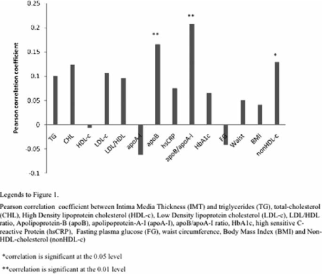
Vascular
84
A0251
Circadian blood pressure variation in patients with type 2 diabetes – relationship between dipper status and measures of arterial stiffness and left ventricular mass
Pär E:son Jennersjö1, Magnus Wijkman1, Toste Länne1, Jan Engvall1, Fredrik Nyström1, Carl Johan Östgren1, Ann-Britt Wiréhn2
1Institutionen för medicin och hälsa, Linköpings universitet, 2Forsknings- och utvecklingsenheten för Närsjukvården i Östergötland
Background. The role for diurnal blood pressure pattern as cardiovascular risk assessment tool in clinical practice is unclear. The aim of this study was to explore the association between nocturnal blood pressure dipper status and measures of arterial stiffness and left ventricular mass in patients with type 2 diabetes.
Material and methods. We analysed data from 414 patients with type 2 diabetes, aged 55–66 years, in the ongoing observational CARDIPP 1 study. Blood samples were taken for analyses of serum lipids and HbA1c. Nurses measured office blood pressure (mean values of 3 measurements in sitting position) and ambulatory blood pressure during twenty-four hours. Left ventricular mass index (LVMI) was determined with echocardiography and aortic pulse wave velocity (PWV) was measured with applanation tonometry over the carotid and femoral arteries.
Results. Dippers were defined as a ≥10% nocturnal reduction in systolic blood pressure. We identified 264 dippers and 150 subjects with a nocturnal non-dipping pattern. There were no differences in office systolic blood pressure (138±16 vs 139±16) or office diastolic blood pressure (80±10 vs 81±11) between dippers and non-dippers, respectively. Non-dippers had higher PWV (11±2.2 vs 10±2.2, p = 0.002) and increased LVMI (127±29 vs 119±29, p = 0.02) compared to dippers. When exploring the strength of the association between dipper status and PWV and LVMI, respectively, in a linear regression adjusted for gender, age, HbA1c, office systolic blood pressure and serum creatinin, the results remained significant for PWV (p = 0.004) and LVMI (p = 0.025).
Conclusion. We conclude that a non-dipping pattern in nocturnal blood pressure was associated with increased arterial stiffness and increased LVMI in middle aged patients with type 2 diabetes. The association was independent of office systolic blood pressure and serum creatinin.
Vascular
85
A0268
A novel prototype-based segmentation for MR angiography which requires only five training cases
Jane Sjögren1, Martin Ugander1, Håkan Arheden1, Einar Heiberg1, Jane Sjögren2, Einar Heiberg2
1Cardiac MR Group, Dept. of Clinical Physiology, Lund University Hospital, Lund, 2Medviso AB, Lund
Background. Image segmentation is an important pre-processing step in medical imaging. In order to obtain accurate segmentation, a priori information often needs to be used. Existing methods often construct a statistical model which typically requires 50–100 manually segmented cases.
The purpose of this work was to develop and assess the accuracy of a novel prototype-based segmentation method where only about five manual segmentations are needed to introduce a priori information.
Methods. Ten healthy volunteers underwent contrast enhanced MR imaging of the aorta at 1.5T(Philips). Imaging employed a steady state free precession sequence, resolution 1.6x1.6x1 mm.
The novel prototype-based segmentation algorithm uses spatial a priori information to restrict the segmentation rather than to govern what to include. A priori information was extracted from five of the cases (training set) and stored into a prototype.
The remaining five cases were used as test set and segmentation of the aorta was done by using the prototype and a level set method. The algorithm was implemented in the cardiac image analysis software Segment, http://segment.heiberg.se/. In the test set the segmentation error was calculated as both a volumetric error and a mean absolute distance between the manual delineation and the prototype-based segmentation.
Results. The segmentation error in the test set was 4±5% (mean±SD) when measured as volumetric error and 0.58±0.06 mm when measured as the error in mean absolute distance. The figure shows 3D surface rendering of the results of the proposed prototype-based segmentation (left) compared to the results of a purely intensity based segmentation (right).
Conclusions. The proposed segmentation algorithm is highly accurate and the segmentations are successfully constrained to only include the aorta. The prototype-based segmentation method only needs five training cases to extract the necessary a priori information and therefore a new prototype can easily be constructed for a new application.
Vascular
86
A0298
Screening for abdominal aortic aneurysm in men in the County of Östergötland, 2007 & 2008
Marie Jonsson1, Pia Bjällander-Stenmark2, Carl-Magnus Arvidsson1, Fredrik Lundgren3
1Fysilologiska kliniken NSÖ, LiÖ, 2Fysiologiska kliniken Hjärtcentrum, LiÖ, 3Thorax-/kärlkirurgen hjärtcentrum, LiÖ
Background. In a recent report (The Swedish Council on Technology Assessment in Health Care, SBU Alert, report 2008–04) SBU has unanimously recommended screening of 65-year-old men for abdominal aortic aneurysm. It was concluded that aneurysm screening reduces aneurysm related mortality, is cost-effective and ethically defensible.
Abdominal aneurysm screening has been extensively studied and discussed in Europe, Australia and United States of America in the late 20th and first part of the 21st Centuries. It's an accepted and recommended technique to reduce mortality due to aneurysm rupture in men in several European Countries and the United States.
Purpose. To evaluate the first two years of abdominal aneurysm screening in the County of Östergötland with respect to yield of aneurysms and compliance.
Method. We used a mobile ultrasound equipment. When the aortic diameter was >= 30 mm the aorta was defined as aneurysmatic. Subjects with an aneurysm were offered an appointment with a vascular surgeon within 14 days. At this appointment they were further informed and offered an operation if the aneurysm was large and regular ultrasoundsurveillance if the aneurysm was small. The screenings in Norrköping and Linköping took place at the departments of Clinical Physiology at Vrinnevisjukhuset, Norrköping and the University Hospital, Linköping. A mobile team screened inhabitants of the rural districts in the County of Östergötland at their local care centre.
Results. During 2007, 2270 65-year-old men were invited for screening and 2070 accepted the invitation and was actually investigated, giving a compliance of 91 percent. During 2008, 2698 65-year-old men and 1110, 70-year-old men were invited and 2430 and 1026 attended, giving a compliance of 90 and 92 percent. Overall compliance was 91 percent. In total 121 aneurysm were found with sizes between 30 and 76 mm (2,2% of the investigated subjects).
Vascular
87
A0332
Soft Tissue Discrimination ex vivo by Dual Energy Computed Tomography
Helene Zachrisson1,2, Elias Engström1, Jan Engvall1,2, Lars Wigström2, Örjan Smedby2, Anders Persson2
1Dept of Clinical Physiology, Linköping University Hospital, 2Center of Medical Image Science and Visualization1, Linköping University Hospital
Background. Compared to examination with a single photon energy, Dual Energy Computed Tomography (DECT) may provide additional information about the chemical composition of tissues. The aim of this in vitro study was to test whether combining two energies may significantly improve the detection of soft tissue components commonly present in arterial plaques.
Material and methods. Tissue samples of myocardial and psoas muscle, venous and arterial thrombus as well as fat from different locations were all scanned using a SOMATOM Definition Dual Source CT system (Siemens AG, Medical Solutions, Forchheim, Germany) with simultaneous tube voltages of 140 and 80kV. The attenuation (Hounsfield units, HU) at 80 and 140kV was measured in representative regions of interest, and the association between measured HU values and tissue types was tested with logistic regression.
Results. The combination of two energy levels (80 kV and 140 kV) significantly improved (p < 0.001) the ability to correctly classify venous thrombus vs arterial thrombus, myocardium or psoas; arterial thrombus vs myocardium or psoas; myocardium vs psoas; as well as the differentiation between fat tissue from various locations. Single energy alone was sufficient for distinguishing fat from other tissues.
Conclusion. DECT offers significantly improved in vitro differentiation between soft tissues occurring in plaques. If this corresponds to better tissue discrimination in vivo needs to be clarified in future studies.
Vascular
88
A0370
Increased pulse pressure is a marker for aortic stiffness independent of mean arterial pressure, age, sex, and other well-known cardiovascular risk factors
Lisa Bellinetto Ford1,2, Olle Melander2, Peter M Nilsson2
1Department of Medical and Health Sciences, Linköping University, Linköping, Sweden, 2Department of Clinical Sciences, Lund University, University Hospital, Malmö, Sweden
Background. This study aims to investigate if peripheral (PP) is a marker for aortic stiffness independent of several well-known cardiovascular risk factors in insulin resistent subjects.
Material and method. A cross-sectional population-based study in Malmö, Sweden, of 348 (42% men) middle-aged subjects, selected for varying degrees of insulin resistance (IR). The prevalence of IR was established by the homeostasis model assessment (HOMA). Aortic stiffness (, inversely related to arterial compliance) was measured non-invasively using echo-tracking sonography.
Results. Mean aortic stiffness was significantly higher in men compared to women (16.9 vs. 12.9; p < 0.001). Multiple linear regression analysis showed that in men PP (p < 0.001) and body mass index (BMI) (p = 0.010) are independently and positively associated with aortic stiffness, independent of mean arterial pressure (MAP). In women, PP (p = 0.001), age (p < 0.001) and HDL cholesterol (p = 0.026) showed independent associations with aortic stiffness, adjusted for MAP. In addition, aortic stiffness correlated in univariate analysis (Pearson's r) with age (r = 0.28, p < 0.001), MAP (r = 0.29, p < 0.001) and triglycerides (r = 0.17, p = 0.044) in men, and with MAP (r = 0.19, p = 0.006), carotid intima media thickness (IMT) (r = 0.19, p = 0.007), BMI (r = 0.18, p = 0.014), and triglycerides (r = 0.15, p = 0.029) in women.
Conclusions. Increased pulse pressure is a marker for aortic stiffness in subjects with insulin resistance (HOMA) independent of mean arterial pressure, age, sex, and additional well-known cardiovascular risk factors.