
 ?Mathematical formulae have been encoded as MathML and are displayed in this HTML version using MathJax in order to improve their display. Uncheck the box to turn MathJax off. This feature requires Javascript. Click on a formula to zoom.
ABSTRACT
While there are many techniques for estimating age-at-death in archaeological dogs, the pulp cavity/tooth width ratio is considered one of the most accurate methods. This study adapts this technique for application to MicroCT imaging, a non-destructive methodology that is rapidly gaining ground in faunal analysis. Mandibular first molar and upper and lower canine teeth recovered from two Late Classic-Early Postclassic period sites in the Maya highlands, Moxviquil and Tenam Puente, were imaged using a SCANCO μCT35 MicroCT scanner. The widths of tooth roots, pulp cavities, cuspal enamel thicknesses, and enamel attrition measurements were then taken using both the scanner’s post-processing system and 3D Slicer, an open-access programme designed for imaging biomedical scans and other 3D files, and pulp cavity infilling ratios were calculated to obtain an age-at-death estimates in months for each specimen. Based on these, this study presents preliminary interpretations of canid age-at-death patterns at Moxviquil and Tenam Puente, including a range of juvenile specimens from a funerary cave context at Moxviquil.
Introduction
As the first non-human animal domesticate, the domestic dog shares an extensive history with human beings resulting in a very complex, nuanced relationship that has developed over the course of more than 15,000 years (Shipman Citation2021; Valadez Azúa et al. Citation2013). The domestic dog has been recovered at archaeological sites the world over, and has fulfilled a variety of social roles and functions from ancient times to the present day (Nomokonova et al. Citation2020, after Bethke and Burtt Citation2020; Clutton-Brock Citation1995; Germonpré et al. Citation2017; Larson et al. Citation2012). Over time, archaeological investigations into the human-dog relationship have broadened our approaches to the study of archaeological domestic dogs, affording archaeologists new ways of assessing the development of this relationship as well as the roles and uses of domestic dogs in human landscapes. While recreating a complete biological profile of any given archaeological specimen, dog or otherwise, is a complex task, zooarchaeological approaches have added to their corpus analysis of domestic dog diet, common behaviours, body sizes and proportions, genetic profiles, trauma and pathologies, and evolutionary history, elucidating more about the human relationship with our earliest canine companions than ever before (Nomokonova et al. Citation2020, after Ameen et al. Citation2019; Drake et al. Citation2017; Frantz et al. Citation2016; Guiry Citation2012; Harcourt Citation1974; Janssens et al. Citation2019; Latham and Losey Citation2019; Lawler et al. Citation2016; Losey et al. Citation2011, Citation2013, Citation2014, Citation2015, Citation2017a, Citation2017b; MacKinnon Citation2010; McManus-Fry et al. Citation2018; Prassack et al. Citation2020; van Asch et al. Citation2013).
An important aspect of understanding ancient domestic dogs and their relationships with the world around them is the assessment of age-at-death estimates. Age-at-death estimates can inform archaeological interpretations related to understanding the long-term implications of the domestication process, the roles of domestic dogs in particular social settings, and the life histories of individual canid specimens (Nomokonova et al. Citation2020, 1). The most commonly employed approaches for determining canid age-at-death includes the assessment of tooth development and eruption patterns, tooth wear, cementum annuli in tooth roots, and epiphyseal fusion of long bones, with cementum annuli understood to be the most accurate method, albeit a destructive one (Landon et al. Citation1998; Mbizah, Steenkamp, and Groom Citation2016). A 2020 study conducted by Tatiana Nomokonova and her colleagues outlines a non-destructive method for estimating age-at-death using radiographic imaging to measure pulp cavity infilling, which they adapted to archaeology from the fields of zoology and wildlife management (Kershaw et al. Citation2005; Knowlton and Whittmore Citation2001; Landon et al. Citation1998; Mbizah, Steenkamp, and Groom Citation2016; Smirnov, Korytin, and Neganov Citation1985; Smirnov Citation1988; Thomson and Rose Citation1992). This method has been demonstrated to provide fairly accurate age-at-death estimates, though not quite as accurate as cementum annuli; however, its non-destructive nature makes it particularly significant to archaeology as an analytical method (Landon et al. Citation1998; Mbizah, Steenkamp, and Groom Citation2016; Nomokonova et al. Citation2020). Prior to Nomokonova et al.’s (Citation2020) study, this approach had not yet been applied to modern dogs and was rarely applied to archaeological ones, having only really been used to provide age estimates for extinct wolves and human beings (Binder, Thompson, and Valkenburgh Citation2002; Cameriere et al. Citation2015; Dehghani et al. Citation2018; De Luca et al. Citation2010; Zaher et al. Citation2011). This study by Nomokonova and colleagues (Citation2020) was the first to systematically apply non-destructive radiographic imaging to the study of age-at-death in archaeological dogs, utilising measurements from an extensive comparative collection of modern dogs, as well as archaeological dog teeth recovered from the arctic Iron Age site of Ust’-Polui in Siberia.
The present study uses microcomputed tomographic imaging (MicroCT) techniques to develop age-at-death estimates for two populations of domestic dogs from two ancient Maya sites, Tenam Puente and Moxviquil, both located in highland Chiapas, Mexico. MicroCT imaging is an important non-destructive imaging technology, which allows for the application of Nomokonova et al.’s (Citation2020) pulp cavity infill models to these canid populations, while also allowing for considerations of cuspal enamel thickness and evidence for tooth wear to compliment these measurements. The age-at-death models for these canid populations are highly significant for the interpretation of archaeological contexts at these sites. While the canid remains include numerous adult individuals, they also include a number of deciduous specimens. In particular, previous analysis of faunal remains from the site of Moxviquil (Paris et al. Citation2020) has identified large numbers of unerupted and unfused juvenile canid cusps, as well as unfused postcranial elements, within the context of a funerary cave, where the canid specimens could plausibly have been interred accompanying human burials.
Background
Domestic Dogs of Mesoamerica
It is generally understood that the first domestic dogs to arrive in Mesoamerica did so in tandem with perhaps multiple early human migrations (Valadez Azúa et al. Citation2013). These domestic dogs proliferated over time and eventually resulted in a variety of morphological and phenotypic dispositions. By the early Colonial period, historical sources from Central Mexico, particularly the Florentine Codex (Sahagún [1569] 1950-82) describe three types of domestic dog breeds: 1) the Techichi or Itzcuintli (non-specialized, coated); 2) the Xoloitzcuintli (also known as the Xolo or Mexican hairless dog (Blanco Padilla et al. Citation2009; Manin et al. Citation2018; Valadez Azúa et al. Citation2009, Citation2013); and 3) the Tlalchichi (coated, short-limbed dog, identifiable based on its achondroplasia condition), thought to be particularly associated with West Mexico (Valadez Azúa et al. Citation2000; Valadez Azúa et al. Citation2013). A fourth type, known exclusively from archaeological remains, is the short-faced dog (Blanco Padilla, Valadez Azúa, and Roderíguez Galacia Citation1999), thought to be particularly associated with the Maya area, with at least three specimens identified from the Yucatan Peninsula, and a fourth set of skeletal remains presenting a brachycephalic condition recovered at the site of Hunchavín in highland Chiapas (Valadez Azúa Citation2014). Most archaeological specimens of domestic dogs are fragmentary remains, which make it extremely difficult to identify types or breeds; identifications of morphotypes or breeds mostly derives from a limited number of offerings or funerary contexts.
Symbolically, the domestic dog was an incredibly important species in ancient Mesoamerica. Among the diverse cultures of southern Mesoamerica, dogs were historically associated with the underworld, reincarnation, and regeneration; the central Mexican deity Xolotl was represented as a dog and was associated with the underworld, death and darkness (White et al. Citation2001; Valadez Azúa and Blanco Padilla Citation2005; Valadez Azúa et al. Citation2013). Ceramic representations of the hairless Xoloitzcuintli and short-legged Tlalchichi breeds are commonly depicted in the Colima pottery tradition of West Mexico during the Classic period (3-4th centuries AD), and as funerary companions in human burial traditions at Tula during the Epiclassic period (7-8th centuries AD); (Valadez Azúa et al. Citation2000). Although domestic dogs played a variety of roles in the cities and towns of ancient Mesoamerica as hunting dogs and companions, contact-period documentary sources also allude to their sacrificial roles. Sahagún alludes to their roles related to Mexica agricultural ceremonies associated with the harvests in July (Tlaxochimaco) and November (Panquetzaliztli; Sahagún 1950–82 [1575-77]: Books 11, 16), while Durán (Citation1971, 278) describes the sacrifice of dogs for feasting events such as weddings and baptisms. The sixteenth century Tlaxcalan chronicler Diego Muñoz Camargo (Citation1986 [1892]) describes the sacrifice and subsequent consumption of Xoloitzcuintlis as a mechanism of soliciting divine intervention during droughts, and Diego de Landa describes the sacrifice of ‘virgin dogs’ in conjunction with New Years festivals, and the sacrifice of a spotted dog (perro manchado) by cacao cultivators during the month of April (Muan; Tozzer Citation1941). Archaeological findings argued to be evidence for canid sacrifices have been found at a number of sites, including Tula (Valadez Azúa et al. Citation2013) and at Xaltocan, where the sacrifice of young domestic dogs and interment in human neonatal burials is thought to relate to their symbolic role as companions for the deceased individual through the underworld (De Lucia Citation2014).
Study Area
The archaeological sample concerned by this study derives from two sites in the Maya highlands of Mesoamerica: Moxviquil and Tenam Puente. Both sites are located in the central highlands of Chiapas, Mexico, which abounds with a variety of geological resources, mountainous landscapes, and has been considered a geographic and cultural frontier zone of the Maya (Paris Citation2012). Moxviquil is located in the tierra fria ecological zone, characterised by higher altitudes above 2,000 masl, colder temperatures, and shorter growing seasons compared to neighbouring lowland areas (Paris Citation2012), while Tenam Puente is located in the tierra templada zone at approximately 1,700 masl (Paris, López Bravo, and Lalo Jacinto Citation2021). Many of the larger monumental zones in highland Chiapas enjoyed continuous occupation from the Late Classic (AD 500-900) through the Early Postclassic (AD 900-1250) periods, while numerous political capitals in many neighbouring lowland areas were abandoned between AD 800 and 900 (Paris Citation2012).
Moxviquil lies on the northern edge of the Jovel Valley at the present-day site of the colonial and modern city of San Cristóbal de las Casas (Paris Citation2012). The site is placed strategically on a defensible hilltop and likely served as an administrative centre to a small, autonomous polity during the Late Classic and Early Postclassic periods, seeing the construction of monumental architecture during the former period and the construction of its civic precinct established around AD 500 (Paris Citation2012; Paris and López Bravo Citation2020; Paris, Taladoire, and Whiting Citation2015; Paris, López Bravo, and Serafin Citation2019), around the same time this occurred at other sites in the region (Bryant Citation1988; Culbert Citation1965). Neighbouring hilltops saw the construction of newer residential settlements clustered on upper terraces and semi-terraces in AD 900, during which time the site saw significant population growth (Paris Citation2012; Paris, López Bravo, and Serafin Citation2019). Original investigations of Moxviquil were conducted in 1952 and 1953 by Frans Blom and Clarence Weiant, and new research was conducted by the Proyecto Económico de los Altos de Chiapas (PEACH, 2009; Paris Citation2012) and Proyecto Interacción Entre Reinos en los Altos de Chiapas (PIERACH, 2015) in outlying residential zones (Paris and López Bravo Citation2021a, Citation2021b; Paris, López Bravo, and Serafin Citation2019). In 1954, Blom identified a cave on the hilltop adjacent to the monumental zone that contained significant human and faunal skeletal remains; detailed excavations at the cave (Operation 7) in 2016 by Paris and López Bravo (Paris, López Bravo, and Serafin Citation2019) recovered the faunal samples included in the present study. In addition to Operation 7, a large portion of the site’s principal residential zone at the apex of the same hilltop, termed Operation 4, was excavated by the Proyecto Interacción Entre Reinos en los Altos de Chiapas in 2015 and 2016, leading to the identification of at least four residential structures and associated sub-structures positioned on artificial terraces (Paris, López Bravo, and Serafin Citation2019). Evidence suggests that the ossuary cave contained the remains of some of Moxviquil’s Operation 4 residents, though unfortunately the feature has been subjected to extensive looting and therefore reflects a likely removal of some funerary goods, damage to osteological remains, disturbance of surface levels, and potential mixing of cultural layers (Paris, López Bravo, and Serafin Citation2019). Overall, 7,000 human osteological elements, diverse funerary offerings, and a wide variety of faunal remains (N = 374) were recovered from Operation 7 (Paris and López Bravo Citation2020; Paris, López Bravo, and Serafin Citation2019; Paris et al. Citation2020). It is suggested that this ossuary cave was likely used as a communal cemetery that served the residents of the Operation 4 hilltop, probably as either a successive primary deposit or a mixture of primary and secondary deposits (Paris, López Bravo, and Serafin Citation2019). The present sample of canid teeth from Operation 7 at Moxviquil includes an NISP value of 116 Canis lupus familiaris specimens from this particular location (Paris et al. Citation2020, Table 1). Of these, 44 specimens were teeth, four of which were teeth suitable for age-at-death assessment. The four specimens included one complete canine and three complete mandibular M1 teeth; all teeth from Moxviquil included in the present study were recovered loose. Other specimens were broken or otherwise damaged, and thus removed from the sample.
The site of Tenam Puente, located on the southeast edge of Chiapas’s Comitán Plateau, is contemporaneous with Moxviquil and lies 13 kilometres southeast of the modern city of Comitán de Domínguez (Laló Jacinto and Alor Jacobo Citation1998; Paris, López Bravo, and Lalo Jacinto Citation2019; Paris, López Bravo, and Lalo Jacinto Citation2021). Evidence for initial settlement of Tenam Puente has been dated to around AD 500, with investment in monumental architecture at the site’s Acropolis and Plaza F during the Late Classic and Early Postclassic periods (Laló Jacinto and de la Luz Aguilar Citation1996; Laló Jacinto Citation2005; Earley Citation2015; Paris, López Bravo, and Lalo Jacinto Citation2021). The planned urban core of the site and earliest monuments were established during the Late Classic, and it remained occupied until around AD 1100 (Earley Citation2015, after Laló Jacinto Citation2005; Paris, López Bravo, and Lalo Jacinto Citation2021). Tenam Puente was first investigated by Eduard Seler in 1901 and again later in 1925 by Frans Blom and Oliver La Farge, with more recent investigations by the Proyecto Arqueológico Tenam Puente (Laló Jacinto Citation2005; Laló Jacinto and de la Luz Aguilar Citation1996; Laló Jacinto and Alor Jacobo Citation1998) and the Proyecto Las Redes Económicas de Tenam Puente (RETP; 2019-present; Paris, López Bravo, and Lalo Jacinto Citation2021), which was established to specifically investigate the site’s economic organisation. Moxviquil and Tenam Puente are contemporaneous in terms of their principal occupation, with continuous occupation through the Late Classic and Early Postclassic periods, and broadly similar architectural patterns where monumental zones are positioned on extensively modified hilltops with large public plazas in the saddle below. Tenam Puente is a much larger city than Moxviquil, spanning over 70 hectares including the residential zone, and while both are located in highland environments, Moxviquil is located nearly 800 m higher in elevation than Tenam Puente, at altitudes of 2,328 masl and 1,714 masl respectively. The domestic dog teeth included in the present study were excavated during the 2019 RETP field season, representing an overall NISP value of 68 canid elements. Of this, 31 specimens were either loose teeth or mandibular fragments containing teeth. For the purposes of the present study, six teeth were considered suitable for age-at-death assessment. This comprises a collection of three canine teeth and three mandibular M1 teeth, all of which were recovered loose. Other specimens were broken or otherwise damaged, including two mandibular M1s missing root tips, and a canine which was split anterior-posteriorly, and were removed from the sample.
Materials and Methods
While several methods may be used in estimating canid age-at-death, measuring the pulp cavity/tooth width ratio is one of the most common and accurate methods (Mbizah, Steenkamp, and Groom Citation2016). Although they are slightly more accurate, most current techniques for estimating cementum annuli are currently destructive (Mbizah, Steenkamp, and Groom Citation2016), which may be undesirable for archaeological specimens due to numerous historic preservation considerations (Nomokonova et al. Citation2020). Both cementum annuli and pulp cavity infill measurements are derived from the same biological principles of development, namely that dental cementum is deposited in a biphasic manner, forming annual rings (Mbizah, Steenkamp, and Groom Citation2016).
In most domestic dogs, juvenile dentition is fully in place by six weeks of age, with deciduous canines in place by the three week mark, and is shed between the ages of three and six months (Arnall Citation1960; Lobprise and Dodd Citation2019: Table 4). Adult dentition then follows, with hard-tissue formation of all permanent teeth occurring within a narrow timeframe of approximately eight weeks starting shortly after birth. While the timing of tooth emergence varies between dog breeds, it is typically estimated from three to four months for incisors, four to six months for canines and premolars, and mandibular M1 crowns erupting at five to seven months of age, with some overlap between the first molar and fourth premolar to be expected (Arnall Citation1960; Hale Citation2005; Lobprise and Dodd Citation2019: Table 4; Shipman Citation2021). At eruption, the pulp cavity of the tooth is very wide, as only primary dentine is present. During the remainder of the tooth’s life, secondary dentine is laid down circumferentially around the pulp and therefore the pulp chamber infills, decreasing in size over time (Mbizah, Steenkamp, and Groom Citation2016). Unlike bone, dentin, or cementum, there is no process of remodelling or repair after enamel formation has been completed, and disruption in its formation leaves a permanent lesion in the ensuing tissue, such as an enamel hypoplasia or enamel hypomineralization (Hillson and Bond Citation1997).
The present study examines a sample of mandibular first molars, maxillary canines, and mandibular canines for pulp cavity assessment following Nomokonova and colleagues’ (Citation2020) study, and enamel thickness assessments following Crossley (Citation1995) for cuspal enamel categorisation. While canine teeth are generally understood to provide the better age-at-death estimate, mandibular first molars are typically better represented in archaeological faunal assemblages and are often recovered more intact than underrepresented and more fragile canines (Nomokonova et al. Citation2020, 3). As such, the present study analyzes both, with the dataset consisting of six mandibular first molars and four canine teeth recovered from Moxviquil and Tenam Puente. It is worth noting here that the small dataset considered by the present study is a product of challenges related to the COVID-19 pandemic, and thus reflects a much smaller dataset than the present study anticipated undertaking.
The present study applies MicroCT techniques to obtain age-at-death estimates for archaeological domestic dogs using pulp cavity infill ratios and enamel thickness. Since becoming commercially available, MicroCT techniques are gaining ground fast in many disciplines on account of their ability to provide very detailed, valuable anatomical and functional information while at the same time standing as a non-destructive, quantitative, fully three-dimensional analysis option (Clark and Badea Citation2021; Guldberg et al. Citation2004). MicroCT has demonstrated particular application to the study of teeth, especially in analyses of human molar morphology, general tooth structure, and relationships between crown contours and pulp chambers in first molars (Campioni, Pecci, and Bedini Citation2020). The technique also allows for accurate and non-destructive 3D mapping and associated quantifications of the mineral content of scanned samples, the measurement of tooth surface areas, and the calculations of volumes and thicknesses of tooth enamel, dentin, and pulp chambers (Davis, Evershed, and Mills Citation2013; Grande et al. Citation2012; Swain and Xue Citation2009). Previous applications of MicroCT imaging to investigate pulp cavity morphology in human teeth have found it useful to utilise post-processing procedures and model manipulation to make the internal pulp chamber and root canal systems of teeth opaque and dental hard tissues transparent, allowing for excellent visualisation of the internal systems (Bjøndal et al. Citation1999). Although MicroCT imaging techniques are relatively new in zooarchaeology, they provide a rapid and non-destructive way to investigate microstructures within faunal dentition, opening up possibilities to investigate new types of research questions, including the growth and development of ancient domestic dogs.
The MicroCT images used in this study were generated at the Hallgrimsson Lab at the University of Calgary’s Foothills Medical Centre using a SCANCO μCT35 MicroCT desktop cone-beam shielded cabinet scanner. Samples were wrapped in protective foam and placed inside a 36 mm tube, which is fitted into the scanner. The scanner then rotates the tube and takes two-dimensional projection images of both internal and external features. These images can then be viewed as individual slices or compiled into manipulable three-dimensional models of the entire tooth. Measurements were then taken using 3D Slicer, an open-access image computing software designed for use with medical, biomedical, and other three-dimensional data. Projection image TIFFs generated by the scanner software were imported into 3D Slicer as image stacks, which were cropped and converted into NRRD files from which a manipulable 3D model can be rendered, while still displaying individual slices, so that the correct slice can be selected for measurement.
Measurements were taken on the slices following methods outlined by Nomokonova et al. (Citation2020) and Mbizah and colleagues (Citation2016) using the line and angle tools. Measurements taken in Slicer automatically scale according to file metadata, but it is worth noting that utilising this or similar programmes necessitates researchers check that voxel spacing and other relevant variables have been imported from the file correctly before beginning analysis; they may be corrected manually in the programme. The lengths of lines and degrees of angles are displayed in 3D Slicer in real time and the zoom functionality is optimised, allowing points to be accurately placed and modified as needed.
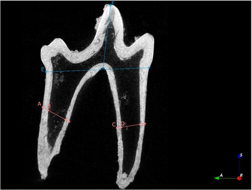
On pulp cavity features, measurements were taken following procedures developed by Nomokonova et al. (Citation2020). Using 3D Slicer, an image slice was selected for measurement along the sagittal plane where the widest point of the pulp cavity appeared to be best reflected. Widths of upper and lower canines and their pulp cavities were measured from the cementoenamel junction (CEJ) at the distal end of the tooth, extending across the tooth at a 90-degree angle relative to the pulp cavity margin (). On mandibular M1 teeth, distal and mesial roots were measured separately at the midpoint between the CEJ and root tip (), following methods established by Nomokonova and colleagues (Citation2020: Figure 3). Samples that were too broken, cracked, or were missing pieces pivotal to this assessment were dropped from the dataset. Two of the specimens were pendant ornaments with drilled perforations through the root. Specimen 181–2 had a perforation through the root that did not impact the CEJ; specimen 230–1 was a pendant ornament with a drilled perforation through the distal root, so only the medial pulp cavity was measured.
Figure 1. Measurements for crown height on canines, shown as A) tooth width at CEJ; B) total pulp cavity width; and C) crown height.
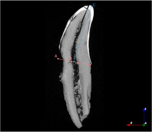
Figure 2. Measurements for pulp cavity closure ratios on mandibular M1s: A) width of the distal root; B) width of the pulp cavity for the distal root; C) width of the pulp cavity for the mesial root; D) width of the mesial root; E) width of the crown at CEJ; and F) crown height.
Measurements taken from the scans in 3D slicer were used to develop bivariate plots. Pulp cavity closure ratios were compared to exponential regression equations from derived values calculated from Nomokonova et al. (Citation2020, Figures 4–7) for age-at-death in modern domestic dogs based on a sample of 751 modern dogs with known life histories. Nomokonova and colleague’s (Citation2020) study established pulp cavity infill ratios for four different assays: upper canines, lower canines, distal roots of mandibular M1, and mesial roots of mandibular M1 based on a dataset of dogs aged between six and 72 months. Exponential regression equations derived from the modern dog teeth were used to estimate age-at-death for the highland Chiapas specimens.
Nomokonova et al. (Citation2020: Table 3) note that the difference in pulp cavity closure between left versus right elements from the same specimen are extremely similar, with a difference range between 0.00 and 0.05 for upper and lower canines, 0.00 and 0.06 for the mandibular M1 distal root, and between 0.00-0.09 for the mandibular M1 mesial root; thus, despite the fragmentary nature of the archaeological assemblage, individual teeth are highly likely to be representative of the specimen age-at-death. Unfortunately, due to the small number of specimens deemed suitable for age-at-death assessment, the fact that the majority of specimens represent isolated teeth, and a disproportionate ratio of right to left teeth in the present sample, a comparison of left and right teeth was not undertaken for this study.
Several considerations and caveats were made based on methodological observations by Nomokonova et al. (Citation2020). They caution that the first molar is not considered as accurate as canine teeth in determining age-at-death from pulp cavity closure ratios due to odontogenetic factors. They also specify that pulp cavity closure ratios more precisely estimate age in juveniles and young adult individuals, and is less accurate in adult and older adult individuals (Nomokonova et al. Citation2020). Mbizah, Steenkamp, and Groom (Citation2016) also provide equations and measurements for the canines, and do not provide equivalent methodologies for the mandibular M1 in African wild dogs, suggesting also that canine data is more accurate with respect to interpretive models for age estimation.
Tooth wear was assessed via enamel thickness measurements (). Enamel thickness measurements were taken in 3D Slicer using the slice view along the sagittal plane of the tooth (). For both mandibular M1 and canine teeth, enamel thickness was measured by orienting the measurement from the crown tip to the interior margin of enamel following methods by Mbizah, Steenkamp, and Groom (Citation2016: Figure 2). Measurements were then ranked on an ordinal scale from 1 to 4, using categories previously determined by Crossley (Citation1995; see ).
Figure 3. Measurement for enamel thickness categorisation.
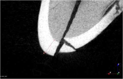
Table 1. Cuspal enamel thickness categories following Crossley (Citation1995).
Once measurements were taken, age-at-death estimates were calculated using the following exponential regression formulas derived from published data by Nomokonova et al. (Citation2020: Figures 4–7; see Figures 5–8). While the derived equations provide useful age-at-death estimates in months, we also acknowledge the uncertainty of these estimates, particularly in older specimens (R-squared values for canines fall around 0.74), so we also used the estimates to classify individuals as juveniles, young adults, prime adults, and senior adults (see ), combining ordinal age categories used by Nomokonova et al. (Citation2020: Table 4) and Mbizah, Steenkamp, and Groom (Citation2016: Figure 12). However, we emphasise that ‘juvenile’ individuals in this context are typically between 5 and 11 months of age, but represented by permanent teeth with fully-developed roots. In the following equations, y is the age-at-death estimate in months, and x is the pulp cavity closure ratio.
Table 2. Age ranges and corresponding categories developed for the purposes of the present study, adapted from Nomokonova et al. Citation2020. In domestic dogs, canines erupt between four and six months, while mandibular M1 crowns erupting at five to seven months of age (Hale Citation2005; Lobprise and Dodd Citation2019). Note that specimens classified as ‘juveniles’ constitute permanent teeth with fully-developed roots, but are estimated to be under 12 months in age.
We suggest that age estimates that consider both pulp cavity ratios and enamel thickness may thus be more accurate than either measure alone. In the present assessment of age-at-death, measurements for pulp cavity closure ratios and enamel thickness were examined using a bivariate plot with a trendline for the negative linear relationship between these factors for canines. This allows for a thoughtful interpretation of individual specimens with unexpectedly thick or thin enamel for particular pulp-cavity closures. Although enamel can be worn down through attrition through everyday hunting and feeding behaviour, enamel thickness can also be affected by other behavioural factors. For example, animals in captivity may exhibit enamel attrition due to stress behaviours such as gnawing restraints or cages (Sugiyama, Somerville, and Schoeninger Citation2015). However, biplot comparisons may not be possible when crown enamel is cracked or chipped, which is unfortunately quite common in archaeological contexts.
We also considered the potential to utilise age estimate equations for enamel thickness developed by Landon et al. (Citation1998) for gray wolves, and by Mbizah, Steenkamp, and Groom (Citation2016: Figure 10) for African wild dogs. Unfortunately, comparisons between these equations and the derived regression formulas suggest that the former equations cannot be used due to the radically different proportions and enamel thickness for these species. We hope that future studies will develop models for age estimates from enamel attrition in domestic dogs.
Results
The results of the present study () provide metric data for pulp cavity closure ratios and enamel attrition in highland Chiapas domestic dogs, allowing us to interpret age-and-death for these specimens. The measurements suggest a diverse set of age categories were represented in this dataset, with the youngest individual estimated at four months in age and the oldest estimated at 35 months in age. As such, preliminary results for age-at-death estimates in the Moxviquil and Tenam Puente samples suggest a significant representation of young dogs. Moxviquil samples included three juveniles and a prime adult, while Tenam Puente’s samples include two juveniles, two young adults, and two prime adults (, , ).
Figure 4. Bivariate plot of pulp cavity closure ratio to enamel thickness for the Moxviquil and Tenam Puente specimens. A linear trendline for the maxillary canine specimens is shown. The three specimens encircled in orange represent likely juvenile specimens from Moxviquil with unusually low pulp cavity closure ratios and unusually thin enamel.
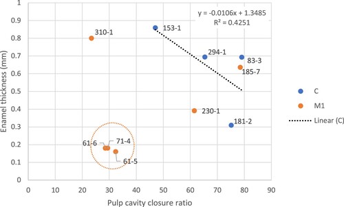
Figure 5. Linear regression of maxillary canine pulp cavity closure ratios to age in months of Moxviquil and Tenam Puente specimens.
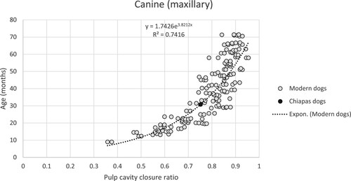
Figure 6. Linear regression of mandibular canine pulp cavity closure ratios to age in months of Moxviquil and Tenam Puente specimens.
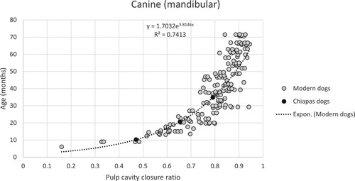
Figure 7. Linear regression of the mesial root of mandibular first molar pulp cavity closure ratios to age in months of Moxviquil and Tenam Puente specimens.
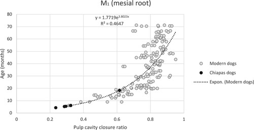
Figure 8. Linear regression of the distal root of mandibular first molar pulp cavity closure ratios to age in months of Moxviquil and Tenam Puente specimens.
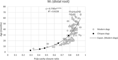
Table 3. Pulp cavity closure ratios and enamel thickness measurements for Moxviquil (PIERACH) and Tenam Puente (RETP) dogs with associated age-at-death. Pulp cavity ratios were calculated using the methods illustrated in and , while enamel thickness was calculated using the methods illustrated in . Pulp cavity age estimates were calculated using the regression equations calculated from Nomokonova et al. Citation2020: Figures 4–7. Cuspal enamel thickness categories follow Crossley Citation1995 (see ). Age categories are assigned by categories outlined in .
Some specimens (61-5, 61-6, 71–4 and 310-1) have very low pulp cavity ratios, to the extent that the regression equations are likely not correctly estimating the ages for particularly young specimens (cf. Nomokonova et al. Citation2020). The mandibular M1 teeth typically erupt between 5 and 7 months, whereas for Specimen 310–1 the mandibular M1 age estimate was 4.3 months, which is inconsistent with a fully-developed M1 permanent tooth. This is reflected in the low R-squared values for the regression lines for the M1 roots. There are several possible contributing factors for this: first, there is high variance in the samples for the M1 pulp cavity ratios; and second, the sample of modern dogs contains relatively few specimens that are 10 months or younger, and thus the archaeological dogs from highland Chiapas are likely typically younger than the majority of the modern dogs used to calculate the regression lines.
In an effort to derive a useful equation for the relationship between pulp cavity closure ratios and enamel thickness, we calculated a linear regression equation for the canines using the archaeological specimens from highland Chiapas, since canines were identified as most accurately predicting age-at death (Nomokonova et al. Citation2020; R2 = 0.74). The line has a negative slope, reflecting attrition over time:
Specimen 153–1 was interpreted as a juvenile, with a low pulp cavity ratio and relatively thick enamel; Specimen 294–1 was interpreted as a young adult, and its pulp cavity ratio and moderate enamel are consistent with a young adult of approximately 21 months (). Prime adult specimens 83–3 and 181–2 did not fit the linear model particularly well; although both had similar pulp cavity ratios, specimen 83–3 had relatively thick enamel, while specimen 181–2 had extremely thin enamel. The thin enamel of specimen 181–2 could have been due to a type of physical restraint that induced gnawing behaviour. Alternatively, specimen 181–2 has a perforation in the root and a thin crack along the length of the crown, which may be affecting our enamel measurements. M1 specimens were also considered with respect to the line, although the specimens for this group were not linearly distributed. Specimen 310–1 was interpreted as a juvenile with low pulp cavity ratios and relatively thick enamel; Specimens 185–7 and 230–1 had relatively thick and thin enamel for their pulp cavity measurements, respectively. The thin enamel of Specimen 230–1 could also have been due to some type of physical restraint; however, like Specimen 181-2, Specimen 230–1 also had a perforated root, and a thin crack along the length of the crown, which may be affecting our enamel measurement.
Three of the Moxviquil specimens were classified as juvenile mandibular M1 specimens. They had very low pulp cavity ratios (0.3–0.5), and also had significantly thinner enamel than most specimens. The teeth had fully developed roots, but were also the smallest teeth with regard to their overall dimensions. This suggests that these specimens were juveniles between 5 and 11 months old, but also raises the possibility of miniaturised breeds.
Discussion
The use of MicroCT imaging adds to the range of non-destructive analytical techniques available for the estimation of age-at-death for archaeological dog specimens. As Nomokonova et al. (Citation2020) and others (Landon et al. Citation1998; Mbizah, Steenkamp, and Groom Citation2016; Van den Broek et al. Citation2022) have shown, calculations of canid age-at-death using pulp cavity closure ratios are fairly accurate, particularly in canines, and can be used to characterise specimens as juveniles (6-11 months), young adults (12-23 months), prime adults (24-60 months), and older adults (60 + months). MicroCT provides a methodological advantage in facilitating non-destructive enamel thickness calculations in conjunction with the pulp cavity ratios, providing secondary data that can refine age estimates and elucidate behavioural and environmental aspects of a particular specimen’s biography.
These discoveries corroborate previous findings from taxonomic and morphological analysis of the Moxviquil funerary cave assemblage, which identified a range of young domestic dog specimens through the documentation of deciduous tooth specimens and unerupted premolar and molar crowns, indicating a significant proportion of juvenile individuals younger than seven months of age (Paris et al. Citation2020). As demonstrated in , three out of four specimens from the Moxviquil sample examined for this study represent juveniles between 5 and 11 months of age and a fourth prime adult individual estimated at around 35 months. This further corroborates our previous hypothesis that many canid specimens recovered at the site were relatively young at death, but other dogs lived into adulthood. Notably, these individuals were recovered from the site’s funerary cave as isolated specimens in conjunction with a diverse range of human and faunal elements, and the domestic dog specimens may be associated with funerary activities in some way (Paris, López Bravo, and Serafin Citation2019). In particular, specimen 83–3 was recovered from the lower strata of a cist burial feature near the cave entrance, while the other three specimens were recovered from the rear of the cave (Paris, López Bravo, and Serafin Citation2019). Additionally, as we have previously reported, the funerary cave assemblage also included a number of younger juvenile dog specimens identified through the presence of isolated, unerupted mandibular PM2, mandibular M1 and maxillary M1 crowns, identified by their lack of roots and their papery texture (Paris et al. Citation2020, 54). Permanent mandibular PM2 teeth erupt at four to six months old, while mandibular M1 crowns erupt at five to seven months old (see Lobprise and Dodd Citation2019: Table 4.1), suggesting that many of Moxviquil’s juvenile dogs were between five and seven months old. The benefit of MicroCT approaches from the present study is that in addition to these younger juvenile dogs, we can now identify juveniles in the 5–11 month range and young adults in the 12–24 month range as well. Identifying 5–11 month juveniles is typically not possible to do using traditional taxonomic techniques, as the phenotypic appearance of the teeth is quite similar to those of young and prime adults. This further adds to the number of young dogs interred in the funerary cave and provides further detail about the wide range of ages-at-death, which apparently included dogs of all ages.
The pattern at Tenam Puente may have been slightly different, and the specimens in this study were attributed to a wide range of ages. Specimens from Tenam Puente were also recovered from more varied contexts at that site and were nearly all dated to the Middle Classic period (Paris, López Bravo, and Lalo Jacinto Citation2021, Table 1). Specimen 153–1 was recovered from fill dated to AD 421–585 beneath the final plaza surface, excavated from a test pit associated with a house located on the southern end of Ballcourt 1. Specimens 310-1, 294-1, and 181–2 were recovered from fill associated with the northwest side of Plaza F-East’s stela platform (Pozo 6); radiocarbon dates associated with the stratigraphic levels for 310–1 and 294–1 were AD 533-629, and a radiocarbon date associated with 181–2 suggest the latter dates from AD 551–643 (Paris, López Bravo, and Lalo Jacinto Citation2021, Table 1). Finally, specimen 185–7 was recovered above the final plaza surface from a midden located to the west of (behind) Tenam Puente’s Structure 50, associated with the western side of Plaza F-West, also identified as this site’s central marketplace (Paris, López Bravo, and Lalo Jacinto Citation2021, Table 1). This specimen is dated slightly later in time than the other specimens, between AD 763 and 889 (Paris, López Bravo, and Lalo Jacinto Citation2021, Table 1). With the exception of specimen 153-1, which likely represents a juvenile, most specimens from Tenam Puente represented young or prime adults. Overall, the specimens from Tenam Puente represent greater variability in their age-at-death estimates than the Moxviquil sample.
Valadez Azúa and Blanco Padilla (Citation2005) have previously suggested that ancient Mesoamericans linked the reproductive cycles of domestic dogs with the agricultural cycles of maize production and the onset of the sub-tropical rainy season, and have argued that some dogs were sacrificed in accordance with yearly agricultural cycle festivals. However, given the range of ages represented in the Moxviquil funerary cave, this seems unlikely, as the sample includes dogs of 4–6 months, 5–7 months, 6–11 months, and 24–48 months, which do not reflect a standard prescribed age-at-death. On the other hand, as a funerary context, these juvenile specimens could more likely have been sacrificed as companions in death; for example, at the Late Postclassic-period household contexts at Xaltocan, puppies were deliberately interred with human neonatal burials (De Lucia Citation2014). The highly disturbed deposits of the Moxviquil funerary cave make this possibility difficult to assess; however, the juvenile dog specimens could plausibly have been interred with any number of juvenile human remains recovered from that deposit (Paris, López Bravo, and Serafin Citation2019).
The present study identified some further areas for extra care or awareness of limitations in this method. The use of MicroCT as opposed to more commercially available radiographic imaging, while presenting immense benefits in the realms of non-destructive high-resolution imaging, has some limitations due to the potential high cost and low accessibility to researchers more broadly. The present study benefits greatly from interdisciplinary collaboration, but we recognise that this is not always available. For this reason, the present study encourages interdisciplinary collaboration wherever possible.
3D Slicer presents its own limitations, including its ability to deal with very large data files – while it is capable, files over two gigabytes will often crash the programme, though this is dependent on the capabilities of the computer being used. One possible solution is to load scans from TIFF image stacks, and to crop large files by selecting a region of interest. It is important to carefully take measurements at the correct location, as features such as the CEJ can be difficult to identify on CT slices. When taking measurements on slices, the researcher needs to carefully select which internal slice to use, which presents some risk for operator error. However, saved measurements in 3D slicer can allow multiple analysts to review the selected measurements, consult with each other, and refine them as needed, which has been our practice on this project. Because slice views can be suboptimal if specimens were not loaded into the scanners appropriately, it is sometimes necessary to improve slice visibility through transformations of the data in post-processing to correctly orient the slices. These features are currently difficult to operate, but we hope that the active and helpful online forums of developers and users will work towards improvements in the associated modules.
Conclusion
The present study provides further validation that pulp cavity closure ratios of canine and mandibular first molar teeth can be used to assess age-at-death in archaeological dogs. Additionally, this study adapts existing methods for measuring these features to the use of MicroCT imaging, which has the added benefit of providing a non-destructive and increasingly available method for obtaining and producing extremely high-resolution images. The results presented here apply MicroCT imaging for estimation of pulp cavity closure ratios and enamel thickness measurements to archaeological domestic dog teeth from the Maya sites of Moxviquil and Tenam Puente in highland Chiapas. Our initial findings corroborate previous evidence that the specimens from Moxviquil’s funerary cave included a significant portion of juvenile individuals, and that the age-at-death for the majority of canids at both sites was between four months and three years of age.
Acknowledgments
The Proyecto Económico de los Altos Chiapas, Proyecto Interacción Entre Reinos en los Altos de Chiapas, Proyecto Las Redes Económicas de Tenam Puente, and Proyecto Tenam Puente were carried out with the permission of the Consejo de Arqueología, Instituto Nacional de Antropología e Historia (INAH). Funding for the Proyecto Económico de los Altos Chiapas for the 2009 season was provided by a National Science Foundation Doctoral Dissertation Improvement Grant (Award ID 0836590) to Paris; support for the Proyecto Interacción Entre Reinos en los Altos de Chiapas for the 2015 season was provided by a Wenner-Gren Foundation International Collaborative Research Grant to Paris and Lopez Bravo. We also thank Universidad de Ciencias y Artes de Chiapas, Centro INAH-Chiapas, ProNatura Chiapas, the Museo Na Bolom and the New World Archaeological Foundation for their support during numerous phases of the original field research. Funding for the Proyecto Las Redes Económicas de Tenam Puente was provided by the Social Science and Humanities Research Council of Canada (SSHRC ref.: 430-2018-00629), the University of Calgary (URGC SSH Faculty Seed Grant RSO Number: 1045964), and the Universidad de Ciencias y Artes de Chiapas, and also thank the community of Ejido Francisco Sarabia. We thank the University of Calgary, particularly the Hallgrimsson Lab, Department of Cell Biology and Anatomy, and the Department of Anthropology and Archaeology for the use of the laboratory equipment and facilities associated with the MicroCT scanning and taxonomic analysis.
Disclosure Statement
No potential conflict of interest was reported by the author(s).
Additional information
Funding
Notes on contributors
Miranda J. George
Miranda George holds a B.Sc in Archaeology from the University of Calgary, at which she is currently an M.A. student in the Department of Anthropology and Archaeology. Her research focuses on zooarchaeological methods, animal agency and network theory, and ancient Maya domestic dogs. Her Master's project is a part of the Proyecto Económico de los Altos Chiapas, Proyecto Interacción Entre Reinos en los Altos de Chiapas, Proyecto Las Redes Económicas de Tenam Puente, and Proyecto Tenam Puente under the supervisión of Dr. Elizabeth H. Paris and in collaboration with Dr. Roberto López Bravo and Ma. Gabriel Laló Jacinto. She is also a consulting field archaeologist with Stantec Consulting's Alberta archaeology office. [email protected].
Elizabeth H. Paris
Elizabeth H. Paris is an archaeologist with a B.A. in Anthropology from the University of Colorado-Boulder and an M.A. and Ph.D. in Anthropology from the University at Albany, SUNY. She is an Associate Professor in the Department of Anthropology and Archaeology at the University of Calgary. Her research focuses on the archaeology of urbanism, craft production, exchange, household archaeology, and materials analysis. She currently directs the Las Redes Económicas de Tenam Puente Project in collaboration with Dr. Roberto López Bravo and Ma. Gabriel Laló Jacinto, and has previously directed archaeological field research in the Jovel Valley of highland Chiapas in collaboration with Dr. Roberto López Bravo. She also collaborates on materials analysis at Mayapan and other sites. Her projects have received external support from the Social Sciences and Humanities Research Council of Canada, the National Science Foundation, the Wenner Gren Foundation, National Geographic, Dumbarton Oaks, and others. She has published over 25 peer-reviewed scientific publications, including numerous collaborations with colleagues and students. [email protected].
Wei Liu
Wei Liu is the MicroCT lab manager at the Hallgrimsson Lab at the University of Calgary's Cumming School of Medicine. He completed his studies at Harbin Medical University before moving to Canada to pursue Software Engineering at the University of Calgary. He specialized in Micro Computed Tomography (MicroCT), and joined the Hallgrimsson Lab in 2005 where he manages and maintains multiple MicroCT systems. Wei has extensive knowledge in 3D X-ray MicroCT scanning, data collection, and deep learning image registration approaches for automated landmarking.
Roberto López Bravo
Roberto López Bravo is a Licenciado en Arqueología from the Escuela Nacional de Antropología e Historia and a Ph.D. in Anthropology from the University of Pittsburgh. He is a tenured profesor at the Escuela de Arqueología of the Universidad de Ciencias y Artes de Chiapas, México. His research focuses on political organization, household archaeology, and exchange in northeast and central Chiapas. He directs archaeological field research in Chiapas at Tenam Puente, in the Jovel Valley, in the Central Depression, and in the Palenque region. He is a Level 1 National Researcher of the Sistema Nacional de Investigadores de México. His projects have received external support from the Wenner Gren Foundation, PIFI/SEP, and the Consejo de Ciencia y Tecnología del Estado de Chiapas. He has published numerous peer-reviewed scientific publications, including numerous collaborations with colleagues and students. [email protected]
Gabriel Lalo Jacinto
Gabriel Laló Jacinto is a Licenciado en Antropología with a specialization in Archaeology from the Universidad Veracruzana, and a M.A. in Estudios Mesoaméricanos from the Faculty of Philosophy and Letters (FFyL), from the Universidad Nacional Autónoma de México (UNAM). He is a Profesor Investigador Titular B at Centro INAH Chiapas and Profesor de Asignatura at the Universidad Intercultural de Chiapas, Las Margaritas. He is the INAH site director of the archaeological sites of Tenam Puente, Chinkultic y Lagartero. He is the director of the Proyecto Tenam Puente and co-director of the Las Redes Económicas de Tenam Puente Project with Dr. Elizabeth Paris and Dr. Roberto López Bravo, as well as numerous projects for the cultural resource management of Mexican national patrimony. As well as published articles and book chapters, he has organized numerous conferences and public talks on archaeological and ethnographic field research, heritage, and cultural patrimony. [email protected].
References
- Ameen, Carly, Tatiana R. Feuerborn, Sarah K. Brown, Anna Linderholm, Ardern Hulme-Beaman, Ophélie Lebrasseur, et al. 2019. “Specialized Sledge Dogs Accompanied Inuit Dispersal Across the North American Arctic.” Proceedings of the Royal Society B, Biological Sciences 286 (1916): 20191929
- Arnall, L. 1960. “Some Aspects of Dental Development in the Dog—II. Eruption and Extrusion.” Journal of Small Animal Practice 1:259–267. https://doi.org/10.1111/j.1748-5827.1960.tb06100.x.
- Bethke, Brandi and Amanda Burtt, eds. 2020. Dogs: Archaeology Beyond Domestication. Gainesville: University Press of Florida.
- Binder, Wendy J., Elicia N. Thompson, and Blaire Van Valkenburgh. 2002. “Temporal Variation in Tooth Fracture among Rancho La Brea Dire Wolves.” Journal of Vertebrate Paleontology 22 (2): 423–428. https://doi.org/10.1671/0272-4634(2002)022[0423:TVITFA]2.0.CO;2.
- Bjøndal, L., O. Carlsen, G. Thuesen, T. Darvann, and S. Kreiborg. 1999. “External and Internal Macromorphology in 3D-Reconstructed Maxillary Molars Using Computerized X-ray Microtomography.” International Endodontic Journal 32:3–9. https://doi.org/10.1046/j.1365-2591.1999.00172.x.
- Blanco Padilla, Alicia, Bernardo Roderíguez, and Raúl Valadez Azúa. 2009. El Estudio de los Cánidos Arqueológicos del México Prehispánico. Instituto de Investigaciones Antropológicos- Instituto Nacional de Antropología e Historia, México.
- Blanco Padilla, Alica, Raúl Valadez Azúa, and B. Roderíguez Galacia. 1999. “Colección Arqueozoológica de Perros del Sitio de Chac-Mool, Punta Pájaros, Quintana Roo.” Arqueología 22:89–106.
- Bryant, Douglas Donne. 1988. Archaeology, ethnohistory, and ethnoarchaeology in the Maya Highlands of Chiapas, Mexico. Papers of the New World Archaeological Foundation No. 54-56. Provo, Utah: Brigham Young University.
- Cameriere, R., S. De Luca, N. Egidi, M. Bacaloni, P. Maponi, L. Ferrante, and M. Cingolani. 2015. “Automatic age Estimation in Adults by Analysis of Canine Pulp/Tooth Ratio: Preliminary Results.” Journal of Forensic Radiology 3:61–66.
- Campioni, Ilaria, Raffaella Pecci, and Rossella Bedini. 2020. “Ten Years of Micro-CT in Dentistry and Maxillofacial Surgery: A Literature Overview.” Applied Sciences 10 (12): 4328. https://doi.org/10.3390/app10124328.
- Clark, D. P., and C. T. Badea. 2021. “Advances in Micro-CT Imaging of Small Animals.” Physica Medica 88:175–192. https://doi.org/10.1016/j.ejmp.2021.07.005.
- Clutton-Brock, Juliet. 1995. “Origins of the Dog: Domestication and Early History.” In The Domestic Dog: Its Evolution, Behaviour and Interactions with People, edited by James Serpell, 7-20. Cambridge: Cambridge University Press.
- Crossley, David A. 1995. “Tooth Enamel Thickness in the Mature Dentition of Domestic Dogs and Cats – Preliminary Study.” Journal of Veterinary Dentistry 12 (3): 111–113. https://doi.org/10.1177/089875649501200302.
- Culbert, T. Patrick. 1965. The Ceramic History of the Central Highlands of Chiapas, Mexico. Papers of the New World Archaeological Foundation No. 19. Provo: Brigham Young University.
- Davis, Graham R., Anthony N. Z. Evershed, and David Mills. 2013. “Quantitative High Contrast X-ray Microtomography for Dental Research.” Journal of Dentistry 41:475–482. https://doi.org/10.1016/j.jdent.2013.01.010.
- Dehghani, Mahdieh, Elaheh Shadkam, Farzaneh Ahrari, and Mahboobe Dehghani. 2018. “Age Estimation by Canines’ Pulp/Tooth Ratio in an Iranian Population Using Digital Panoramic Radiography.” Forensic Science International 285:44–49. https://doi.org/10.1016/j.forsciint.2018.01.016.
- De Luca, Stedano, Inmaculada Alemán, Francesca Bertoldi, Luigi Ferrante, Paola Mastrangelo, Mariano Cingolani, and Roberto Cameriere. 2010. “Age Estimation by Tooth/Pulp Ratio in Canines by Peri-Apical X-Rays: Reliability in age Determination of Spanish and Italian Medieval Skeletal Remains.” Journal of Archaeological Science 37:3048–3058. https://doi.org/10.1016/j.jas.2010.06.034.
- De Lucia, Kristin. 2014. “Everyday Practice and Ritual Space: The Organization of Domestic Ritual in Pre-Aztec Xaltocan, Mexico.” Cambridge Archaeological Journal 24 (3): 379–403. https://doi.org/10.1017/S0959774314000511.
- Drake, Abby Grace, Michael Coquerelle, Pavel A. Kosintsev, Olga P. Bachura, Mikhail Sablin, Andrei V. Gusev, Lacey S. Fleming, and Robert J. Losey. 2017. “Three-Dimensional Geometric Morphometric Analysis of Fossil Canid Mandibles and Skulls.” Scientific Reports 7 (1): Article 9508. https://doi.org/10.1038/s41598-017-10232-1
- Durán, Diego. 1971[1588]. Book of the Gods and Rites and the Ancient Calendar. 1st ed. Norman: University of Oklahoma Press.
- Earley, Caitlin Cargile. 2015. At the edge of the Maya world: power, politics, and identity in monuments from the Comitán Valley, Chiapas, Mexico. PhD dissertation, Department of Art History, University of Texas, Austin. https://repositories.lib.utexas.edu/handle/2152/39686.
- Germonpré, Mietje, Sergey Fedorov, Petr Danilov, Patrik Galeta, Elodie-Laure Jimenez, Mikhail Sablin, and Robert J. Losey. 2017. “Palaeolithic and Prehistoric Dogs and Pleistocene Wolves from Yakutia: Identification of Isolated Skulls.” Journal of Archaeological Science 78: 1–19. https://doi.org/10.1016/j.jas.2016.11.008
- Grande, Nicola M., Gianluca Plotino, Gianluca Gambarini, Luca Testarelli, Ferdinando D’Ambrosio, Raffaella Pecci, and Rossella Bedini. 2012. “Present and Future in the use of Micro-CT Scanner 3D Analysis for the Study of Dental and Root Canal Morphology.” Annali Dell’Istituto Superior di Sanità 48 (1): 26–34.
- Guiry, Eric J. 2012. “Dogs as Analogs in Stable Isotope-Based Human Paleodietary Reconstructions: A Review and Considerations for Future Use.” Journal of Archaeological Method Theory 19 (3): 351–376. https://doi.org/10.1007/s10816-011-9118-z.
- Guldberg, Robert E., Angela S. P. Lin, Rhima Coleman, Galen Robertson, and Craig Duvall. 2004. “Microcomputed Tomography Imaging of Skeletal Development and Growth.” Birth Defects Research Part C 72:250–259. https://doi.org/10.1002/bdrc.20016.
- Hale, Fraser A. 2005. “Juvenile Veterinary Dentistry.” Veterinary Clinics of North America: Small Animal Practice 35 (4): 789–817. https://doi.org/10.1016/j.cvsm.2005.02.003.
- Harcourt, R. A. 1974. “The Dog in Prehistoric and Early Historic Britain.” Journal of Archaeological Science 1 (2): 151–175. https://doi.org/10.1016/0305-4403(74)90040-5.
- Hillson, Simon, and Sandra Bond. 1997. “Relationship of Enamel Hypoplasia to the Pattern of Tooth Crown Growth: A Discussion.” American Journal of Physical Anthropology 104 (1): 89–103.
- Janssens, Luc August, Timothy Vitales, and Dennis Lawler. 2019. “Are Spinal Deformities in Ancient Dogs Related to Weight Bearing? The Importance of Fluctuating Asymmetry.” International Journal of Osteoarchaeology 29 (1): 168–173. https://doi.org/10.1002/oa.2720.
- Kershaw, K., L. Allen, A. Lisle, and K. Withers. 2005. “Determining the age of Adult Wild Dogs (Canis lupus Dingo, C. l. Domesticus and Their Hybrids). I. Pulp Cavity:Tooth Width Ratios.” Wildlife Research 32:581–585. https://doi.org/10.1071/WR03109.
- Knowlton, Frederick F., and Susan L. Whittmore. 2001. “Pulp Cavity – Tooth Width Ratios from Known-age and Wild-Caught Coyotes Determined by Radiography.” Wildlife Social Bulletin 29 (1): 239–244.
- Laló Jacinto, Gabriel. 2005. “Exploraciones arqueológicas en Tenam Puente, Chiapas.” In IV Coloquio Pedro Bosch Gimpera: Arqueología Mexicana, edited by Ernesto Vargas, 755–774. México: Universidad Nacional Autónoma de México, Instituto de Investigaciones Antropológicas.
- Laló Jacinto, Gabriel, and María de la Luz Aguilar. 1996. “El Postclásico Temprano en Tenam Puente.” In Quinto Foro de Arqueología de Chiapas, 23–37. Tuxtla Gutiérrez: Gobierno del Estado de Chiapas (Serie Memorias).
- Laló Jacinto, Gabriel, and Omar Alor Jacobo. 1998. “Notas del Clásico Tardío y Posclásico Temprano en Tenam Puente, Chiapas, México.” In XI Simposio de Investigaciones Arqueológicas en Guatemala, 827–836. Guatemala: Museo Nacional de Arqueología y Etnología.
- Landon, David B., Carol A. Waite, Rolf O. Peterson, and David L. Mech. 1998. “Evaluation of Age Determination Techniques for Gray Wolves.” The Journal of Wildlife Management 62 (2): 674–682. https://doi.org/10.2307/3802343.
- Laurent A. F. Franz, Victoria E. Mullin, Maud Pionnier-Capitan, Ophélie Lebrasseur, Morgane Ollivier, Angela Perri, et al. 2016. “Genomic and Archaeological Evidence Suggest a Dual Origin of Domestic Dogs.” Science (American Association for the Advancement of Science) 352 (6290): 1228–1231. https://doi.org/10.1126/science.aaf3161.
- Latham, Katherine J. and Robert J. Losey. 2019. “Spondylosis Deformans as an Indicator of Transport Activities in Archaeological Dogs: A Systematic Evaluation of Current Methods for Assessing Archaeological Specimens.” PloS One 14 (4): e0214575. https://doi.org/10.1371/journal.pone.0214575
- Larson, Greger, Elinor K. Karlsson, Angela Perri, Matthew T. Webster, Simon Y. W. Ho, Joris Peters, et al. 2012. “Rethinking Dog Domestication by Integrating Genetics, Archaeology, and Biogeography.” Proceedings of the National Academy of Sciences – PNAS 109 (23): 8878–8883. https://doi.org/10.1073/pnas.1203005109.
- Lawler, Dennis F., Chris Widga, David A. Rubin, Jennifer A. Reetz, Richard H. Evans, Basil P. Tangredi, et al. 2016. “Differential Diagnosis of Vertebral Spinous Process Deviations In Archaeological And Modern Domestic Dogs.” Journal of Archaeological Science, Reports 9: 54–63. https://doi.org/10.1016/j.jasrep.2016.06
- Lobprise, Heidi B., and Johnathon R. Dodd. 2019. Wiggs’s Veterinary Dentistry: Principles and Practice. Second edition. Hoboken: John Wiley & Sons, Inc.
- Losey, Robert J., Vladimir I. Bazaliiskii, Sandra Garvie-Lok, Mietje Germonpré, Jennifer A. Leonard, Andrew L. Allen, Anne M. Katzenberg, and Mikhail V. Sablin. 2011. “Canids as Persons: Early Neolithic Dog And Wolf Burials, Cis-Baikal, Siberia.” Journal of Anthropological Archaeology 30 (2): 174–189. https://doi.org/10.1016/j.jaa.2011.01.001.
- Losey, Robert J., Sandra Garvie-Lok, Jennifer A. Leonard, Anne M. Katzenberg, Mietje Germonpré, Tatiana Nomokonova, Mikhail V. Sablin, Olga I. Goriunova, Natalia E. Berdnikova, and Nikolai A. Savel’ev. 2013. “Burying Dogs in Ancient Cis-Baikal, Siberia: Temporal Trends and Relationships with Human Diet and Subsistence Practices.” PloS One 8 (5): e63740. https://doi.org/10.1371/journal.pone.0063740.
- Losey, R. J., E. Jessup, T. Nomokonova, and M. Sablin. 2014. “Craniomandibular Trauma and Tooth Loss in Northern Dogs and Wolves: Implications for The Archaeological Study of Dog Husbandry and Domestication.” PloS One 9 (6): e99746. https://doi.org/10.1371/journal.pone.0099746.
- Losey, R. J., B. Osipov, R. Sivakumaran, T. Nomokonova, E. V. Kovychev, and N. G. Diatchina. 2015. “Estimating Body Mass in Dogs and Wolves Using Cranial and Mandibular Dimensions: Application to Siberian Canids.” International Journal of Osteoarchaeology 25 (6): 946–959. https://doi.org/10.1002/oa.2386.
- Losey, R. J., L. S. Fleming, T. Nomokonova, A. V. Gusev, N. V. Fedorova, S. Garvie-Lok, O. Bachura, P. A. Kosintsev, and M. V. Sablin. 2017a. “Human and Dog Consumption of Fish on the Lower ob River of Siberia: Evidence for a Major Freshwater Reservoir Effect at the Ust’-Polui site”. Radiocarbon 60 (1): 239–260. https://doi.org/10.1017/RDC.2017.77.
- Losey, R. J., K. McLachlin, T. Nomokonova, K. Latham, and L. Harrington. 2017b. “Body Mass Estimates in Dogs and North American Gray Wolves Using Limb Element Dimensions.” International Journal of Osteoarchaeology 27 (2): 180–191. https://doi.org/10.1002/oa.2528.
- MacKinnon, Michael. 2010. “‘Sick as a Dog’: Zooarchaeological Evidence for Pet Dog Health and Welfare in the Roman world.” World Archaeology 42 (2): 290–309. https://doi.org/10.1080/00438241003673011
- Manin, Aurélie, Morgane Ollivier, Fabiola Bastian, Antoine Zazzo, Olivier Tombret, Juan Carlos Equihua Manrique, and Christine Lefèvre. 2018. “Can we Identify the Mexican Hairless dog in the Archaeological Record? Morphological and Genetic Insights from Tizayuca, Basin of Mexico.” Journal of Archaeological Science 98:128–136. https://doi.org/10.1016/j.jas.2018.08.008.
- Mbizah, Moreangels M., Gerhard Steenkamp, and Rosemary J. Groom. 2016. “Evaluation of the Applicability of Different Age Determination Methods for Estimating Age of the Endangered African Wild Dog (Lycaon Pictus).” PLoS One 11 (10): e0164676.
- McManus-Fry, Ellen, Rick Knecht, Keith Dobney, Michael P. Richards, and Kate Britton. 2018. "Dog-human dietary relationships in Yup’Ik western Alaska: the stable isotope and zooarchaeological evidence from pre-contact Nunalleq." Journal of Archaeological Science, Reports 17: 964–972. https://doi.org/10.1016/j.jasrep.2016.04.007.
- Muñoz Camargo, Diego, and Germán Vázquez. 1986 [1892]. Historia de Tlaxcala. 1st ed. Madrid: Historia 16.
- Nomokonova, Tatiana, Robert J. Losey, Kira McLachlin, Olga P. Bachura, Andrei V. Gusev, Pavel A. Kosintsev, Natalia V. Fedorova, and Mikhail V. Sablin. 2020. “Age Estimation of Archaeological Dogs Using Pulp Cavity Closure Ratios.” Journal of Archaeological Science 123 (105252): 1–10.
- Paris, Elizabeth H. 2012. “Political Economy on the Postclassic Western Maya Frontier.” PhD Dissertation, State University of New York at Albany, Department of Anthropology. Proquest Dissertations Publishing, 3523364.
- Paris, Elizabeth H., and Roberto López Bravo. 2020. “Gulf Coast Influence at Moxviquil, Chiapas, Mexico.” Cambridge Archaeological Journal 30 (2): 183–199. https://doi.org/10.1017/S0959774319000465.
- Paris, Elizabeth H., and Roberto López Bravo. 2021a. “Obsidian Exchange Networks in the Jovel Valley, Chiapas, Mexico: A Compositional Analysis Approach.” Journal of Archaeological Science: Reports 35 (102773).
- Paris, Elizabeth H., and Roberto López Bravo. 2021b. “6 Urban Commerce in the Jovel Valley of Highland Chiapas.” Archaeological Papers of the American Anthropological Association 32 (1): 80–94. http://doi.org/10.1111/apaa.v32.1.
- Paris, Elizabeth H., Roberto López Bravo, and Gabriel Laló Jacinto. 2019. “An Archaic Period Stemmed and Barbed Point from Tenam Puente, Chiapas, Mexico.” Archaeologia Iberoamericana 43:62–66.
- Paris, Elizabeth H., Roberto López Bravo, and Gabriel Laló Jacinto. 2021. “The Making of a Plaza: Public Spaces and Marketplaces at Tenam Puente, Chiapas, Mexico.” Estudios de Cultura Maya 58:45–83. https://doi.org/10.19130/iifl.ecm.2021.58.23862.
- Paris, Elizabeth H., Roberto López Bravo, Ellen Pacheco, and Miranda George. 2020. “Hunting, Husbandry, Exchange and Ritual: Animal use and Meaning at Moxviquil, Chiapas Mexico.” Anthropozoologica 55 (4): 43–72. https://doi.org/10.5252/anthropozoologica2020v55a4.
- Paris, Elizabeth H., Roberto López Bravo, and Stanley Serafin. 2019. “A Funerary Cave at Moxviquil, Chiapas, Mexico.” Journal of Field Archaeology 45 (2): 86–105. https://doi.org/10.1080/00934690.2019.1693091.
- Paris, Elizabeth H., Eric Taladoire, and Thomas A. Lee Whiting. 2015. “Return to Moxviquil: Form and Function in a Small Maya City.” Ancient Mesoamerica 26 (1): 81–112. https://doi.org/10.1017/S0956536115000048.
- Prassack, Kari A., Josephine DuBois, Martina Lázničková-Galetová, Mietje Germonpré, and Peter S. Ungar. 2020. “Dental Microwear as a Behavioral Proxy for Distinguishing Between Canids at the Upper Paleolithic (Gravettian) Site of Předmostí, Czech Republic.” Journal of Archaeological Science 115:105092. https://doi.org/10.1016/j.jas.2020.105092.
- Sahagún, Fray Bernardino de. 2013 [1959]. Florentine Codex: General History of the Things of New Spain, Translated from the Aztec Into English, with Notes and Illustrations. Santa Fe: N. M. School of American Research.
- Shipman, Pat. 2021. Our Oldest Companions: The Story of the First Dogs. Cambridge.: The Belknap Press of Harvard University Press.
- Smirnov, V. S. 1988. “Issledovanie Dinamiki Chidlennosti Volka s Ispol’zovaniem Dvukh Metodov Opredeliniia Vozrasta.” In Analiz Vozrastnoi Struktury Populiastii Pozvonochnykh, edited by V. S. Bezel, 114–132. Sverdlovsk.: UrO AN SSSR.
- Smirnov, V. S., N. S. Korytin, and V. G. Neganov. 1985. Kotrol za dinamikoi chislennosti volka po vozrastnomy sostavy dobyvaemykh zhivotnykh (metodicheskie rekomendatsii). Sverdlovsk.: UNTS AN SSSR.
- Sugiyama, Nawa, Andrew D. Somerville, and Margaret J. Schoeninger. 2015. “Stable Isotopes and Zooarchaeology at Teotihuacan, Mexico Reveal Earliest Evidence of Wild Carnivore Management in Mesoamerica.” PLoS One 10 (9): e0135635.
- Swain, Michael V., and Jing Xue. 2009. “State of the Art of Micro-CT Applications in Dental Research.” International Journal of Oral Science 1 (4): 177–188. https://doi.org/10.4248/IJOS09031.
- Thomson, P. C., and K. Rose. 1992. “Age Determination of Dingoes from Characteristics of Canine Teeth.” Wildlife Research 19:597–599. https://doi.org/10.1071/WR9920597.
- Tozzer, Alfred M. 1941. Landa’s Relación De Las Cosas De Yucatán. Papers of the Peabody Museum of American Archaeology and Ethnology 18. Cambridge, MA: Harvard University.
- Valadez Azúa, Raul, Alicia Blanco Padilla, Bernardo Rodríguez Galacia, and Christopher. M. Götz. 2009. “Perros pelones del México prehispánico.” Arqueobios 3 (1): 5–18.
- Valadez Azúa, Raul. 2014. “Un perro de raza desconocida de Hunchavin.” Antropológicas Boletín 3 (52): 1–11.
- Valadez Azúa, Raul, and Alicia Blanco Padilla. 2005. “Perro y maíz en el México prehispánico.” Revista AMMVEPE 16 (2): 63–70.
- Valadez Azúa, Raul, Alicia Blanco Padilla, Bernardo Rodríguez Galacia, and Gilberto Perez Roldan. 2013. “Chapter 18: The Dog in the Mexican Archaeozoological Record.” In The Archaeology of Mesoamerican Animals, no. 1, edited by Christopher M. Gotz, and Kitty F. Emery, 557–720. Atlanta.: Lockwood Press.
- Valadez Azúa, Raul, Alicia Blanco Padilla, Fernando Viniegra Rodríguez, Katiuska Olmos Jiménez, and Bernardo Rodríguez Galacia. 2000. “El tlalchichi, perro de patas cortas del occidente mesoamericano.” Revista AMMVEPE 11 (2): 49–57.
- van Asch, Barbara, Ai-Bing Zhang, Mattias C. R. Oskarsson, Cornelya F. C. Klutsch, Antonio Amorim, and Peter Savolainen. 2013. “Pre-Columbian Origins of Native American dog Breeds, with Only Limited Replacement by European Dogs, Confirmed by mtDNA Analysis.” Proceedings of the Royal Society B: Biological Sciences 1766:20131142. https://doi.org/10.1098/rspb.2013.1142.
- Van den Broek, Martine, Emmelie Stock, Yoni Vermeiren, Leen Verhaert, Luc Duchateau, and Pieter Cornillie. 2022. “Age Estimation in Young Dogs by Radiographic Assessment of the Canine Pulp Cavity/Tooth Width Ratio.” Anatomia, Histologia, Embryologia 51 (2): 269–279. https://doi.org/10.1111/ahe.12787.
- White, Christine D., Mary E. D. Pohl, Henry P. Schwarcz, and Fred J. Longstaffe. 2001. “Isotopic Evidence for Maya Patterns of Deer and Dog Use at Preclassic Colha.” Journal of Archaeological Science 28:89–107. https://doi.org/10.1006/jasc.1999.0560.
- Zaher, Jaklin Fekri, Irene Atef Fawzy, Sahar Refaat Habib, and Magdy Mohamed Ali. 2011. “Age Estimation from Pulp/Tooth Area Ratio in Maxillary Incisors among Egyptians Using Dental Radiographic Images.” Journal of Forensic and Legal Medicine 18:62–65. https://doi.org/10.1016/j.jflm.2010.12.004.