
ABSTRACT
Like the proposal of nanotechnology by Richard Feynman, the nanoarchitectonics concept was initially proposed by Masakazu Aono. The nanoarchitectonics strategy conceptually fuses nanotechnology with other research fields including organic chemistry, supramolecular chemistry, micro/nanofabrication, materials science, and bio-related sciences, and aims to produce functional materials from nanoscale components. In this review article, bio-interactive nanoarchitectonics and two-dimensional materials and environments are discussed as a selected topic. The account gives general examples of nanoarchitectonics of two-dimensional materials for energy storage, catalysis, and biomedical applications, followed by explanations of bio-related applications with two-dimensional materials such as two-dimensional biomimetic nanosheets, fullerene nanosheets, and two-dimensional assemblies of one-dimensional fullerene nanowhiskers (FNWs). The discussion on bio-interactive nanoarchitectonics in two-dimensional environments further extends to liquid–liquid interfaces such as fluorocarbon–medium interfaces and viscous liquid interfaces as new frontiers of two-dimensional environments for bio-related applications. Controlling differentiation of stem cells at fluidic liquid interfaces is also discussed. Finally, a conclusive section briefly summarizes features of bio-interactive nanoarchitectonics with two-dimensional materials and environments and discusses possible future perspectives.
Graphical
1. Introduction: background stories
1.1. From nanotechnology to nanoarchitectonics
As one of the remarkable advancements of science and technology in recent decades, nanotechnology and related research fields have attracted much attention. These approaches enable us to precisely observe, evaluate, and manipulate nanoscale objects even at molecular and atomic levels. Novel findings and discoveries made possible by exploring phenomena specifically observable in nanoscale regimes have been continuously reported [Citation1–3]. These advancements are not limited to observation and evaluation. Materials synthesis and fabrication of functional systems based on nanotechnological investigations are also being developed [Citation4–6].
As Nakamura, Harano, and co-workers recently demonstrated, sophisticated electron microscopy techniques (single-molecule atomic-resolution real-time electron microscopy, SMART-EM) enable us to observe shuttling, rotating, and interacting motions of a single fullerene (C60) molecule entrapped within a vibrating carbon nanotube in real-time with sub-angstrom resolution and sub-millisecond precision [Citation7]. The SMART-EM techniques can be used to observe single-molecule-level chemical reactions with the aid of the fishhook technique [Citation8]. Introduction of aromatic groups onto the graphitic surface of carbon nanohorns with positive and negative curvature was detected. Immobilization of molybdenum dioxo catalyst at the graphitic surfaces of carbon nanohorns was directly confirmed by the SMART-EM technique [Citation9]. The nanoarchitected catalyst showed high activities in several reactions including N-oxide reductions, polyethylene terephthalate hydrogenolysis, and reductive carbonyl coupling. Visualization of plasmon-induced reactions with single-molecule-level precision was enabled by a scanning tunnelling microscope (STM) as reported by Kazuma [Citation10]. With aid of density functional theory calculations, bond dissociation reactions caused by plasmon excitation at a nanoscale gap between a metal substrate and the STM tip with light irradiation can be analysed. Narita, Müllen, and co-workers are actively working on on-surface synthesis of large polycyclic aromatic hydrocarbon and graphene nanoribbons from well-defined molecular sources [Citation11]. This series of approaches are supported by high-resolution observation through noncontact atomic force microscopy [Citation12]. As recently demonstrated by Kawai and co-workers, coupling reactions directly mediated by small tip motion is possible; additional reactions with bromine atoms and fullerene molecules to specific surface positions using a scanning probe tip were realized with single-molecule-level precision [Citation13]. Device operation with molecular and atom-level phenomena have been demonstrated as atom switches [Citation14,Citation15] and molecular memory [Citation16,Citation17], respectively, by Aono and co-workers. In a recent review article by Yamashita, confinement of single-molecule magnets into designed nanospaces formed by single-walled carbon nanotube and metal-organic framework (MOF) is suggested for application in advanced devices such as high-density memory devices and quantum computing [Citation18].
On the other hand, methodologies based on well-established sciences such as organic chemistry [Citation19–21], supramolecular chemistry [Citation22–24], coordination chemistry [Citation22–27], bio-related sciences [Citation28–30], and other materials sciences [Citation31–33] have continuously contributed to the production of functional material systems. Undoubtedly, syntheses and fabrications of functional materials are indispensable keys to satisfy various social demands in energy [Citation34–36], environmental [Citation37–39], and biomedical targets [Citation40–42]. As seen in solar cells [Citation43–45], fuel cells [Citation46–48], batteries [Citation49–51], photocatalytic conversions [Citation52–54], and supercapacitors [Citation55–57] for energy demands, sensing/detection [Citation58–60], removal [Citation61–63], and degradation [Citation64–66] of problematic materials for environmental issues, drug delivery [Citation67–69] and various therapies [Citation70–72] in biomedical applications, control over precise structures and organizations is as important as intrinsic properties of materials themselves. Therefore, the introduction of nanotechnological fundamentals to materials science is crucial for further developments of materials science to meet recent social demands. A new paradigm and concept to couple nanotechnology and other research fields in materials science with nanostructure regulation needs to be established as a post-nanotechnology concept [Citation73]. This task is assigned to an emerging concept: nanoarchitectonics [Citation74,Citation75].
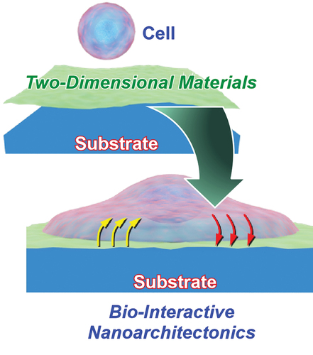
Like the proposal of nanotechnology by Richard Feynman [Citation76,Citation77], the nanoarchitectonics concept was initially proposed by Masakazu Aono [Citation78,Citation79]. The nanoarchitectonics strategy conceptually fuses nanotechnology with other research fields including organic chemistry, supramolecular chemistry, micro/nano-fabrication, materials science, and bio-related sciences () [Citation80,Citation81]. This methodology aims to produce functional materials from nanoscale components by combining and selecting individual processes such as atom/molecule manipulation, chemical/physical conversion, self-assembly/self-organization, various fabrication techniques, and bio-related processes [Citation82]. Because the nanoarchitectonics concept is very general, related approaches can be applied to a wide range of materials. Even in recent publications, it can be found that the nanoarchitectonics concept is utilized for rather traditional materials targets such as organic modification of oxide-based layered materials [Citation83] and composite materials for removal of cesium ions from water [Citation84] as well as forefront areas like coordination asymmetry [Citation85] and DNA/RNA-based molecular machines [Citation86]. In an analogy to the theory of everything in physics, nanoarchitectonics can be regarded as the strategy of everything in materials science [Citation87]. Applications of the nanoarchitectonics approach have been proposed in a wide variety of functional targets including functional material production [Citation88,Citation89], structural regulations [Citation90,Citation91], catalysts [Citation92,Citation93], sensors [Citation94,Citation95], devices [Citation96,Citation97], energy-related applications [Citation98,Citation99], solutions for environmental problems [Citation100,Citation101], basic bioscience [Citation102,Citation103], and biomedical applications [Citation104,Citation105].
Figure 1. Outline of nanoarchitectonics strategy.
However, nanoarchitectonics strategies may include features different from conventional materials fabrications. Unlike macroscopic and microscopic phenomena, nanoscale interactions between objects (atoms, molecules, and materials) are exposed to various factors of uncertainty such as thermal fluctuations, statistic distributions, complicated mutual interactions, and quantum effects. Summed effects in materials are not simply additive but can also be synergistic. Materials nanoarchitectonics may need to consider effects of such uncertainties in their constructions and functions [Citation106]. This situation is reminiscent of biological systems, in which various functional elements work together under thermal fluctuations [Citation107,Citation108]. Another distinct feature of nanoarchitectonics processes would be its suitability for forming hierarchical structures [Citation109]. Unlike self-assembly processes at equilibrium, nanoarchitectonics can combine various construction processes. Formation of hierarchical structures with asymmetrical and/or anisotropic organization becomes possible in nanoarchitectonics-based materials constructions. In most biological functional systems, hierarchical organizations are fundamental for creating rational flows of energy, electrons, and information. Therefore, nanoarchitectonics approaches have the potential to form bio-like highly functional materials systems.
1.2. Two-dimensional materials and environments
Among various examples in materials nanoarchitectonics, approaches specific to two-dimensional materials and two-dimensional environments (interfacial environments) exhibit exciting possibilities [Citation110–112]. One of the motivations for active research on two-dimensional nanoarchitectonics is rapid advancements in two-dimensional materials. Various two-dimensional materials including graphene families [Citation113,Citation114], two-dimensional semiconductors [Citation115,Citation116], various nanosheets [Citation117,Citation118], two-dimensional organic polymers [Citation119,Citation120], two-dimensional coordination polymers [Citation121,Citation122], and organic thin films [Citation123–125] have been widely researched. They are promising components for materials nanoarchitectonics. In recent reports, various functions are demonstrated by materials constructed from two-dimensional materials as structural components. For example, Tajima et al. reported long lifetimes of charge-separate states in mixed-dimensional (zero-dimensional/two-dimensional) van der Waals heterojunctions of anthracene physically adsorbed on few-layer MoS2 nanosheets [Citation126]. The observed properties would be useful for efficient photocatalysts, photovoltaics, and optoelectronics applications. Vinu and co-workers successfully synthesized hybrids of two-dimensional mesoporous fullerene and carbon materials for energy-related applications such as Li-ion batteries and supercapacitors [Citation127]. For environment-oriented applications, photodegradation of picric acid by two-dimensional materials made from germananes terminated with hydrogen and methyl groups was demonstrated by Sturala, Sofer, and co-workers [Citation128]. Surface plasmon resonance sensors were nanoarchitected from two-dimensional MoS2 nanosheets modified with the supramolecule calix[4]arene as demonstrated by Hu, Chen, and co-workers [Citation129]. This sensor system was utilized for detection of bovine serum albumin antibodies. As summarized in a recent review article by Choi and co-workers, two-dimensional materials can be used in various imaging technologies including magnetic resonance imaging, computed tomography and positron emission tomography as well as conventional optical imaging [Citation130]. These imaging methods are expected to be utilized in image-guided and precision therapy. Free-standing conductive two-dimensional polymer nanosheets were recently reported by Fujie and co-workers [Citation131]. The nanoarchitected polymer nanosheets can make good contact with unevenly structured surfaces, such as the veins of plant leaves. Integration of a Bluetooth system into the polymer nanosheets enables wireless biopotential measurement in plants. As Li and co-workers summarized in a recent review article, stimuli-responsive drug delivery systems can be nanoarchitected from various two-dimensional nanosheets [Citation132]. As Oaki and Igarashi recently demonstrated, application of materials informatics for predicting exfoliation processes to prepare two-dimensional materials has also been proposed [Citation133].
Interfacial environments as two-dimensional working media also promote understanding of various materials phenomena and nanoarchitectonics-based materials synthesis [Citation134,Citation135]. Particularly, dynamic interfaces such as an air–water interface provide unusual opportunities for molecular controls including molecular machine handling by hand-motion-like macroscopic mechanical input [Citation136–138] and mechanical tuning of molecular receptor performances [Citation139,Citation140]. These unique functions are created by extremely different motional freedoms between lateral and transverse directions of the fluidic interfacial media. In addition, the heterogeneous dielectric nature of the air–water interface induces drastic enhancements in molecular recognition capability of aqueous guest molecules [Citation141,Citation142]. Although hydrogen-bond-based molecular recognition is highly suppressed in bulk water, an incredible increase in binding efficiency (106 − 107 times in some cases) is observed at the interfacial media [Citation143]. This leads to efficient sensing system designs and a true understanding of biological phenomena [Citation144]. Although molecular recognition in biological systems occur in high dielectric aqueous media, aqueous media are not ideal media for hydrogen bonding for external guests. This seemingly contradictory situation can be resolved by considering interfacial environments. Molecular recognition in biological systems mostly occur at interfaces with aqueous medium as is the case for the surface of cell membranes, inner surfaces of proteins, and macromolecular interfaces of nucleic acids. Two-dimensional interfacial media can be regarded as powerful biomimetic environments for bio-inspired nanoarchitectonics [Citation145,Citation146].
Accordingly, there has been a lot of interest in analysing and understanding various phenomena at interfacial environments as seen in recent literature. Investigations on two-dimensional lipid membranes formed at interfacial environments have been progressing continuously. For example, Ishibashi and co-workers elucidated diffusion coefficients of trans-stilbene embedded within a lipid bilayer membrane of dimyristoylphosphatidylcholine formed at a silica–water interface [Citation147]. Two different observed values of effective viscosity imply local inhomogeneity of the lipid membrane as a fluidic two-dimensional medium. Oxidation of unsaturated phospholipids with various head groups under low-level ozone environments at the air–water interface was systematically investigated using heterodyne-detected sum frequency generation spectroscopy as reported by Inoue, Ye, and co-workers [Citation148]. Makiura et al. investigated two-dimensional assemblies of amphiphilic mesogens with different peptide chains at the air–water interface [Citation149]. The knowledge obtained from these model studies is useful for understanding the ordering of liquid crystalline materials at various interfaces. Kinjo et al. reported theoretical approaches for molecular dynamics simulation of fractures at interfaces between polyphenylene sulfide and aluminum oxide under tensile force [Citation150]. It was revealed that fracturing between the polymer and solid is initiated by small voids forming within the polymer layer.
Two-dimensional interfacial media have been used for materials nanoarchitectonics to synthesize new types of two-dimensional materials and related functional materials systems. In fact, nanoarchitectonics synthesis and organization of ultrathin two-dimensional materials have been demonstrated in one-dimensional supramolecular polymerization of two-dimensional DNA origami nanosheets [Citation151], integrated fabrication of optically regulated one-dimensional supramolecular fibers [Citation152], carbon nanosheet synthesis from carbon nanoring molecular precursors by vortex LB method [Citation153], and highly oriented two-dimensional nanofilms of polymeric semiconductors by super-high-temperature LB method [Citation154,Citation155]. As summarized in a recent review article by Wan and co-workers, various two-dimensional interfacial media such as gas–liquid, liquid–liquid, and liquid–solid interfaces are useful for synthesizing two-dimensional covalent organic frameworks (COFs) without limitations associated with their poor solubility [Citation156]. Particularly, interfaces formed with insoluble solvents (liquid–liquid interfaces) provide opportunities to form small shape-defined crystals arising from drastic changes in material solubility. For example, the dynamic liquid–liquid interfacial precipitation method results in macaroni-shaped fullerene (C60) crystals that can be converted into mesoporous carbon tubes upon high-temperature heat treatment at 900°C [Citation157]. The formed carbon materials exhibited nice supercapacitor performances with excellent stability even after 10,000 charging–discharging cycles. Niidome, Kurawaki, and co-workers demonstrated the formation of gold-silver bimetallic thiolate complexes at the interface between octanethiol and water accompanied with luminescence conversion from red to blue [Citation158].
These examples demonstrate the strikingly important roles of two-dimensional materials and environments in materials nanoarchitectonics. Anisotropic structures and dielectrically heterogeneous natures of such two-dimensional matter are surely beneficial for various applications including popular energy-related applications and bio-related investigations.
1.3. Objectives of this review article
These background descriptions suggested several important points for producing functional systems in current science and technology of advanced materials. Following sufficient increase in control over nanostructure in functional materials, a novel methodology, nanoarchitectonics, has emerged as the post-nanotechnology concept. In the nanoarchitectonics approach, functional material systems are architected from nanoscale unit-components using various processes including self-assembly/self-organization. Nanoarchitectonics processes have features reminiscent of biological systems, where synergistic material interactions under nanoscale fluctuations play important roles in organizing hierarchical structures for advanced functions. It may even be said that real biological systems are products of natural nanoarchitectonics processes. Therefore, creation of bio-like highly functional material systems would be an ultimate goal of nanoarchitectonics approaches.
From the viewpoint of producing new artificial materials systems with biological-level high functionality, bio-interactive nanoarchitectonics must be considered. Bio-related functional material systems are usually based on bioactive interactions as seen in recent examples such as ultrasound-activated therapeutics [Citation159], molecular hybridization for detection and imaging of biomarkers inside brains afflicted with Alzheimer’s disease [Citation160], interactive materials systems for detecting microcystin-leucine arginine as one of the most toxic and harmful freshwater toxins [Citation161], nanostructured materials systems with localized surface plasmon resonance for sensing cytokine [Citation162], and structurally designed peptide nucleic acids for interactive detection of specific forms and sites of DNA and RNA [Citation163].
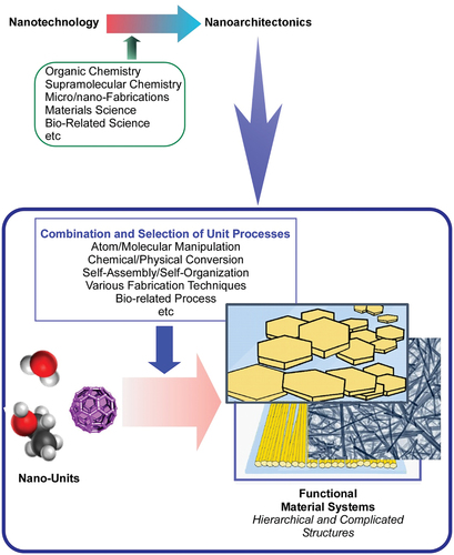
Although various nanoscale units are architected in the bio-interactive functional materials in the previous examples, this review focuses especially on two-dimensional materials and environments (). As described in the previous section, two-dimensional materials are star-players in recent nanomaterials sciences, and two-dimensional (interfacial) environments are advantageous media for molecular interactions including biological interactions. In this review article, several examples are selected to explain bio-interactive nanoarchitectonics coupled with two-dimensional materials and environments. Following general examples of nanoarchitectonics of two-dimensional materials for energy storage, catalysis, and biomedical applications (section 2), bio-related applications with emerging two-dimensional materials such as two-dimensional biomimetic nanosheets, fullerene nanosheets, and two-dimensional assemblies of one-dimensional fullerene nanowhiskers (FNWs) are explained (section 3). Further discussions on bio-interactive nanoarchitectonics in two-dimensional environments extend to liquid–liquid interfaces such as fluorocarbon–medium interfaces and viscous liquid interfaces as new frontiers of two-dimensional environments for bio-related applications (section 4). In the latter section, controlling differentiation in stem cells at fluidic liquid interfaces is discussed. Finally, a conclusive section briefly summarizes features of bio-interactive nanoarchitectonics with two-dimensional materials and environments and discusses possible future perspectives.
Figure 2. Objectives of this review article: bio-interactive nanoarchitectonics with two-dimensional materials and/or at two-dimensional environments.
2. General applications of nanoarchitectonics of two-dimensional materials
Following the discovery of graphene, many types of two-dimensional nanomaterials with monolayer thickness have been developed and explored because of their unique properties endowed by the monolayer conformation [Citation164,Citation165]. With the excitement of discovering these novel properties, earlier research mainly focused on the discrete properties of nanosheets. However, as the subject of two-dimensional nanomaterials became further established, the potential for assembling these two-dimensional nanosheets into three-dimensional shapes and hybrid structures gathered interest. Here, we summarise the main areas of application for such assemblies with focus on commonly used two-dimensional nanomaterials.
2.1. Energy storage and catalysis
Two-dimensional nanomaterials have shown promise for energy storage due to their good electrical conductivity and large surface area providing abundant active sites for ions to intercalate [Citation166,Citation167]. Nonetheless, it was also discovered that a number of factors limit their efficacy such as their tendency to aggregate and electrochemical degradation of active sites [Citation168,Citation169]. Hetero-structures made up of two-dimensional nanosheets have been developed to eliminate or minimize limiting factors and fabricate enhanced energy storage devices.
A sandwich-like assembly of two-dimensional monolayer MoS2 nanosheets with mesoporous carbon between each layer of nanosheet (MoS2/m-C) was engineered for lithium-ion storage () [Citation170]. The MoS2/m-C structure was obtained by amidating dopamine onto oleic-acid-protected monolayer MoS2, followed by self-polymerizing dopamine and finally annealing the obtained structure at 850°C for 2 h. Engineered MoS2/m-C was shown to solve some of the common problems in MoS2-based anode electrode materials for lithium-ion batteries such as low conductivity of MoS2 in the c-direction and aggregation and restacking of MoS2 nanosheets. This hybrid structure also provided the largest interfacial contact for Li ion storage among MoS2-based anode electrode materials. Another sandwich-like structure consisting of carbon nanotubes (CNTs) and graphene nanosheets was engineered for lithium-sulfur (Li-S) batteries () [Citation169]. Vertically aligned CNTs were anchored onto the graphene nanosheets (ACNT/G), creating a hierarchical structure. The obtained architecture provided effective ion diffusion channels and three-dimensional electron transfer pathways. Nitrogen doping (N-ACNT/G) enhanced the electrochemical performance by introducing additional active and defect sites. Engineered N-ACNT/G demonstrated improved cyclic and rate performance when used as the cathode in Li-S batteries.
Figure 3. Assemblies of two-dimensional nanomaterials are good anode or cathode candidates for lithium batteries. a) Schematic illustrating the concept of the rational design of MoS2/m-C nanosheet superstructure for creating ideal MoS2/c atomic interfaces to enhance lithium-ion storage. Reproduced with permission [Citation168] 2015, Wiley-VCH. b) Conceptual scheme of the design of N-ACNT/G hybrids with graphene and aligned CNTs as building blocks. (i) Structural hybridization of aligned CNTs and graphene via catalytic growth on bifunctional natural catalysts; (ii) in situ nitrogen doping for moderating chemical modification of the carbon scaffolds. Reproduced with permission [Citation169] 2014, Wiley-vch.
![Figure 3. Assemblies of two-dimensional nanomaterials are good anode or cathode candidates for lithium batteries. a) Schematic illustrating the concept of the rational design of MoS2/m-C nanosheet superstructure for creating ideal MoS2/c atomic interfaces to enhance lithium-ion storage. Reproduced with permission [Citation168] 2015, Wiley-VCH. b) Conceptual scheme of the design of N-ACNT/G hybrids with graphene and aligned CNTs as building blocks. (i) Structural hybridization of aligned CNTs and graphene via catalytic growth on bifunctional natural catalysts; (ii) in situ nitrogen doping for moderating chemical modification of the carbon scaffolds. Reproduced with permission [Citation169] 2014, Wiley-vch.](/cms/asset/96d67aef-cdb4-43cf-8d12-82e42e5af192/tsta_a_2054666_f0003_oc.jpg)
As is the case for energy storage applications, the true potential of two-dimensional materials as catalysts is hindered by a number of factors. Using strategic architectures of hybrid structures, better catalysts can be obtained from two-dimensional nanomaterials [Citation170,Citation171]. Zn/Cr-layered double hydroxide (Zn/Cr-LDH) and lead niobate (HPb2Nb3O10) nanosheets were self-assembled into layer-by-layer stacked formation (LDH-PHO) for photocatalytic oxygen generation () [Citation172]. In a spontaneous electrostatic process, negatively charged HPb2Nb3O10 nanosheets self-assembled alternately with positively charged Zn/Cr-LDH nanosheets. The assembled hetero-structure demonstrated enhanced photocatalytic activity compared to lone Zn/Cr-LDH nanosheets due to suitable band alignment and large contact area between the layered nanosheets.
Figure 4. Schematic illustrating the synthesis of LDH-PNO for the photocatalytic oxygen generation. Reproduced with permission [Citation172] 2021, Elsevier. Hybrid structure was self-assembled by layer-by-layer stacking of Zn/Cr-layered double hydroxide (Zn/cr-LDH) and lead niobate (HPb2Nb3O10) nanosheets.
![Figure 4. Schematic illustrating the synthesis of LDH-PNO for the photocatalytic oxygen generation. Reproduced with permission [Citation172] 2021, Elsevier. Hybrid structure was self-assembled by layer-by-layer stacking of Zn/Cr-layered double hydroxide (Zn/cr-LDH) and lead niobate (HPb2Nb3O10) nanosheets.](/cms/asset/feadef6c-0e7d-4400-8a67-56a7ff3dd086/tsta_a_2054666_f0004_oc.jpg)
2.2. Biomedical applications
Two-dimensional nanomaterials have greatly contributed to the development of diagnostic and therapeutic tools such as biosensors, drug delivery systems and photothermal and photodynamic therapy agents [Citation173,Citation174]. Exploration into the assemblies and hybrid structures of these two-dimensional nanomaterials has widened the potential biomedical applications of these materials.
Three-dimensional assemblies and hybrid structures involving two-dimensional nanosheets were mainly explored for the fabrication of tissue engineering scaffolds. Besides good photothermal and conductive properties, two-dimensional nanosheets were impactful especially due to their ability to induce cell differentiation without adding differentiation inducers in the culture media [Citation175,Citation176]. The potential of MoS2 and reduced graphene oxide (rGO) nanosheets as electrically conductive agents and nanotopographical stimuli for fabricating cardiac tissue engineering scaffolds was evaluated. The scaffold was constructed by introducing MoS2 and rGO nanosheets into silk fibroin nanofibers. Induced pluripotent stem cells (IPSCs) transfected with TBX18 gene were cultured on this scaffold. Through a series of experiments assessing the mechanical properties of the structure and cultured IPSCs, it was determined that the scaffold could be a good candidate for cardiac tissue engineering [Citation175]. In another study, GO nanosheet coated polycaprolactone (PCL) nanofiber scaffold was utilised for neural tissue engineering () [Citation176]. Differentiation of neural stem cells (NSCs) into oligodendrocytes was observed to be dependent on GO concentration. It was shown that the GO coating provided a suitable surface for cell adhesion and promoted neuronal electrophysiology due to its good conductivity.
Figure 5. Scaffolds for tissue engineering applications can be constructed by the assembly of two-dimensional nanomaterials. a) Schematic diagram depicting the fabrication and application of graphene-nanofiber hybrid scaffolds. Polymeric nanofibers (composed of polycaprolactone) generated using electrospinning were subsequently coated with GO and seeded with NSCs. NSCs cultured on the graphene-nanofiber hybrid scaffolds show enhanced differentiation into oligodendrocyte lineage cells. Reproduced with permission [Citation176] 2014, Wiley-VCH. b) Schematic illustration of the preparation of the therapeutic hydrogel from a GelMA prepolymer and BP nanosheets. Reproduced with permission [Citation178] 2019, Royal Society of chemistry.
![Figure 5. Scaffolds for tissue engineering applications can be constructed by the assembly of two-dimensional nanomaterials. a) Schematic diagram depicting the fabrication and application of graphene-nanofiber hybrid scaffolds. Polymeric nanofibers (composed of polycaprolactone) generated using electrospinning were subsequently coated with GO and seeded with NSCs. NSCs cultured on the graphene-nanofiber hybrid scaffolds show enhanced differentiation into oligodendrocyte lineage cells. Reproduced with permission [Citation176] 2014, Wiley-VCH. b) Schematic illustration of the preparation of the therapeutic hydrogel from a GelMA prepolymer and BP nanosheets. Reproduced with permission [Citation178] 2019, Royal Society of chemistry.](/cms/asset/9d93faae-fec3-47c3-94ed-bd87e888c2ea/tsta_a_2054666_f0005_oc.jpg)
Two-dimensional black phosphorus (BP) is especially preferred for bone regeneration applications due to its phosphorus (P) content. BP is easily broken down into phosphorus (P), a vital element in bones, upon interacting with oxygen, visible light and water. Hence, different structures were combined with two-dimensional BP for bone tissue engineering. In one such example, a 3D printed bioglass scaffold was modified with two-dimensional BP nanosheets for photothermal therapy of osteosarcoma and ensuing bone regeneration [Citation177]. In another study, the photothermal capability of two-dimensional BP was utilised for its antibacterial effect, while the two-dimensional BP hydrogel nanocomposite promoted mineralization and supported crosslinking networks for osteogenesis and bone tissue formation () [Citation178]. Another use of the photothermal capability of two-dimensional BP in bone tissue engineering applications was demonstrated through the fabrication of an osteoimplant; two-dimensional BP nanosheets were combined with poly(lactic-co-glycolic acid) (PLGA) and implanted into the hind legs of rats. Osteogenesis was observed upon periodic near-infrared (NIR) laser irradiation [Citation179].
In addition to tissue engineering applications, other biomedical uses of two-dimensional assemblies, such as drug delivery and gene delivery, have been explored. Li et al. designed a layer-by-layer assembly of MoS2 nanosheets mediated by DNA oligonucleotides for the delivery and stimuli-responsive release of the chemotherapeutic drug Doxorubicin (DOX) () [Citation180]. Thiol-terminated single-stranded DNA (ssDNA) was anchored onto the sulphur vacancies on the MoS2 nanosheets. Self-assembly and stimuli-responsive capabilities were achieved using DNA-based aptamers as linkers. DOX was loaded into the layered structure through its ability to preferentially bind GC base-pair duplexes. The chosen ATP-responsive aptamer enabled the release of carried DOX at the ATP-rich tumour site by dissociating from the DNA oligonucleotides and preferentially binding to ATP molecules instead. MoS2 nanosheets acted as a protective shield for the cargo while allowing small ATP molecules to infiltrate for stimuli-responsive effect. Paul et al. utilised GO incorporated into injectable low-modulus methacrylated gelatin (GelMA) hydrogel for gene delivery to myocardial tissue [Citation181]. The cargo of pro-angiogenic human vascular endothelial growth factor plasmid DNA (pDNAVEGF) was bound to the surface of GO prior to its combination with GelMA. In vitro and in vivo studies showed enhanced cardiac activity in cells transfected by the delivered gene therapy.
Figure 6. Stimuli-Responsive anti-cancer drug delivery system is formed by layer-by-layer self-assembly of MoS2 nanosheets. Schematic illustration of formation of multilayer Dox/D2/MoS2-NS and their intracellular Dox release process. (I) ATP-aptamer-induced LbL assembly; (II) Dox loading in multilayer nanostructures; (III) in vitro treatment of Dox/D2/MoS2-NS; (IV) cell uptake through endocytosis; (V) endosomal/lysosomal escape; (VI) ATP-induced Dox release in the cytosol. Reproduced with permission [Citation180] 2017, American Chemical Society.
![Figure 6. Stimuli-Responsive anti-cancer drug delivery system is formed by layer-by-layer self-assembly of MoS2 nanosheets. Schematic illustration of formation of multilayer Dox/D2/MoS2-NS and their intracellular Dox release process. (I) ATP-aptamer-induced LbL assembly; (II) Dox loading in multilayer nanostructures; (III) in vitro treatment of Dox/D2/MoS2-NS; (IV) cell uptake through endocytosis; (V) endosomal/lysosomal escape; (VI) ATP-induced Dox release in the cytosol. Reproduced with permission [Citation180] 2017, American Chemical Society.](/cms/asset/653360f0-cb4c-49ca-9330-b4fc42ab2600/tsta_a_2054666_f0006_oc.jpg)
3. Emerging two-dimensional materials in bio-related applications
3.1. Two-dimensional biomimetic nanosheets
As a new subcategory of two-dimensional nanomaterials, biomimetic nanosheets are being developed by layering biologically obtained nanomaterials with inorganic nanomaterials in two-dimensional conformations. In this technique, naturally soft bio-nanomaterials such as cell membrane fragments are stiffened to maintain two-dimensional shape through added support from hard inorganic nanoparticles.
In one earlier example, researchers layered red blood cell (RBC) membranes with MoSe2 nanosheets for cancer immunotherapy [Citation182]. RBC membranes increased circulation time of nanoparticles by providing immune evasion. MoSe2 provided near-infrared (NIR) laser-induced ablation of the primary tumour by photothermal therapy (PTT), while immunotherapy was achieved through activating the inherent immune system through antigens released from ablated tumour cells.
In another example, the advantage of two-dimensional bio-nanomaterials over their spherical counterparts in anti-tumour applications was demonstrated () [Citation183]. For this purpose, two-dimensional cell membrane nanoparticles (NS-M) were prepared by layering gold nanostars (NS) with cell membrane, while three-dimensional cell membrane nanoparticles (NS@M) were prepared by encapsulating NS into cell membrane vesicles. Through comparing the two modalities, it was shown that NS-M has superior anti-tumour efficacy. Due to its flattened shape, NS-M could more effectively accumulate in the tumour site and attach to tumour cells strongly, which in turn increased its anti-tumour efficacy. Similarly, structured nanosheets were prepared in another work by combining E. coli membrane nanosheets with gold nanoparticles (AuMNs) [Citation184]. AuMNs were successful in PTT-induced killing of tumours and worked as immunologic adjuvants to promote anti-PD-L1 efficacy.
Figure 7. Combining biomimetic two-dimensional nanomaterials with inorganic nanomaterials is an emerging technique for designing new diagnostic and therapeutic nanomaterials. a) Schematic illustration of the stiffened nanomembrane and the three-dimensional nanosphere’s interaction with target cell surface. b) Schematic illustration of the drug-loaded stiffened nanomembrane for tumor ablation and chemotherapy. Reproduced with permission [Citation183] 2020, American Chemical Society. c) Schematic illustration of the design and preparation of FePse3@app@ccmnss. Reproduced under terms of the CC-BY license [Citation185].
![Figure 7. Combining biomimetic two-dimensional nanomaterials with inorganic nanomaterials is an emerging technique for designing new diagnostic and therapeutic nanomaterials. a) Schematic illustration of the stiffened nanomembrane and the three-dimensional nanosphere’s interaction with target cell surface. b) Schematic illustration of the drug-loaded stiffened nanomembrane for tumor ablation and chemotherapy. Reproduced with permission [Citation183] 2020, American Chemical Society. c) Schematic illustration of the design and preparation of FePse3@app@ccmnss. Reproduced under terms of the CC-BY license [Citation185].](/cms/asset/f036e6ff-5031-428e-888c-c5fd6d9d8cb7/tsta_a_2054666_f0007_oc.jpg)
In a more recent study, two-dimensional FePSe3 metal phosphorous trichalcogenides were combined with CT26 cell membrane and loaded with anti-PD-1 peptide (APP) for imaging and therapy on cancer cells () [Citation185]. FePSe3 acted as magnetic resonance imaging (MRI) and photoacoustic imaging (PAI) agents while also contributing to anti-tumour activity by inducing PTT under NIR laser irradiation. The anti-tumour effect was enhanced by T cell activation through the loaded APP. CT26 cell membrane worked as a targeting agent, helping the constructed two-dimensional nanoparticle to homotypically locate cancer cells.
3.2. Fullerene nanosheets
C60 fullerene is an allotrope of sp2 carbons arranged in a spherical network [Citation186,Citation187]. In solution, C60 fullerenes self-assemble into crystal structures defined by intermolecular forces. C60 fullerene’s size (1 nm diameter) and zero-dimensional structure make it a versatile molecular building block. Using liquid–liquid interfacial precipitation (LLIP) to control structure, various one-dimensional to three-dimensional assemblies have been demonstrated. Cell culture substrates made from fullerene assemblies are desirable for their well-defined structure and strong protein adsorption.
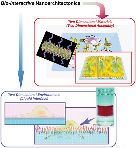
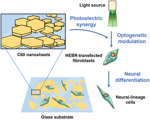
Fullerene nanosheets are two-dimensional crystalline assemblies of C60 fullerene shaped like thin hexagonal prisms. Fullerene nanosheets are prepared using LLIP at the interface between carbon tetrachloride (CCl4) and an alcohol. The size of nanosheets depends on the alcohol used; the hexagonal face of nanosheets produced with methanol, ethanol, and isopropyl alcohol (IPA) has diameters between 500–700 nm, 2–3 µm, and 7–9 µm, respectively, [Citation188]. Luo et al. reprogrammed human fibroblasts to neural lineages through optogenetic modulation on a fullerene nanosheet platform [Citation189] (). Human fibroblasts were transfected with highly expressible bacteriorhodopsin (HEBR) genes, which produce photo-responsive proton pumps with the potential to trigger neural activity. C60 nanosheets prepared through LLIP (CCl4/benzene and IPA) were formed into Langmuir-Blodgett (LB) films on glass and polyurethane substrates. Transfected fibroblasts cultured on C60 nanosheet substrates and stimulated with light significantly upregulated neural lineage-related genes GFAP, β-tubulin, MAP2 and corresponding marker proteins; transfected fibroblasts differentiated toward neural lineages without induction medium. Optogenetic modulation is synergistically enhanced by photoelectric properties of C60. Fullerene nanosheet substrates are promising for neural therapies based on optogenetics.
Figure 8. C60 nanosheets are used as a substrate for HEBR-transfected fibroblasts. the nanosheet substrate synergistically amplified optogenetic modulation of fibroblasts expressing the HEBR photon pump, resulting in increased differentiation to neural-lineage cells (as characterized by neural gene and marker expression). See text for details.
3.3. Two-dimensional assembly of one-dimensional fullerene nanowhiskers
Fullerene nanowhiskers (FNWs) are one-dimensional crystalline fibers assembled from C60 fullerene. FNWs are prepared using LLIP at the interface between toluene (or m-xylene) and an alcohol (usually IPA) [Citation190–192]. FNWs used in cell culturing typically have cross-sectional diameter between 300 and 500 nm; FNW lengths range between 1 and 500 µm depending on fabrication parameters, representing a wide range of aspect ratios.
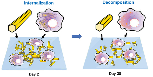
Nudejima et al. co-cultured macrophage-like cells with FNWs, observing phagocytosis and decomposition into fullerene molecules [Citation193,Citation194] (). FNWs (toluene and IPA) were dispersed in culture medium (10 µg/mL). After 2 days of exposure, >70% of cells internalized FNWs. After 28 days, short FNWs became more numerous, and granular crystallizations appeared in cell membranes. This suggests that cell interactions with FNWs may involve internalization and decomposition, as opposed to simply adhesion. The result also demonstrates a potential for FNWs in drug delivery systems and tissue engineering. In a follow-up study, Okuda-Shimazaki et al. found very weak cytotoxicity associated with macrophage-like cells internalizing FNWs, lower than multiwall carbon nanotubes (MWCNTs) at the same concentration [Citation195].
Figure 9. Macrophage-Like cells internalize FNWs within 2 days. Within 28 days, average fullerene nanowhisker length became shorter, and granular crystallizations appeared in cell membranes, indicating decomposition by macrophages. Cell interactions with fullerene nanowhiskers induced internalization and decomposition.
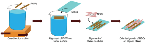
Methods to organize one-dimensional FNWs into two-dimensional arrays of controlled geometry at air–water interfaces were also developed. The organized assemblies are easily transferred to solid substrates. As cell culture substrates, FNW arrays exert effective control over cell adhesion, morphology, and differentiation. Krishnan et al. used vortex-aligned FNWs to control the orientation of human osteoblast MG63 cells [Citation196]. FNWs (toluene and IPA) were added to a water surface in a uniform layer. Mechanical stirring causes vortex flow that orients FNWs with the motion. FNWs are transferred to a substrate during motion. Length and packing density of FNWs, distance from the center of rotation (during transfer), rotation rate, and substrate hydrophobicity tune the geometry of FNWs arrays. MG63 cells preferentially adhered to FNWs, growing along the axes of alignment; this strongly influenced cell morphology. Cell adhesion favours FNWs due to enhanced adsorption of extracellular matrix (ECM) proteins and increased surface roughness. Cell proliferation on FNWs and glass was comparable, indicating low toxicity. These results demonstrate potential for two-dimensional assembled FNW substrates to promote adhesion and guide cell growth. In a similar approach, Hsieh et al. used cyclic linear motion to produce aligned FNWs (toluene and IPA; m-xylene and IPA), which promoted differentiation in murine NSCs () [Citation197].
Figure 10. FNWs were aligned with one-direction motion by the lab shaker and captured on a glass slide to produce aligned FNWs scaffolds. When used as substrates for NSCs culture, NSCs tend to orient along the direction FNWs. Morphological changes of NSCs triggered neural differentiation.
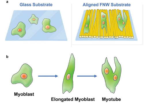
Lateral compression in the LB technique spontaneously aligns FNWs parallel to barriers. Minami et al. used the LB approach to fabricate aligned FNWs, which stimulated myogenic differentiation in murine skeletal myoblasts [Citation198] (). FNWs (toluene and IPA) were compressed against and transferred to glass slides, forming LB films of aligned FNWs achieving high surface coverage of 79%; the films display consistent geometry independent of substrate properties (i.e., hydrophobicity). Myoblasts took on elongated morphologies (aspect ratio 1.78-fold that on glass), with growth direction and orientation highly correlated to FNW alignment. Myoblasts significantly upregulated myogenic genes MyoD and Myogenin. Myoblast fused to form multinucleated phenotypes characteristic of mature myotubes. The topography of aligned FNWs regulated cell morphology and growth direction, which induced myogenic differentiation.
Figure 11. A) on aligned FNW substrates fabricated with the LB technique, myoblasts elongated and oriented along FNW alignment. Over time, some cells fused, becoming multinucleated, a characteristic representative of mature myotubes. b) Elongation induces an early stage of differentiation; fusion of elongated cells occurs at the late stages of differentiation.
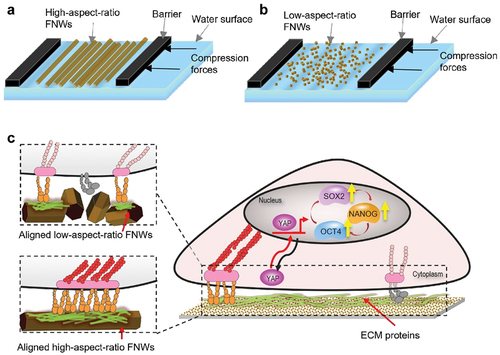
Song et al. effected tunable alignment of FNWs assembled through the LB approach. Improved control over nanoarchitecture enabled more detailed correlation between FNW alignment and cell behaviours. High alignment promoted self-renewal and retention of multipotency in human mesenchymal stem cells (hMSCs) () [Citation199]. FNWs (m-xylene and IPA) of different aspect ratios were fabricated by controlling addition rate (of IPA to C60/m-xylene solution). Adjusting the composition of FNWs (high aspect ratio: low aspect ratio) tuned their alignment in the resulting LB film. With increasing alignment, hMSCs exhibited increasing unidirectional orientation (along FNWs) and elongation. Upregulation of stemness genes OCT4, SOX2, NANOG and corresponding markers correlated with increasing alignment. Hallmarks of stemness persisted over 2 weeks, indicating long-term multipotency retention through symmetrical self-renewal. This result may become significant for in vitro stem cell expansion, which is desirable in tissue engineering and clinical applications. The hydrophobic and nano-topographic surface of aligned FNWs provided appropriate cues to restrict focal adhesion growth and activate mechanotransducive Yes-Associated Protein (YAP) signalling pathways; YAP nuclear translocation promoted upregulation of stemness genes, modulating cell fate.
Figure 12. Tuneable aligned FNWs scaffolds regulate stemness genes expression of hMSCs. a) LB technique aligns high-aspect ratio FNWs parallel to barriers, forming aligned FNWs arrays. b) Alignment decreases with low-aspect ratio FNWs. Adjusting the ratio of different FNWs tunes alignment on a continuous range. c) Aligned FNWs provide appropriate cues for mechanotransductive upregulation of stemness genes through the YAP signalling pathway.
Cell substrates made from two-dimensional FNW assemblies modulate cell morphology and differentiation, as shown with osteo, neural, muscular, and stem cells in featured reports. Novel self-assembly processes progressively improved control over nano-topography, producing well-controlled cell experiments and meaningful results for tissue engineering applications.
4. New frontier of two-dimensional environment for bio-related applications: liquid-liquid interface
Artificial cell substrates are almost exclusively solids (glass, metals, and polymers). Still, cells growing and maintaining vital functions on liquid substrates has been demonstrated. Water-immiscibility allows a liquid to form a phase boundary with tissue culture medium. If the liquid is also denser than water, then it will form the bottom phase. Liquid–liquid interface culture (LIC) grows cells at the phase boundary. Cells are supported on the bottom phase (water-immiscible liquid) and receive nutrients from the top phase (medium). Liquid–liquid interface culture offers unique insights into cell-substrate interactions and unique conditions for modulating cell behavior. Elastic stress is nearly absent as compared to solid substrates. Viscous stress ranges widely depending on liquid properties. These factors create atypical cell environments, where cell behaviors can be strongly correlated to viscoelastic substrate properties.
4.1. Fluorocarbon–medium interface
Cell culture at the interface between perfluorocarbon (PFC) oil and culture medium was first described by Rosenberg in 1964 [Citation200–202]. Perfluorocarbons are inert hydrophobic liquids; the immiscible interface between perfluorocarbon and medium can support anchorage-dependent cells. The interface also sustains cells, owing to perfluorocarbons’ high gas solubility and low toxicity. It is important to note that cells do not adhere directly to the interface, but instead to serum proteins (recruited from medium) or artificial microcarriers adsorbed at the interface [Citation202–205]. Due to weak interactions, proteolytic enzymes or chelating agents are unnecessary to detach cells. Hence, passaging becomes simple and preserves intact membrane proteins [Citation206]. This forms a defining advantage over solid substrates. Nonetheless, cells grown on perfluorocarbon-medium tend to aggregate and exhibit reduced growth and spreading [Citation207]. Initially, the perfluorocarbon-medium liquid interface culture was done exclusively on fibroblasts because they facilitate their own attachment and growth by secreting large quantities of collagen and fibronectin [Citation208]. Sanfillipo et al. enabled fibroblast and epithelial cell growth on perfluorocarbon with transforming growth factors (TGFs), but cell aggregation and poor spreading remained significant issues [Citation209].
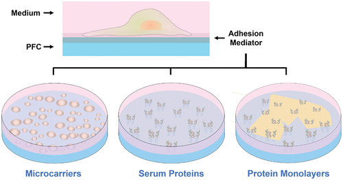
In 1983, Giaever and Keese made a breakthrough in introducing cell culture on pre-assembled protein monolayers at the perfluorocarbon–medium interface () [Citation210]. The monolayer self-assembles after simply depositing matrix protein solutions on perfluorocarbon and incubating. The monolayers are further enriched by adsorbing serum proteins from medium. Giaever and Keese cultured murine and human fibroblasts on poly-L-lysine (PLL) monolayer, successfully growing confluent cell sheets [Citation210]. Ando et al. created collagen type IV (COL), laminin (LN), fibronectin (FN), fibrinogen (FG), and gelatin (GN) monolayers, observing endothelial cell growth comparable to that on polystyrene [Citation211]. Kwon et al. noted significantly improved fibroblast growth and spreading on PLL and GN monolayers [Citation212]. Improvements in spreading may be related to a protein monolayer’s tensile strength [Citation210–212]; aggregation has been associated with monolayer fracturing caused by cell-exerted tension [Citation202,Citation210].
Figure 13. Ostensibly, liquid-liquid interface culture (LIC) involves cell adhesion and growth at the interface between a perfluorocarbon (PFC) and culture medium; but cells actually adhere to molecules adsorbed at the interface. Reported adhesion mediators include emulsion-based microcarriers, serum proteins recruited from medium, and pre-assembled protein monolayers.
Perfluorocarbon composition leads to significant differences in cell spreading and morphology. FC70 [Citation202,Citation207,Citation210], FC72 [Citation202,Citation210], FC40 [Citation205,Citation207,Citation213], FC43 [Citation202,Citation208,Citation209,Citation211], and perfluorodecalin (PFD) [Citation207,Citation214,Citation215] support palpable cell growth and spreading, while FC-77[Citation202,Citation204,Citation210,Citation212], L-series [Citation202], and KPF-series [Citation207] do not. High-purity perfluorocarbons tend to inhibit spreading, suggesting endogenous contaminants may act as surfactants modulating protein adsorption ability [Citation207,Citation209,Citation210]. This has been further evidenced in studies using exogenous surfactants [Citation202,Citation216]. Perfluorocarbon structure may also influence cell morphology. Minami et al. tested a linear aliphatic perfluorocarbon (perfluorooctane (PFO)), a cyclic perfluorocarbon (undecafluoro-tryifluoromethylcyclohexane), an aromatic perfluorocarbon (hexafluorobenzene), and various mixtures of these perfluorocarbons, finding cell spreading only on neat PFO [Citation217]. Some perfluorocarbons also exhibit cell-specific inhibitory effects or toxicity [Citation201].
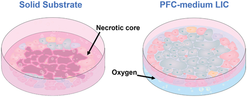
Perfluorocarbon-medium liquid interface culture has significant implications for tissue engineering because it enhances respiratory gas transport, which is an issue for high-density cell clusters. For example, large islets cultured on plastic substrates tend to suffer hypoxic damage due to reduced oxygen supply, leading to necrosis. Culture at the perfluorocarbon–medium interface offers unique advantages in pan-directional oxygen delivery (). Juszczak et al. cultured islets on PFD, preserving murine islet viability for 72 h and improving post-transplant insulin secretion [Citation214]. The 72 h viability of liquid–liquid interface culture islets reached 79% compared to 53.4% on plastic; islets on plastic contained large necrotic cores, which were absent in liquid interface culture islets. Liquid–liquid interface culture islets secreted significantly more insulin in response to glucose stimulation. This was also reflected post-transplantation, as 50% of liquid–liquid interface culture islet recipients became nonglycemic, compared to 14.3% of plastic-cultured islet recipients, and 31.3% of fresh islet recipients. Besides islets, liquid–liquid interface culture oxygen delivery is found to benefit chondrocyte clusters on polylactide scaffolds [Citation218].
Figure 14. High-Density cell clusters such as islets are prone to hypoxia. Oxygen diffusion to the islet core is limited, particularly on solid substrates; central necrosis ensues. Perfluorocarbons (PFCs) have high gas solubility. PFC-medium liquid-liquid interface culture (LIC) offers pan-directional oxygen delivery, encouraging islet survival and improved function.
Substrate viscoelasticity strongly influences cell adhesion, morphology, and differentiation [Citation219–223]. Effective absence of viscoelastic stress in perfluorocarbon-medium liquid interface culture has interesting implications. Minami et al. cultured C2C12 myoblasts at perfluorocarbon–medium interfaces, finding suppression of myogenic differentiation even in differentiation medium (DM) [Citation216]. Myoblasts cultured on polystyrene upregulated myogenic genes myoD, myf5, myogenin, and muscle-specific gene MHC, indicating differentiation into myotubes. Myoblasts cultured on PFO-DM retained high viability and spread but only upregulated myoD; absent viscoelastic stress in liquid–liquid interface culture weakened cellular traction force (CTF), causing mechanotransducive myf5 and myogenin downregulation, which suppressed myogenic differentiation.
The behavior of stem cells in liquid–liquid interface culture is of interest for tissue engineering and clinical applications. Hanga et al. cultured hMSCs using liquid–liquid interface culture, citing benefits of membrane protein preservation for stem cell expansion [Citation213]. FC40 was pre-conditioned by incubating with growth medium. This was sufficient for good transient attachment; within 2 h, hMSCs assumed their characteristic spindle-like morphology, as they would on plastic substrates; hMSCs were well spread and highly viable (>90%). However, over 7 days, liquid–liquid interface culture cell numbers and growth kinetics were lower (>2.5 fold) than on polystyrene, indicating suppressed cell activity. Importantly, liquid–liquid interface culture hMSCs retained their stemness markers, pluripotency, and differentiation potential post-expansion.
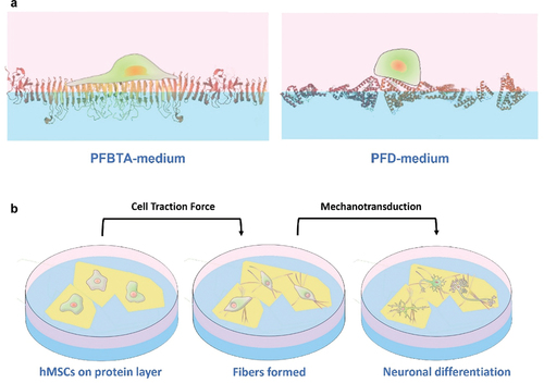
Jia et al. discovered that the behavior of hMSCs on pre-assembled protein monolayers depends heavily on cell-perceived stiffness () [Citation224]. Proteins assembled on different perfluorocarbons exhibit different packing densities; resistance to deformation by CTF is perceived by cells as stiffness. Stiff protein layers (128 kPa) formed on perfluorotributylamine (PFBTA) promoted cell spreading, strong focal adhesions, and YAP nuclear translocation (evidencing mechanotransduction). Soft protein layers (92 kPa) formed on PFD impaired focal adhesion growth and cell spreading. Previous studies reported fractured protein layers caused by cell-exerted tension, which in turn caused cell aggregation [Citation202,Citation210]. In Jia et al.’s follow-up work, hMSCs significantly deformed protein monolayers formed on PFO, triggering a feedback mechanism that altered cell morphology and fate () [Citation225]. The protein layer (96 kPa) transformed into elongated one-dimensional fibers under CTF-induced interfacial jamming. The elongated fibers coincided with elongated focal adhesions, followed by hMSCs adopting neuronal morphology. Significant upregulation of neuronal genes MAP2 and TUBB3 and corresponding markers further evidence neuronal differentiation. This mutual modulation between cells and protein layers is reminiscent of feedback mechanisms in tissue morphogenesis. Biomimetic stem cell regulation using liquid–liquid interface culture may be an interesting pursuit for tissue engineering.
Figure 15. a) Stronger protein denaturation on PFBTA (compared to PFD) creates a densely packed protein monolayer; the monolayer resists CTF, so it is perceived by cells as a stiffer substrate. on PFBTA, hMSCsspread, formed strong focal adhesions, and activated mechanotransducive signalling. on PFD, hMSCs only formed dynamic adhesions. b) on PFO, CTF exerted by hMSCs deformed protein monolayers into one-dimensional fibers. the fibers influenced hMSC morphology, activating mechanotransducive neuronal differentiation.
4.2. Viscous liquid interface
Formation of short bonds between polymer chains, known as crosslinking, is an effective way to modulate material viscoelasticity. Some weakly crosslinked or non-crosslinked polymers are effectively viscous liquids [Citation216]. Cell culture at the interface between medium and such viscous liquids has been demonstrated. Kong et al. studied the rheology of weakly crosslinked and non-crosslinked poly(dimethyl siloxane) (PDMS), observing full stress relaxation (i.e. classical viscous liquid rheology). HaCaT epidermal cells grew and spread on non-crosslinked PDMS; adhesion appears to be mediated by adsorbed serum proteins, as is the case for perfluorocarbons [Citation216]. Sylgard 184 PDMS supported cell adhesion, while neat PDMS with comparable viscosity did not. Additives in Sylgard 184 PDMS may have acted as surfactants mediating protein adsorption to the medium-PDMS interface.
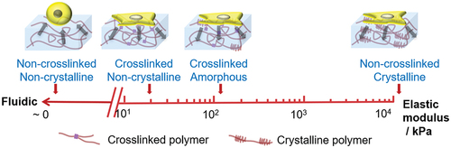
Uto et al. created a copolymer of caprolactone (CL) and D,L-lactide (DLLA) with adjustable viscosity and elasticity; substrate viscosity played a bigger role in cell spreading than previously understood [Citation223] (). Elasticity of CL-DLLA copolymer was controlled by adjusting the CL/DLLA composition. Viscosity was controlled with crosslinking. Non-crosslinked CL-DLLA was immiscible with medium and exhibited viscous liquid rheology. The interface was primed with FN coating before seeding with NIH3T3 fibroblasts. Cells remained spherical on liquid-like CL-DLLA, becoming increasingly well spread as viscosity and elasticity increased. On liquid substrates, dissipation of CTF prevents formation of focal adhesions and actin stress fibers. Fibroblast spreading increased significantly with crosslinking, and more gradually with elasticity; cells may be significantly more sensitive to substrate viscosity than elasticity, contrary to established notions [Citation219–222]. In the CL-DLLA substrate, relative viscous and elastic properties are easily adjusted. Cell responses to viscosity can be isolated and contrasted with responses to elasticity, uncovering new dimensions in biomaterials.
Figure 16. CL-DLLA copolymer has adjustable viscosity and elasticity. Non-crosslinked polymers exhibit viscous liquid rheology. Viscosity increases with crosslinking, and elasticity arises from crystalline regions. As a cell substrate, CL-DLLA copolymer’s adjustable viscosity and elasticity regulates cell spreading. Fibroblast spreading increases significantly with crosslinking, and more gradually with elasticity. This leads to the conclusion that cells may be significantly more sensitive to substrate viscosity than elasticity. See text for details.
5. Short-term perspectives
In this review article, several examples of bio-interactive nanoarchitectonics with two-dimensional materials and two-dimensional environments (interfaces) are presented. Although these examples cannot cover all related aspects, general trends and insights can be found therein. Interfaces are important conduits for transmitting nanoarchitected material/structural information to bioactive systems including living cells. Extending beyond intrinsic properties of the interfacial environment, two-dimensional materials and two-dimensional material assemblies effectively modulate bio-interactive capabilities at the interface. A distinct feature is information relay with incredible functional amplification and scaling. A small interactive cue created at the interface can propagate to trigger vital functions such as cell differentiation. Simple material interactions can decide the fates of living creatures. This excellent information processing is built upon sophisticated organization of functional molecules and their rational signalling pathways. In many ways, it is a brilliant demonstration of the ultimate goal of nanoarchitectonics approaches for functional materials systems. Production of highly advanced functional materials based on bio-like sophisticated component organization is a quintessential task for materials nanoarchitectonics [Citation226], a task enabled by architecting nanoscale components as the post-nanotechnology methodology. Unlike naturally occurring systems, nanoarchitectonics processes benefit from much wider component selections from non-bio materials such as two-dimensional materials. Therefore, there is even greater potential to create functional materials. Emerging fields such as materials informatics [Citation227–229] could support the development of functional materials systems from such a wide pool of components and selection freedoms.
While nanomaterials possess much potential for interesting applications through nanoarchitectonics, consumer and work-related exposure to nanomaterials have some safety concerns. Some of these common nanomaterials like silica, titanium dioxide and zinc oxide nanoparticles are added to consumer products like powdered food products and sunscreens. These nanomaterials have been shown to elicit pro-cytotoxic, inflammatory, pro-carcinogenic and dysfunctional outcomes in a variety of cell types present in various major organs [Citation230–238]. It would then be critical that with our growing understanding of the negative effects of nanomaterials, we can then use nanoarchitectonics design principles and tools to avoid the negative imprinting of nanomaterialistic features into next-generation nanomaterials to build functional bionanomaterials that are safe for the humans and the environment.
Nanoarchitectonics has mostly focused on engineering new materials up to this date, and it is on an exponential trajectory of growth. Much of this growth is based on wet laboratory experimentation studying interactions between molecules. Experimental parameters accumulated over decades of nanoarchitectural synthesis research can be fed as machine learning datasets. Machine learning algorithms can data-mine for patterns to propose combinations of parameters that may have been overlooked by real bench synthesis studies. These in-silico methods will accelerate the development cycles for new materials from design to actual proofs-of-concept. As a general outlook, metaphysical tools together with bench-based synthesis methods may propel nanoarchitectonics to the next stage of growth as we strive to search, design, apply and produce new nanomaterials for societally beneficial applications.
Disclosure statement
No potential conflict of interest was reported by the author(s).
Additional information
Funding
Notes on contributors

Xuechen Shen
Xuechen Shen is working on a Ph.D. degree at The University of Tokyo under Professor Ariga’s guidance, while researching with the Supermolecules Group at the World Premier International (WPI) Research Centre for Materials Nanoarchitectonics (MANA), NIMS.

Jingwen Song
Jingwen Song received her Ph.D. degree from The University of Tokyo under the guidance of Professor Katsuhiko Ariga in 2021. She was also studied in the Supermolecules Group at the World Premier International (WPI) Research Centre for Materials Nanoarchitectonics (MANA), NIMS from 2018.

Cansu Sevencan
Cansu Sevencan received her Ph.D. degree from the National University of Singapore (NUS) under the guidance of Associate Professor David Tai Leong in 2020. Her research interests include cell membrane nanotechnologies and biological effects and applications of nanomaterials.

David Tai Leong
David Tai Leong is an Associate Professor at the Department of Chemical and Biomolecular Engineering, National University of Singapore (NUS). He obtained his PhD in Biology and Bachelor degrees in Chemical Engineering from NUS and received his post-doctoral training at the Howard Hughes Medical Institute, University of Pennsylvania. He is also an elected Fellow of the Royal Society of Chemistry. His research interests span across fundamental understanding of biological effects of nanomaterials to their applications in nanomedicine, biosensing and nanotoxicology.

Katsuhiko Ariga
Katsuhiko Ariga received his Ph.D. degree from the Tokyo Institute of Technology in 1990. He joined the National Institute for Materials Science (NIMS) in 2004 and is currently the leader of the Supermolecules Group and principal investigator of the World Premier International (WPI) Research Centre for Materials Nanoarchitectonics (MANA), NIMS. He is also appointed as a professor in The University of Tokyo.
References
- Kimura K, Miwa K, Imada H, et al. Selective triplet exciton formation in a single molecule. Nature. 2019;570(7760):210–224. DOI:10.1038/s41586-019-1284-2
- Gohda Y. First-Principles determination of intergranular atomic arrangements and magnetic properties in rare-earth permanent magnets. Sci Technol Adv Mater. 2021;22(1):113–123.
- Yamamoto HM. Phase-Transition devices based on organic Mott insulators. Bull Chem Soc Jpn. 2021;94(10):2505–2539.
- Trinh TT, Kim J, Sato R, et al. Synthesis of mesoscopic particles of multi-component rare earth permanent magnet compounds. Sci Technol Adv Mater. 2021;22(1):37–54. DOI:10.1080/14686996.2020.1862630
- Kasuya N, Tsurumi J, Okamoto T, et al. Two-Dimensional hole gas in organic semiconductors. Nat Mater. 2021;20(10):1401–1406. DOI:10.1038/s41563-021-01074-4
- Hosono N, Uemura T. Development of functional materials via polymer encapsulation into metal–organic frameworks. Bull Chem Soc Jpn. 2021;94(8):2139–2148.
- Shimizu T, Lungerich D, Stuckner J, et al. Real-Time video imaging of mechanical motions of a single molecular shuttle with sub-millisecond sub-angstrom precision. Bull Chem Soc Jpn. 2020;93(9):1079–1085. DOI:10.1246/bcsj.20200134
- Kamei K, Shimizu T, Harano K, et al. Aryl radical addition to curvatures of carbon nanohorns for single-molecule-level molecular imaging. Bull Chem Soc Jpn. 2020;93(12):1603–1608. DOI:10.1246/bcsj.20200232
- Kratish Y, Nakamuro T, Liu Y, et al. Synthesis and characterization of a well-defined carbon nanohorn-supported molybdenum dioxo catalyst by SMART-EM imaging surface structure at the atomic level. Bull Chem Soc Jpn. 2021;94(2):427–432. DOI:10.1246/bcsj.20200299
- Kazuma E. Real-Space studies of plasmon-induced dissociation reactions with an STM. Bull Chem Soc Jpn. 2020;93(12):1552–1557.
- Xu X, Kinikar A, Di Giovannantonio M, et al. On-Surface synthesis of dibenzohexacenohexacene and dibenzopentaphenoheptaphene. Bull Chem Soc Jpn. 2021;94(3):997–999. DOI:10.1246/bcsj.20200382
- Xu X, Müllen K, Narita A. Syntheses and characterizations of functional polycyclic aromatic hydrocarbons and graphene nanoribbons. Bull Chem Soc Jpn. 2020;93(4):490–506.
- Kawai S, Krejčí O, Nishiuchi T, et al. Three-Dimensional graphene nanoribbons as a framework for molecular assembly and local probe chemistry. Sci Adv. 2020;6(9):eaay8913. DOI:10.1126/sciadv.aay8913
- Waser R, Aono M. Nanoionics-Based resistive switching memories. Nat Mater. 2007;6(11):833–840.
- Ohno T, Hasegawa T, Tsuruoka T, et al. Short-Term plasticity and long-term potentiation mimicked in single inorganic synapses. Nat Mater. 2011;10(8):591–595. DOI:10.1038/nmat3054
- Nakaya M, Tsukamoto S, Kuwahara Y, et al. Molecular scale control of unbound and bound C60 for topochemical ultradense data storage in an ultrathin C60 film. Adv Mater. 2010;22(14):1622–1625. DOI:10.1002/adma.200902960
- Nakaya M, Aono M, Nakayama T. Ultrahigh-Density data storage into thin films of fullerene molecules. Jpn J Appl Phys. 2016;55(11):1102B4.
- Yamashita M. Next generation multifunctional nano-science of advanced metal complexes with quantum effect and nonlinearity. Bull Chem Soc Jpn. 2021;94(1):209–264.
- Povie G, Segawa Y, Nishihara T, et al. Synthesis of a carbon nanobelt. Science. 2017;356:172–175.
- Sun Z, Ikemoto K, Fukunaga TM, et al. Finite phenine nanotubes with periodic vacancy defects. Science. 2019;363(6423):151–155. DOI:10.1126/science.aau5441
- Stockdale TP, Lam NYS, Anketell MJ, et al. The stereocontrolled total synthesis of polyketide natural products: a thirty-year journey. Bull Chem Soc Jpn. 2021;94(2):713–731. DOI:10.1246/bcsj.20200309
- van Bommel KJC, Friggeri A, Shinkai S. Organic templates for the generation of inorganic materials. Angew Chem Int Ed. 2003;42(9):980–999.
- Hashim PK, Bergueiro J, Meijer EW, et al. Supramolecular polymerization: a conceptual expansion for innovative materials. Prog Polym Sci. 2020;105:101250.
- Percec V, Xiao Q. Helical self-organizations and emerging functions in architectures, biological and synthetic macromolecules. Bull Chem Soc Jpn. 2021;94(3):900–928.
- Lee JSM, Otake K, Kitagawa S. Transport properties in porous coordination polymers. Coord Chem Rev. 2020;421:213447.
- Hanikel N, Prévot MS, Yaghi OM. MOF water harvesters. Nat Nanotechnol. 2020;15(5):348–355.
- Hosono N. Design of porous coordination materials with dynamic properties. Bull Chem Soc Jpn. 2021;94(1):60–69.
- Yew YP, Shameli K, Miyake M, et al. Green biosynthesis of superparamagnetic magnetite Fe3O4 nanoparticles and biomedical applications in targeted anticancer drug delivery system: a review. Arab J Chem. 2020;13(1):2287–2308. DOI:10.1016/j.arabjc.2018.04.013
- Sasaki J, Abe GL, Li A, et al. Large three-dimensional cell constructs for tissue engineering. Sci Technol Adv Mater. 2021;22(1):571–582. DOI:10.1080/14686996.2021.1945899
- Kamimura YR, Kanai M. Chemical insights into liquid-liquid phase separation in molecular biology. Bull Chem Soc Jpn. 2021;94(3):1045–1058.
- Shawky A, Mohamed RM, Mkhalid IA, et al. One-Pot synthesis of Mn3O4-coupled Ag2WO4 nanocomposite photocatalyst for enhanced photooxidative desulfurization of thiophene under visible light irradiation. Appl Nanosci. 2020;10(5):1545–1554. DOI:10.1007/s13204-019-01212-0
- Li Y, Henzie J, Park T, et al. Fabrication of flexible microsupercapacitors with Binder-Free ZIF-8 derived carbon films via electrophoretic deposition. Bull Chem Soc Jpn. 2020;93(1):176–181. DOI:10.1246/bcsj.20190298
- Takano S, Tsukuda T. Chemically modified gold/silver superatoms as artificial elements at nanoscale: design principles and synthesis challenges. J Am Chem Soc. 2021;143(4):1683–1698.
- Yoon HJ, Kwak SS, Kim SM, et al. Aim high energy conversion efficiency in triboelectric nanogenerators. Sci Technol Adv Mater. 2020;21(1):683–688. DOI:10.1080/14686996.2020.1800366
- Wang Z, Song H, Liu H, et al. Coupling of solar energy and thermal energy for carbon dioxide reduction: status and prospects. Angew Chem Int Ed. 2020;59(21):8016–8035. DOI:10.1002/anie.201907443
- Sakaushi K. Science of electrode processes in the 21st century: fundamental understanding of microscopic mechanisms towards advancing electrochemical technologies. Bull Chem Soc Jpn. 2021;94(10):2423–2434.
- Nakayama SF, Yoshikane M, Onoda Y, et al. Worldwide trends in tracing poly- and perfluoroalkyl substances (PFAS) in the environment. TrAc-Trends Anal Chem. 2019;121:115410.
- Johnson AC, Jin X, Nakada N. Learning from the past and considering the future of chemicals in the environment. Science. 2010;367(6476):384–387.
- Singh B, Na J, Konarova M, et al. Functional mesoporous silica nanomaterials for catalysis and environmental applications. Bull Chem Soc Jpn. 2020;93(12):1459–1496. DOI:10.1246/bcsj.20200136
- Mi P, Cabral H, Kataoka K. Ligand-Installed nanocarriers toward precision therapy. Adv Mater. 2020;32(13):1902604.
- Saleem M, Rasheed S, Yougen C. Silk fibroin/hydroxyapatite scaffold: a highly compatible material for bone regeneration. Sci Technol Adv Mater. 2020;21(1):242–266.
- Podder A, Lee HJ, Kim BH. Fluorescent nucleic acid systems for biosensors. Bull Chem Soc Jpn. 2021;94(3):1010–1035.
- Miyasaka T, Kulkarni A, Kim GM, et al. Perovskite solar cells: can we go organic-free, lead-free, and dopant-free? Adv Energy Mater. 2020;10(13):1902500. DOI:10.1002/aenm.201902500
- Al-Attafi K, Nattestad A, Qutaish H, et al. Solvothermally synthesized anatase TiO2 nanoparticles for photoanodes in dye-sensitized solar cells. Sci Technol Adv Mater. 2021;22(1):100–112. DOI:10.1080/14686996.2021.1873059
- Matsuo Y. Creation of highly efficient and durable organic and perovskite solar cells using nanocarbon materials. Bull Chem Soc Jpn. 2021;94(3):1080–1089.
- Guo D, Shibuya R, Akiba C, et al. Active sites of nitrogen-doped carbon materials for oxygen reduction reaction clarified using model catalysts. Science. 2016;351(6271):61–365. DOI:10.1126/science.aad0832
- Yoshimune W, Harada M. Impact of nonadsorbed ionomer on viscosity of catalyst inks for polymer electrolyte fuel cells. Bull Chem Soc Jpn. 2020;93(2):302–307.
- Kodama K, Nagai T, Kuwaki A, et al. Challenges in applying highly active Pt-based nanostructured catalysts for oxygen reduction reactions to fuel cell vehicles. Nat Nanotechnol. 2021;16(2):140–147. DOI:10.1038/s41565-020-00824-w
- Hosaka T, Kubota K, Hameed AS, et al. Research development on K-ion batteries. Chem Rev. 2020;120(14):6358–6466. DOI:10.1021/acs.chemrev.9b00463
- Liu J, Gu T, Sun X, et al. Synthesis of MnO/C/Co3O4 nanocomposites by a Mn2+-oxidizing bacterium as a biotemplate for lithium-ion batteries. Sci Technol Adv Mater. 2021;22(1):429–440. DOI:10.1080/14686996.2021.1927175
- Li P, Li C, Guo X, et al. Metal-Iodine and metal-bromine batteries: a review. Bull Chem Soc Jpn. 2021;94(8):2036–2042. DOI:10.1246/bcsj.20210182
- Maeda K. Z-Scheme water splitting using two different semiconductor photocatalysts. ACS Catal. 2013;3(7):1486–1503.
- Wang Q, Hisatomi T, Jia Q, et al. Scalable water splitting on particulate photocatalyst sheets with a solar-to-hydrogen energy conversion efficiency exceeding 1%. Nat Mater. 2016;15(6):611–615. DOI:10.1038/nmat4589
- Lang X, Gopalan S, Fu W, et al. Photocatalytic water splitting utilizing electrospun semiconductors for solar hydrogen generation: fabrication, modification and performance. Bull Chem Soc Jpn. 2021;94(1):8–20. DOI:10.1246/bcsj.20200175
- Shrestha RG, Maji S, Shrestha LK, et al. Nanoarchitectonics of nanoporous carbon materials in supercapacitors applications. Nanomaterials. 2020;10(4):639. DOI:10.3390/nano10040639
- Raman V, Rhee D, Selvaraj AR, et al. High-Performance flexible transparent micro-supercapacitors from nanocomposite electrodes encapsulated with solution processed MoS2 nanosheets. Sci Technol Adv Mater. 2021;22(1):875–884. DOI:10.1080/14686996.2021.1978274
- Liu H, Chen W, Zhang R, et al. Naturally O-N-S Co-doped carbon with multiscale pore architecture derived from lotus leaf stem for high-performance supercapacitors. Bull Chem Soc Jpn. 2021;94(6):1705–1714. DOI:10.1246/bcsj.20210027
- Mori T, Akamatsu M, Okamoto K, et al. Micrometer-Level naked-eye detection of caesium particulates in the solid state. Sci Technol Adv Mater. 2013;14(1):015002. DOI:10.1088/1468-6996/14/1/015002
- Kitamura K, Sadamasu K, Muramatsu M. Efficient detection of SARS-CoV-2 RNA in the solid fraction of wastewater. Sci Total Environ. 2021;763:144587.
- Sasaki Y, Lyu X, Tang W, et al. Polythiophene-Based chemical sensors: toward on-site supramolecular analytical devices. Bull Chem Soc Jpn. 2021;94(11):2613–2622. DOI:10.1246/bcsj.20210265
- Robinson MR, Coustel R, Abdelmoula M, et al. As(V) and As(III) sequestration by starch functionalized magnetite nanoparticles: influence of the synthesis route onto the trapping efficiency. Sci Technol Adv Mater. 2020;21(1):524–539. DOI:10.1080/14686996.2020.1782714
- Xu GR, An ZH, Xu K, et al. Metal organic framework (MOF)-based micro/nanoscaled materials for heavy metal ions removal: the cutting-edge study on designs, synthesis, and applications. Coord Chem Rev. 2021;427:213554.
- Singh G, Lee JM, Kothandam G, et al. A review on the synthesis and applications of nanoporous carbons for the removal of complex chemical contaminants. Bull Chem Soc Jpn. 2021;94(4):1232–1257. DOI:10.1246/bcsj.20200379
- Kawai F, Kawabata T, Oda M. Current knowledge on enzymatic PET degradation and its possible application to waste stream management and other fields. Appl Microbiol Biotechnol. 2019;103(11):4253–4268.
- Hunge YM, Yadav AA, Khan S, et al. Photocatalytic degradation of bisphenol a using titanium dioxide@nanodiamond composites under UV light illumination. J Colloid Interface Sci. 2021;582:1058–1066.
- Yuan G, Li F, Li K, et al. Research progress on photocatalytic reduction of Cr(VI) in polluted water. Bull Chem Soc Jpn. 2021;94(4):1142–1155. DOI:10.1246/bcsj.20200317
- Cabral H, Kataoka K. Progress of drug-loaded polymeric micelles into clinical studies. J Control Release. 2014;190:465–476.
- Cui G, Zhao K, You K, et al. Synthesis and characterization of phenylboronic acid-containing polymer for glucose-triggered drug delivery. Sci Technol Adv Mater. 2020;21(1):1–10. DOI:10.1080/14686996.2019.1700394
- Castanheira EJ, Correia TR, Rodrigues JMM, et al. Novel biodegradable laminarin microparticles for biomedical applications. Bull Chem Soc Jpn. 2020;93(6):713–719. DOI:10.1246/bcsj.20200034
- Asamitsu S, Obata S, Yu Z, et al. Recent progress of targeted G-quadruplex-preferred ligands toward cancer therapy. Molecules. 2019;24:429.
- Liao W, Du Y, Zhang C, et al. Exosomes: the next generation of endogenous nanomaterials for advanced drug delivery and therapy. Acta Biomater. 2019;86:1–14.
- Parthiban V, Yen PYM, Uruma Y, et al. Designing synthetic glycosylated photosensitizers for photodynamic therapy. Bull Chem Soc Jpn. 2020;93(8):978–984. DOI:10.1246/bcsj.20200079
- Ariga K. Nanoarchitectonics: what’s coming next after nanotechnology? Nanoscale Horiz. 2021;6(5):364–378.
- Ariga K, Ji Q, Nakanishi W, et al. Nanoarchitectonics: a new materials horizon for nanotechnology. Mater Horiz. 2015;2(4):406–413. DOI:10.1039/C5MH00012B
- Ariga K, Minami K, Ebara M, et al. What are the emerging concepts and challenges in NANO? Nanoarchitectonics, hand-operating nanotechnology and mechanobiology. Polym J. 2016;48(4):371–389. DOI:10.1038/pj.2016.8
- Feynman RP. There’s plenty of room at the bottom. Eng Sci. 1960;23:22–36.
- Roukes M. Plenty of room, indeed. Sci Am. 2001;285(3):48–51.
- Ariga K, Ji Q, Hill JP, et al. Forming nanomaterials as layered functional structures toward materials nanoarchitectonics. NPG Asia Mater. 2012;4(5):e17. DOI:10.1038/am.2012.30
- Ariga K. Nanoarchitectonics revolution and evolution: from small science to big technology. Small Sci. 2021;1(1):2000032.
- Ariga K, Nishikawa M, Mori T, et al. Self-Assembly as a key player for materials nanoarchitectonics. Sci Technol Adv Mater. 2019;20(1):51–95. DOI:10.1080/14686996.2018.1553108
- Song J, Jia X, Ariga K. Methods with nanoarchitectonics for small molecules and nanostructures to regulate living cells. Small Methods. 2020;4:2000500.
- Ariga K, Li J, Fei J, et al. Nanoarchitectonics for dynamic functional materials from atomic-/molecular-level manipulation to macroscopic action. Adv Mater. 2016;28(6):1251–1286. DOI:10.1002/adma.201502545
- Tirayaphanitchkul C, Imwiset K, Ogawa M. Nanoarchitectonics through organic modification of oxide based layered materials; concepts, methods and functions. Bull Chem Soc Jpn. 2021;94(2):678–693.
- Boukhalfa N, Darder M, Boutahala M, et al. Composite nanoarchitectonics: alginate beads encapsulating sepiolite magnetite/Prussian blue for removal of cesium ions from water. Bull Chem Soc Jpn. 2021;94(1):122–132. DOI:10.1246/bcsj.20200247
- Ariga K, Shionoya M. Nanoarchitectonics for coordination asymmetry and related chemistry. Bull Chem Soc Jpn. 2021;94(3):839–859.
- Liang X, Li L, Tang J, et al. Dynamism of supramolecular DNA/RNA nanoarchitectonics: from interlocked structures to molecular machines. Bull Chem Soc Jpn. 2020;93(4):581–603. DOI:10.1246/bcsj.20200012
- Ariga K. There’s still plenty of room at the bottom: if nanoarchitectonics reaches its full potential, we could precisely build any material structure. Chemistry World. 2021;18:5.
- Ramanathan M, Shrestha LK, Mori T, et al. Amphiphile nanoarchitectonics: from basic physical chemistry to advanced applications. Phys Chem Chem Phys. 2013;15(26):10580–10611. DOI:10.1039/c3cp50620g
- Pham TA, Qamar A, Dinh T. Nanoarchitectonics for wide bandgap semiconductor nanowires: toward the next generation of nanoelectromechanical systems for environmental monitoring. Adv Sci. 2020;7(21):2001294.
- Rydzek G, Ji Q, Li M, et al. Electrochemical nanoarchitectonics and layer-by-layer assembly: from basics to future. Nano Today. 2015;10:138–167.
- Ariga K, Mori T, Kitao T, et al. Supramolecular chiral nanoarchitectonics. Adv Mater. 2020;32(41):1905657. DOI:10.1002/adma.201905657
- Abe H, Liu J, Ariga K. Catalytic nanoarchitectonics for environmentally compatible energy generation. Mater Today. 2019;19(1):12–18.
- Wang C, Wang H, Na J, et al. 0D–1D hybrid nanoarchitectonics: tailored design of FeCo@N–C yolk–shell nanoreactors with dual sites for excellent Fenton-like catalysis. Chem Sci. 2021;12(46):15418–15422. DOI:10.1039/D1SC05000A
- Ishihara S, Labuta J, Van Rossom W, et al. Porphyrin-Based sensor nanoarchitectonics in diverse physical detection modes. Phys Chem Chem Phys. 2014;16(21):9713–9746. DOI:10.1039/c3cp55431g
- Liu J, Zhou H, Yang W, et al. Soft nanoarchitectonics for enantioselective biosensing. Acc Chem Res. 2020;53(3):644–653. DOI:10.1021/acs.accounts.9b00612
- Ariga K, Ito M, Mori T, et al. Atom/molecular nanoarchitectonics for devices and related applications. Nano Today. 2019;28:100762.
- Terabe K, Tsuchiya T, Tsuruoka T. A variety of functional devices realized by ionic nanoarchitectonics, complementing electronics components. Adv Electron Mater. 2021;2100645.
- Kim J, Kim JH, Ariga K. Redox-Active polymers for energy storage nanoarchitectonics. Joule. 2017;1:739–768.
- Giussi JM, Cortez ML, Marmisollé WA, et al. Practical use of polymer brushes in sustainable energy applications: interfacial nanoarchitectonics for high-efficiency devices. Chem Soc Rev. 2019;48(3):814–849. DOI:10.1039/C8CS00705E
- Ariga K, Ishihara S, Abe H, et al. Materials nanoarchitectonics for environmental remediation and sensing. J Mater Chem. 2012;22(6):2369–2377. DOI:10.1039/C1JM14101E
- Sciortino F, Sanchez-Ballester NM, Mir SH, et al. Functional elastomeric copolymer membranes designed by nanoarchitectonics approach for methylene blue removal. J Inorg Organomet Polym. 2021;31(5):1967–1977. DOI:10.1007/s10904-021-01971-w
- Ariga K, Ji Q, Mori T, et al. Enzyme nanoarchitectonics: organization and device application. Chem Soc Rev. 2013;42(15):6322–6345. DOI:10.1039/c2cs35475f
- Ariga K, Fakhrullin R. Nanoarchitectonics on living cells. RSC Adv. 2021;11(31):18898–18914.
- Banerjee S, Pillai J. Solid lipid matrix mediated nanoarchitectonics for improved oral bioavailability of drugs. Expert Opin Drug Metab Toxicol. 2019;15(6):499–515.
- Sharma S, Masud MK, Kaneti YV, et al. Extracellular vesicle nanoarchitectonics for novel drug delivery applications. Small. 2021;17:2102220.
- Aono M, Ariga K. The Way to nanoarchitectonics and the way of nanoarchitectonics. Adv Mater. 2016;28(6):989–992.
- Turlier H, Betz T. Unveiling the active nature of living-membrane fluctuations and mechanics. Annu Rev Condens Matter Phys. 2019;10(1):213–232.
- Huber GA, McCammon JA. Brownian dynamics simulations of biological molecules. Trends Chem. 2019;1(8):727–738.
- Ariga K, Jia X, Song J, et al. Nanoarchitectonics beyond self-assembly: challenges to create bio-like hierarchic organization. Angew Chem Int Ed. 2020;59(36):15424–15446. DOI:10.1002/anie.202000802
- Ariga K, Lee MV, Mori T, et al. Two-Dimensional nanoarchitectonics based on self-assembly. Adv Colloid Interface Sci. 2010;154(1–2):20–29. DOI:10.1016/j.cis.2010.01.005
- Govindaraju T, Avinash MB. Two-Dimensional nanoarchitectonics: organic and hybrid materials. Nanoscale. 2012;4:6102–6117.
- Xu J, Zhang J, Zhang W, et al. Interlayer nanoarchitectonics of two-dimensional transition-metal dichalcogenides nanosheets for energy storage and conversion applications. Adv Energy Mater. 2017;7(23):1700571. DOI:10.1002/aenm.201700571
- Toko K, Murata H. Layer exchange synthesis of multilayer graphene. Nanotechnology. 2021;32:472005.
- Matsumoto I, Sekiya R, Haino T. Nanographenes from distinct carbon sources. Bull Chem Soc Jpn. 2021;94(4):1394–1399.
- Rahmanian E, Malekfar R, Pumera M. Nanohybrids of two-dimensional transition-metal dichalcogenides and titanium dioxide for photocatalytic applications. Chem: Eur J. 2018;24(1):18–31.
- Sridharan K, Shenoy S, Kumar SG, et al. Advanced two-dimensional heterojunction photocatalysts of stoichiometric and non-stoichiometric bismuth oxyhalides with graphitic carbon nitride for sustainable energy and environmental applications. Catalysts. 2021;11(4):426. DOI:10.3390/catal11040426
- He Y, Zhang Z, Chen G, et al. Silicon nanosheets derived from silicate minerals: controllable synthesis and energy storage application. Nanoscale. 2021;13(44):18410–18420. DOI:10.1039/D1NR04667E
- Yasutake H, Islam MS, Rahman MA, et al. High proton conductivity from titanium oxide nanosheets and their variation based on crystal phase. Bull Chem Soc Jpn. 2021;94(7):1840–1845. DOI:10.1246/bcsj.20210139
- Nagao Y. Progress on highly proton-conductive polymer thin films with organized structure and molecularly oriented structure. Sci Technol Adv Mater. 2020;21(1):79–91.
- Kim W, Hwang W, Kim NH, et al. Permselective two-dimensional polymer film-based chemical sensors. Bull Chem Soc Jpn. 2021;94(3):869–871. DOI:10.1246/bcsj.20200346
- Makiura R, Motoyama S, Umemura Y, et al. Surface nano-architecture of a metal–organic framework. Nature Mater. 2010;9(7):565–571. DOI:10.1038/nmat2769
- Takahashi M. Oriented films of metal-organic frameworks on metal hydroxides via heteroepitaxial growth. Bull Chem Soc Jpn. 2021;94(11):2602–2612.
- Ariga K. Don’t forget Langmuir–Blodgett films 2020: interfacial nanoarchitectonics with molecules, materials, and living objects. Langmuir. 2020;36(26):7158–7180.
- Ariga K, Lvov Y, Decher G. There is still plenty of room for layer-by-layer assembly for constructing nanoarchitectonics-based materials and devices. Phys Chem Chem Phys. in press. DOI:10.1039/D1CP04669A
- Ariga K. Langmuir-Blodgett nanoarchitectonics, out of the box. Acc Mater Res. in press. DOI:10.1021/accountsmr.1c00240
- Tajima T, Okabe S, Takaguchi Y. Photoinduced electron transfer in a MoS2 /anthracene mixed-dimensional heterojunction in aqueous media. Bull Chem Soc Jpn. 2020;93(6):745–750.
- Baskar AV, Ruban AM, Davidraj JM, et al. Single-Step synthesis of 2D mesoporous C60/carbon hybrids for supercapacitor and Li-ion battery applications. Bull Chem Soc Jpn. 2021;94:133–140.
- Bouša D, Marvan P, Kosina J, et al. Picric acid violet light assisted photodegradation mediated by germanene-based materials. Bull Chem Soc Jpn. 2021;94(6):1695–1701. DOI:10.1246/bcsj.20210056
- Chen J, Hu R, Zhu H, et al. Supramolecule stripped MoS2 nanosheets for enhanced surface plasmon resonance spectroscopy application. Bull Chem Soc Jpn. 2021;94(10):2402–2409. DOI:10.1246/bcsj.20210241
- Eom S, Choi G, Nakamura H, et al. 2-Dimensional nanomaterials with imaging and diagnostic functions for nanomedicine; a review. Bull Chem Soc Jpn. 2020;93(1):1–12. DOI:10.1246/bcsj.20190270
- Taniguchi H, Akiyama K, Fujie T. Biopotential measurement of plant leaves with ultra-light and flexible conductive polymer nanosheets. Bull Chem Soc Jpn. 2020;93(8):1007–1013.
- Li BL, Li R, Zou HL, et al. Engineered functionalized 2D nanoarchitectures for stimuli-responsive drug delivery. Mater Horiz. 2020;7(2):455–469. DOI:10.1039/C9MH01300H
- Oaki Y, Igarashi Y. Materials informatics for 2D materials combined with sparse modeling and chemical perspective: toward small-data-driven chemistry and materials science. Bull Chem Soc Jpn. 2021;94(10):2410–2422.
- Ariga K, Mori T, Li J. Langmuir nanoarchitectonics from basic to frontier. Langmuir. 2019;35:3585–3599.
- Ariga K, Ishii M, Mori T. 2D nanoarchitectonics: soft interfacial media as playgrounds for microobjects, molecular machines, and living cells. Chem Eur J. 2020;26(29):6461–6472.
- Ariga K, Yamauchi Y, Mori T, et al. 25th anniversary article: what can be done with the Langmuir-Blodgett method? Recent developments and its critical role in materials science. Adv Mater. 2013;25(45):6477–6512. DOI:10.1002/adma.201302283
- Ariga K, Mori T, Hill JP. Mechanical control of nanomaterials and nanosystems. Adv Mater. 2012;24(2):158–176.
- Ariga K. The evolution of molecular machines through interfacial nanoarchitectonics: from toys to tools. Chem Sci. 2020;11(39):10594–10604.
- Mori T, Okamoto K, Endo H, et al. Mechanical tuning of molecular recognition to discriminate the single-methyl-group difference between thymine and uracil. J Am Chem Soc. 2010;132(37):12868–12870. DOI:10.1021/ja106653a
- Ariga K. Molecular tuning nanoarchitectonics for molecular recognition and molecular manipulation. ChemNanomat. 2020;6(6):870–880.
- Ariga K, Kunitake T. Molecular recognition at air−water and related interfaces: complementary hydrogen bonding and multisite interaction. Acc Chem Res. 1998;31(6):371–378.
- Ariga K, Ito H, Hill JP, et al. Molecular recognition: from solution science to nano/materials technology. Chem Soc Rev. 2012;41(17):5800–5835. DOI:10.1039/c2cs35162e
- Onda M, Yoshihara K, Koyano H, et al. Molecular recognition of nucleotides by the guanidinium unit at the surface of aqueous micelles and bilayers. a comparison of microscopic and macroscopic interfaces. J Am Chem Soc. 1996;18:8524–8530.
- Ariga K. Molecular recognition at the air–water interface: nanoarchitectonic design and physicochemical understanding. Phys Chem Chem Phys. 2020;22(43):24856–24869.
- Song J, Jia X, Ariga K. Interfacial nanoarchitectonics for responsive cellular biosystems. Mater Today Bio. 2020;8:100075.
- Ariga K, Tsai KC, Shrestha LK, et al. Life science nanoarchitectonics at interfaces. Mater Chem Front. 2021;5(3):1018–1032. DOI:10.1039/D0QM00615G
- Kondoh M, Moritani H, Ishibashi T. Observation of translational diffusion in a planer supported lipid bilayer membrane by total internal reflection-transient grating method. Bull Chem Soc Jpn. 2020;93(5):671–675.
- Inoue K, Fujimoto K, Takada C, et al. Effect of head group on low-level ozone oxidation of unsaturated phospholipids on a water surface. Bull Chem Soc Jpn. 2021;94(2):486–489. DOI:10.1246/bcsj.20200250
- Makiura R, Niwa A, Eimura H, et al. Air/water interfacial monolayer assembly of peptide-conjugated liquid-crystalline molecules. Bull Chem Soc Jpn. 2021;94(8):2060–2067. DOI:10.1246/bcsj.20210166
- Kinjo T, Yoneyama H, Umemoto K. Molecular dynamics simulation of fracture at a polymer and metal oxide joining interface. Bull Chem Soc Jpn. 2021;94(5):1563–1570.
- Yonamine Y, Cervantes-Salguero K, Minami K, et al. Supramolecular 1-D polymerization of DNA origami through a dynamic process at the 2-dimensionally confined air–water interface. Phys Chem Chem Phys. 2016;18(18):12576–12581. DOI:10.1039/C6CP01586G
- Sakakibara K, Chithra P, Das B, et al. Aligned 1-D Nanorods of a π-Gelator exhibit molecular orientation and excitation energy transport different from entangled fiber networks. J Am Chem Soc. 2014;136(24):8548–8551. DOI:10.1021/ja504014k
- Mori T, Tanaka H, Dalui A, et al. Carbon nanosheets by morphology-retained carbonization of two-dimensional assembled anisotropic carbon nanorings. Angew Chem Int Ed. 2018;57(31):9679–9683. DOI:10.1002/anie.201803859
- Ito M, Yamashita Y, Tsuneda Y, et al. 100 °C-Langmuir–Blodgett method for fabricating highly oriented, ultrathin films of polymeric semiconductors. ACS Appl Mater Interfaces. 2020;12(50):56522–56529. DOI:10.1021/acsami.0c18349
- Ito M, Yamashita Y, Mori T, et al. Hyper 100 °C Langmuir−Blodgett (Langmuir−Schaefer) technique for organized ultrathin film of polymeric semiconductors. Langmuir. in press. DOI:10.1021/acs.langmuir.1c02596.
- Bai B, Wang D, Wan LJ. Synthesis of covalent organic framework films at interfaces. Bull Chem Soc Jpn. 2021;94(3):1090–1098.
- Maji S, Shrestha RG, Lee J, et al. Macaroni fullerene crystals-derived mesoporous carbon tubes as a high rate performance supercapacitor electrode material. Bull Chem Soc Jpn. 2021;94(5):1502–1509. DOI:10.1246/bcsj.20210059
- Tameike M, Niidome T, Niidome Y, et al. Novel Photoluminescent gold complexes prepared at Octanethiol–water interfaces: control of optical properties by addition of silver ions. Bull Chem Soc Jpn. 2021;94(7):1875–1881. DOI:10.1246/bcsj.20210064
- Paris JL, Vallet-Regí M. Ultrasound-Activated nanomaterials for therapeutics. Bull Chem Soc Jpn. 2020;93(2):220–229.
- Arora H, Ramesh M, Rajasekhar K, et al. Molecular tools to detect alloforms of Aβ and Tau: implications for multiplexing and multimodal diagnosis of Alzheimer’s disease. Bull Chem Soc Jpn. 2020;93(4):507–546. DOI:10.1246/bcsj.20190356
- Pang P, Lai Y, Zhang Y, et al. Recent advancement of biosensor technology for the detection of microcystin-LR. Bull Chem Soc Jpn. 2020;93(5):637–646. DOI:10.1246/bcsj.20190365
- Luo X, Zhu C, Saito M, et al. Cauliflower-Like nanostructured localized surface plasmon resonance biosensor chip for cytokine detection. Bull Chem Soc Jpn. 2020;93(9):1121–1126. DOI:10.1246/bcsj.20200088
- Liang X, Liu M, Komiyama M. Recognition of target site in various forms of DNA and RNA by peptide nucleic acid (PNA): from fundamentals to practical applications. Bull Chem Soc Jpn. 2021;94(6):1737–1756.
- Ma R, Sasaki T. Two-Dimensional oxide and hydroxide nanosheets: controllable high-quality exfoliation, molecular assembly, and exploration of functionality. Acc Chem Res. 2015;48(1):136–143.
- Li SL, Tsukagoshi K, Orgiu E, et al. Charge transport and mobility engineering in two-dimensional transition metal chalcogenide semiconductors. Chem Soc Rev. 2016;45(1):118–151. DOI:10.1039/C5CS00517E
- Liu B, Zhang JG, Shen G. Pursuing two-dimensional nanomaterials for flexible lithium-ion batteries. Nano Today. 2016;11(1):82–97.
- Zang X, Wang T, Han Z, et al. Recent advances of 2D nanomaterials in the electrode materials of lithium-ion batteries. Nano. 2019;14(02):1930001. DOI:10.1142/S1793292019300019
- Jiang H, Ren D, Wang H, et al. 2D Monolayer MoS2-Carbon interoverlapped superstructure: engineering ideal atomic interface for lithium ion storage. Adv Mater. 2015;27(24):3687–3695. DOI:10.1002/adma.201501059
- Tang C, Zhang Q, Zhao MQ, et al. Nitrogen-Doped aligned carbon nanotube/graphene sandwiches: facile catalytic growth on bifunctional natural catalysts and their applications as scaffolds for high-rate lithium-sulfur batteries. Adv Mater. 2014;26(35):6100–6105. DOI:10.1002/adma.201401243
- Zhu Y, Peng L, Fang Z, et al. Structural engineering of 2D nanomaterials for energy storage and catalysis. Adv Mater. 2018;30(15):1706347. DOI:10.1002/adma.201706347
- Yang Y, Wu M, Zhu X, et al. 2020 Roadmap on two-dimensional nanomaterials for environmental catalysis. Chin Chem Lett. 2019;30(12):2065–2088. DOI:10.1016/j.cclet.2019.11.001
- Zheng B, Mao L, Shi J, et al. Facile layer-by-layer self-assembly of 2D perovskite niobate and layered double hydroxide nanosheets for enhanced photocatalytic oxygen generation. Int J Hydrogen Energy. 2021;46(69):34276–34286. DOI:10.1016/j.ijhydene.2021.07.233
- Rohaizad N, Mayorga-Martinez CC, Fojtů M, et al. Two-Dimensional materials in biomedical, biosensing and sensing applications. Chem Soc Rev. 2021;50(1):619–657. DOI:10.1039/D0CS00150C
- Bolotsky A, Butler D, Dong C, et al. Two-dimensional materials in biosensing and healthcare: from in vitro diagnostics to optogenetics and beyond. ACS Nano. 2019;13(9):9781–9810. DOI:10.1021/acsnano.9b03632
- Nazari H, Heirani-Tabasi A, Hajiabbas M, et al. Incorporation of two-dimensional nanomaterials into silk fibroin nanofibers for cardiac tissue engineering. Polym Adv Technol. 2020;31(2):248–259. DOI:10.1002/pat.4765
- Shah S, Yin PT, Uehara TM, et al. Guiding stem cell differentiation into oligodendrocytes using graphene-nanofiber hybrid scaffolds. Adv Mater. 2014;26(22):3673–3680. DOI:10.1002/adma.201400523
- Yang B, Yin J, Chen Y, et al. 2D-Black-Phosphorus-Reinforced 3D-printed scaffolds: a stepwise countermeasure for osteosarcoma. Adv Mater. 2018;30(10):1705611. DOI:10.1002/adma.201705611
- Miao Y, Shi X, Li Q, et al. Engineering natural matrices with black phosphorus nanosheets to generate multifunctional therapeutic nanocomposite hydrogels. Biomater Sci. 2019;7(10):4046–4059. DOI:10.1039/C9BM01072F
- Tong L, Liao Q, Zhao Y, et al. Near-Infrared light control of bone regeneration with biodegradable photothermal osteoimplant. Biomaterials. 2019;193:1–13.
- Li BL, Setyawati MI, Chen L, et al. Directing assembly and disassembly of 2D MoS2 nanosheets with DNA for drug delivery. ACS Appl Mater Interfaces. 2017;9(18):15286–15296. DOI:10.1021/acsami.7b02529
- Paul A, Hasan A, Al Kindi H, et al. Injectable graphene oxide/hydrogel-based angiogenic gene delivery system for vasculogenesis and cardiac repair. ACS Nano. 2014;8:8050–8062.
- He L, Nie T, Xia X, et al. Designing bioinspired 2D MoSe2 nanosheet for efficient photothermal-triggered cancer immunotherapy with reprogramming tumor-associated macrophages. Adv Funct Mater. 2019;29(30):1901240. DOI:10.1002/adfm.201901240
- Zhu J, Sevencan C, Zhang M, et al. Increasing the potential interacting area of nanomedicine enhances its homotypic cancer targeting efficacy. ACS Nano. 2020;14(3):3259–3271. DOI:10.1021/acsnano.9b08798
- Li X, Liu J, Zhang W, et al. Biogenic hybrid nanosheets activated photothermal therapy and promoted anti-PD-L1 efficacy for synergetic antitumor strategy. ACS Appl Mater Interfaces. 2020;12(26):29122–29132. DOI:10.1021/acsami.0c09111
- Fang X, Wu X, Li Z, et al. Biomimetic anti-PD-1 peptide-loaded 2D FePSe3 nanosheets for efficient photothermal and enhanced immune therapy with multimodal MR/PA/thermal imaging. Adv Sci. 2021;8(2):2003041. DOI:10.1002/advs.202003041
- Kurotobi K, Murata Y. A single molecule of water encapsulated in fullerene C60. Science. 2011;333:613–616.
- Nakanishi W, Minami K, Shrestha LK, et al. Bioactive nanocarbon assemblies: nanoarchitectonics and applications. Nano Today. 2014;9(3):378–394. DOI:10.1016/j.nantod.2014.05.002
- Sathish M, Miyazawa K. Size-tunable hexagonal fullerene (C60) nanosheets at the liquid−liquid interface. J Am Chem Soc. 2007;129(45):13816–13817.
- Luo PW, Han HW, Yang CS, et al. Optogenetic modulation and reprogramming of bacteriorhodopsin-transfected human fibroblasts on self-assembled fullerene C60 nanosheets. Adv Biosyst. 2019;3(2):e1800254. DOI:10.1002/adbi.201800254
- Miyazawa K. Synthesis of fullerene nanowhiskers using the liquid–liquid interfacial precipitation method and their mechanical, electrical and superconducting properties. Sci Technol Adv Mater. 2015;16(1):013502.
- Miyazawa K, Kuwasaki Y, Obayashi A, et al. C60 nanowhiskers formed by the liquid–liquid interfacial precipitation method. J Mater Res. 2002;17(1):83–88. DOI:10.1557/JMR.2002.0014
- Miyazawa K, Hamamoto K, Nagata S, et al. Structural investigation of the C60/C70 whiskers fabricated by forming liquid–liquid interfaces of toluene with dissolved C60/C70 and isopropyl alcohol. J Mater Res. 2003;18(5):1096–1103. DOI:10.1557/JMR.2003.0151
- Nudejima S, Miyazawa K, Okuda-Shimazaki J, et al. Observation of phagocytosis of fullerene nanowhiskers by PMA-treated THP-1 cells. J Phys Conf Ser. 2009;159:012008.
- Nudejima S, Miyazawa K, Okuda-Shimazaki J. Biodegradation of C60 fullerene nanowhiskers by macrophage-like cells. Adv Biomed Res. 2010;89–94.
- Okuda-Shimazaki J, Nudejima S, Takaku S, et al. Effects of fullerene nanowhiskers on cytotoxicity and gene expression. Health. 2010;02(12):1456–1459. DOI:10.4236/health.2010.212216
- Krishnan V, Kasuya K, Ji Q, et al. Vortex-Aligned fullerene nanowhiskers as a scaffold for orienting cell growth. ACS Appl Mater Interfaces. 2015;7(28):15667–15673. DOI:10.1021/acsami.5b04811
- Hsieh FY, Shrestha LK, Ariga K, et al. Neural differentiation on aligned fullerene C60 nanowhiskers. Chem Commun. 2017;53(80):11024–11027. DOI:10.1039/C7CC06395D
- Minami K, Kasuya Y, Yamazaki T, et al. Highly ordered 1D fullerene crystals for concurrent control of macroscopic cellular orientation and differentiation toward large-scale tissue engineering. Adv Mater. 2015;27(27):4020–4026. DOI:10.1002/adma.201501690
- Song J, Jia X, Minami K, et al. Large-Area aligned fullerene nanocrystal scaffolds as culture substrates for enhancing mesenchymal stem cell self-renewal and multipotency. ACS Appl Nano Mater. 2020;3(7):6497–6506. DOI:10.1021/acsanm.0c00973
- Rosenburg MD. Cell surface interactions and interfacial dynamics. In: Emmelot P, Muhlbock O, editors. Cellular control mechanisms and cancer. Amsterdam: Elsevier; 1964. p. 146–164.
- King A, Mulligan B, Lowe K. Perfluorochemicals and cell culture. Nat Biotechnol. 1989;7(10):1037–1042.
- Keese CR, Giaever I. Cell growth on liquid interfaces: role of surface active compounds. Proc Natl Acad Sci, USA. 1983;80(18):5622–5626.
- van Wezel AL. Growth of cell-strains and primary cells on micro-carriers in homogeneous culture. Nature. 1967;216:64–65.
- Keese CR, Giaever I. Cell growth on liquid microcarriers. Science. 1983;219(4591):1448–1449.
- Murasiewicz H, Nienow AW, Hanga MP, et al. Engineering considerations on the use of liquid/liquid two‐phase systems as a cell culture platform. J Chem Technol Biotechnol. 2017;92(7):1690–1698. DOI:10.1002/jctb.5166
- Terada S, Sato M, Katayama R, et al. Recovery of intact membrane proteins from adherent animal cells grown in a liquid-liquid interface. J Ferment Bioeng. 1992;74(5):330–332. DOI:10.1016/0922-338X(92)90070-B
- Shiba Y, Ohshima T, Sato M. Growth and morphology of anchorage-dependent animal cells in a liquid/liquid interface system. Biotechnol Bioeng. 1998;57(5):583–589.
- Sobel S, Rosenberg MD. Characterization of cellular attachment and spreading molecules at liquid-liquid interfaces. Anal Biochem. 1984;140(2):486–489.
- Sanfilippo B, Ciardiello F, Salomon DS, et al. Growth of cells on a perfluorocarbon-medium interphase: a quantitative assay for anchorage-independent cell growth. Vitr Cell Dev Biol. 1988;24:71–78.
- Giaever I, Keese CR. Behavior of cells at fluid interfaces. Proc Natl Acad Sci, USA. 1983;80(1):219–222.
- Ando J, Albelda SM, Levine EM. Culture of human adult endothelial cells on liquid-liquid interfaces: a new approach to the study of cell-matrix interactions. Vitr Cell Dev Biol - Anim. 1991;27:525–532.
- Kwon YJ, Yu H, Peng CA. Enhanced retroviral transduction of 293 cells cultured on liquid-liquid interfaces. Biotechnol Bioeng. 2001;72(3):331–338.
- Hanga MP, Murasiewicz H, Pacek AW, et al. Expansion of bone marrow-derived human mesenchymal stem/stromal cells (hMSCs) using a two-phase liquid/liquid system. J Chem Technol Biotechnol. 2017;92(7):1577–1589. DOI:10.1002/jctb.5279m
- Juszczak MT, Elsadig A, Kumar A, et al. Use of perfluorodecalin for pancreatic islet culture prior to transplantation: a liquid-liquid interface culture system—preliminary report. Cell Transplant. 2011;20(2):323–332. DOI:10.3727/096368910X514189
- Pilarek M, Grabowska I, Ciemerych MA, et al. Morphology and growth of mammalian cells in a liquid/liquid culture system supported with oxygenated perfluorodecalin. Biotechnol Lett. 2013;35(9):1387–1394. DOI:10.1007/s10529-013-1218-2
- Kong D, Nguyen KDQ, Megone W, et al. The culture of HaCat cells on liquid substrates is mediated by a mechanically strong liquid–liquid interface. Faraday Discuss. 2017;204:367–381.
- Minami K, Mori T, Nakanishi W, et al. Suppression of myogenic differentiation of mammalian cells caused by fluidity of a liquid–liquid interface. ACS Appl Mater Interfaces. 2017;9(36):30553–30560. DOI:10.1021/acsami.7b11445
- Pilarek M, Grabowska I, Senderek I, et al. Liquid perfluorochemical-supported hybrid cell culture system for proliferation of chondrocytes on fibrous polylactide scaffolds. Bioprocess Biosyst Eng. 2014;37(9):1707–1715. DOI:10.1007/s00449-014-1143-3
- Engler AJ, Sen S, Sweeney HL, et al. Matrix elasticity directs stem cell lineage specification. Cell. 2006;126:677–689.
- Dupont S, Morsut L, Aragona M, et al. Role of YAP/TAZ in mechanotransduction. Nature. 2011;474:179–183.
- Hoffman BD, Grashoff C, Schwartz MA. Dynamic molecular processes mediate cellular mechanotransduction. Nature. 2011;475(7356):316–323.
- Shih YRV, Tseng KF, Lai HY, et al. Matrix stiffness regulation of integrin-mediated mechanotransduction during osteogenic differentiation of human mesenchymal stem cells. J Bone Miner Res. 2011;26(4):730–738. DOI:10.1002/jbmr.278
- Uto K, Mano SS, Aoyagi T, et al. Substrate fluidity regulates cell adhesion and morphology on poly(ε-caprolactone)-based materials. ACS Biomater Sci Eng. 2016;2(3):446–453. DOI:10.1021/acsbiomaterials.6b00058
- Jia X, Minami K, Uto K, et al. Modulation of mesenchymal stem cells mechanosensing at fluid interfaces by tailored self-assembled protein monolayers. Small. 2019;15(5):1804640. DOI:10.1002/smll.201804640
- Jia X, Minami K, Uto K, et al. Adaptive liquid interfacially assembled protein nanosheets for guiding mesenchymal stem cell fate. Adv Mater. 2020;32(4):1905942. DOI:10.1002/adma.201905942
- Ariga K, Yamauchi Y. Nanoarchitectonics from atom to life. Chem – Asian J. 2020;15(6):718–728.
- Fujinami M, Seino J, Nakai H. Quantum chemical reaction prediction method based on machine learning. Bull Chem Soc Jpn. 2020;93(5):685–693.
- Iwasaki T, Maruyama M, Niwa T, et al. Design of peptides with strong binding affinity to poly(methyl methacrylate) resin by use of molecular simulation-based materials informatics. Polym J. 2021;53(12):1439–1449. DOI:10.1038/s41428-021-00543-6
- Chaikittisilp W, Yamauchi Y, Ariga K. Material evolution with nanotechnology, nanoarchitectonics, and materials informatics: what will be the next paradigm shift in nanoporous materials? Adv Mater. 2022;34(7):2107212.
- Falk A, Buckley B, Calusine G, et al. Polytype control of spin qubits in silicon carbide. Nat Commun. 2013;4(1):1819. DOI:10.1038/ncomms2854
- Setyawati MI, Tay CY, Leong DT. Effect of zinc oxide nanomaterials-induced oxidative stress on the p53 pathway. Biomaterials. 2013;34:10133–10142.
- Tay CY, Cai P, Setyawati MI, et al. Nanoparticles strengthen intracellular tension and retard cellular migration. Nano Lett. 2014;14(1):83–88. DOI:10.1021/nl4032549
- Tay CY, Fang W, Setyawati MI, et al. Nano-Hydroxyapatite and nano-titanium dioxide exhibit different subcellular distribution and apoptotic profile in human oral epithelium. ACS Appl Mater Interfaces. 2014;6(9):6248–6256. DOI:10.1021/am501266a
- Setyawati MI, Yuan X, Xie J, et al. The influence of lysosomal stability of silver nanomaterials on their toxicity to human cells. Biomaterials. 2014;35(25):6707–6715. DOI:10.1016/j.biomaterials.2014.05.007
- Chia SL, Tay CY, Setyawati MI, et al. Biomimicry 3D gastrointestinal spheroid platform for the assessment of toxicity and inflammatory effects of zinc oxide nanoparticles. Small. 2015;11:702–712.
- Setyawati MI, Tay CY, Leong DT. Mechanistic investigation of the biological effects of SiO2, TiO2, and ZnO nanoparticles on intestinal cells. Small. 2015;11(28):3458–3468.
- Setyawati MI, Sevencan C, Bay BH, et al. Nano-TiO2 drives epithelial-mesenchymal transition in intestinal epithelial cancer cells. Small. 2018;14(30):1800922. DOI:10.1002/smll.201800922
- Lee M, Ni N, Tang H, et al. A framework of paracellular transport via nanoparticles‐induced endothelial leakiness. Adv Sci. 2021;8(21):2102519. DOI:10.1002/advs.202102519