
 ?Mathematical formulae have been encoded as MathML and are displayed in this HTML version using MathJax in order to improve their display. Uncheck the box to turn MathJax off. This feature requires Javascript. Click on a formula to zoom.
ABSTRACT
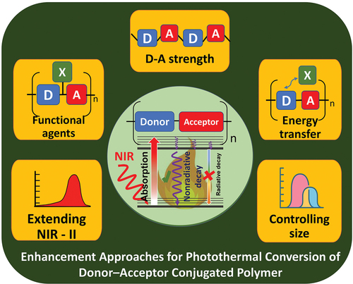
Conjugated polymer (CP)-based photothermal materials have been widely acknowledged as a promising class of photothermal agents for biomedical applications. This is because of their light-harvesting ability, photothermal conversion efficiency, photostability, and favorable biocompatibility. Donor–acceptor (D–A) CPs, which are based on the evolution of CPs, have attracted considerable interest in this field because of their tunable absorption in the near-infrared biological window. This property enables their deep penetration into cancer sites, improving the efficiency of anti-cancer treatment. This review mainly focuses on the potential of D–A CP to achieve improved and efficient photothermal conversion, exploring its optimized advantages for photothermal therapy applications. Based on the general insight provided by the Jablonski diagram, the mechanism and related principles for activating photothermal conversion in CPs and D–A CP are proposed and discussed. This provides an overall understanding of the correlation between molecular CP nanomaterials and heat generation. This review presents the details of methodologies for the rational design of CP nanoparticles with efficient photothermal conversion ability, thus facilitating the use of CPs in biomedical diagnostic and therapeutic applications.
Highlights
Photothermal conversion mechanism of organic materials and influencing elements based on Jablonski’s diagram.
Enhanced approaches for photothermal conversion of conjugated polymer nanoparticles.
Improved methodologies in photothermal conversion of donor–acceptor conjugated polymer nanoparticles.
Potential and future direction of donor–acceptor conjugated polymer development in photothermal therapy and biomedical applications.
Graphical abstract
1. Introduction
Cancer is a major health problem that causes morbidity and mortality worldwide, requiring immediate treatment. Currently, the traditional clinical treatments for cancer are surgery, radiotherapy, and chemotherapy. However, their side effects and unsatisfactory outcomes (such as lack of specificity, drug resistance, pain experienced by patients, low recovery, and metastatic disease) have limited their advantages [Citation1,Citation2]. Over the last decade, among the cancer therapies, photothermal therapy (PTT) has been acknowledged as a promising effective method and thus attracted enormous interest [Citation3,Citation4]. The technique is based on the conversion of light into thermal energy through the function of photothermal agents (PTAs). These agents elevate temperature to disrupt cell membranes and nuclear processes, thereby triggering cancer cell death. They are activated by external laser irradiation, especially in the near-infrared (NIR) biological therapy window ranges, NIR I (750–1000 nm) and NIR II (1000–1350 nm). The distinct advantages of using this range irradiation are deep penetration, negligible scattering, and low tissue absorption, which can minimize damage to surrounding healthy tissues [Citation5,Citation6]. The technique of PTT is a highly effective and noninvasive treatment that can be used to eradicate various types of cancer. Moreover, in terms of nonradiative dissipation, PTAs facilitate the creation of acoustic waves that can be detected by photoacoustic imaging (PAI). Hence, accurate cancer treatment and diagnosis by a multifunctional methodology are supported [Citation3,Citation7].
In PTT, the development of exogenous PTAs with considerable accumulation in tumors is expected to be key to assessing the efficiency of the treatment [Citation3]. The photothermal conversion efficiency (PCE) of PTAs is a calculated value representing the transformation of absorbed light energy into heat. An ideal PTA must have strong NIR absorbance, high mass absorption, high PCE, excellent optical stability, biocompatibility, and tumor-targeting ability [Citation8]. Accordingly, the advancement of PTT studies has accelerated the production of a variety of PTAs. In the last decade, many inorganic and organic nanomaterials with promising therapeutic applications have been widely investigated. In particular, inorganic PTAs (including gold nanomaterials, carbon nanomaterials, metal chalcogenide materials, and two-dimensional materials) have attracted intense interest owing to their excellent NIR absorption. However, concerns regarding difficult catabolism and long-term retention in the biological body have been raised. In contrast, organic PTAs, such as small molecules and conjugated polymers (CPs), offer additional advantages in terms of biosafety [Citation9–11]. Small molecule dyes (e.g. cyanine, BODIPY, thiocyanine, and porphyrin) have been reported as alternatives with tunable absorption ability and higher biological compatibility than metal materials [Citation12,Citation13]. However, low PCE, photobleaching, and poor water solubility are among the problems that restrict their further application.
To overcome the aforementioned problems, the use of a CP with a delocalized electronic structure and long π-conjugation in its backbone can be an outstanding solution. This polymer was originally developed to enhance the energy generation at longer wavelengths in organic electronics and photovoltaic cell applications [Citation14–16]. It has remarkable and unique properties, such as light-harvesting ability, photostability, high brightness, and favorable biocompatibility. Consequently, CPs have been investigated for biomedical applications, such as biosensing [Citation17], bioimaging [Citation18], phototherapy [Citation19], and photoactivation [Citation20]. Organic thermal CPs (including polyaniline (PANI), polydopamine (PDA), polypyrrole (PPy), and poly(ethylenedioxythiophene):polystyrene (PEDOT:PSS)) are widely explored in the PTT field [Citation21–25]. Donor–acceptor CPs (D–A CPs), which are based on the evolution of CPs, have recently attracted considerable attention in PTT studies. In D–A CPs, each repeat unit is composed of an alternative combination of electrical donor and acceptor segments. The π-orbitals overlapping between donors and acceptors result in the formation of new positions, from the highest occupied molecular orbital (HOMO) to the lowest unoccupied molecular orbital (LUMO), inducing the related electronic bandgap of the entire D–A CP. Owing to the push–pull structure, the low bandgap can be controlled, allowing the CP to absorb energy at a longer wavelength in NIR region with better penetration and low side effects; this renders it a promising region for PTT applications [Citation26]. In this regard, many considerable efforts have been devoted to study bandgap modulation, which can be tuned by the strength of D–A molecules, D–A ratio, and molecular weight (MW) of polymer [Citation27,Citation28]. Upon the activation of the D–A unit by photon energy, the intramolecular charge in the unit is induced and transferred along the backbone. Consequently, the charge recombination between electrons and holes is suppressed, reducing light emission [Citation29]. Based on this feature, the absorbed energy in the D–A CP can be released by the nonradiative pathway in the competitive photophysical process [Citation4], leading to the enhancement of heat generation based on D–A CP [Citation30,Citation31].
At first, this review aims briefly to summarize recent engineering progress in CP-based organic nanomaterials for enhancing photothermal conversion, which has considerable potential in PTT. And then, the prospect of D–A CP is emphasized by focusing on recent molecular engineering and nanoengineering approaches that can optimize the advantages of this class of materials. To clarify this aspect, the photothermal conversion mechanism and relevant factors for improving the PCE of organic molecules were discussed. The mention includes the adjustment of absorption peak as well as the enhancement of light absorption ability and efficiency of nonradiative decay. The study shows the significant correlation between structure and photothermal property in designing CP nanoparticles (CPNs). The relation among factors indicates the dominantly nonradiative decay resulting in heat generation, consistently observed in the competitive decay process based on the photophysical transition. Finally, the challenges and perspectives of D–A CPN for PTT applications are elaborated to facilitate understanding as well as stimulate the interest of researchers in the fabrication and utilization of photothermal D–A CPNs for photothermal medicine.
2. General mechanism, efficiency, and enhancement strategy of organic PA
2.1. Photothermal conversion mechanism (Jablonski diagram)
The interaction of a photon impinging on a chromophore can be classified into three processes: transmission, scattering, and absorption (). Light transmission occurs when no interaction occurs between photons and molecules. Light scattering (including inelastic or elastic scattering) occurs when incident light is reflected in various directions with different intensities. The remaining process, absorption, depends on complementing (matching) the electromagnetic frequency of light and nature of matter; this transfers the absorbed energy in various energy forms. The electronically and vibrationally excited state transition of a chromophore is derived from the absorption of a photon. It is controlled through a photophysical process following the Jablonski diagram. Therein, the arrangement of vibrational and electronic energy is built along the vertical direction, and the molecular spin multiplicity is managed in horizontal groups. The diagram clarifies the tendency between the radiative and nonradiative decays of a chromophore, enabling the exploration of the fundamental principle for the design of organic phototheranostic agents. As shown in , radiative transitions are indicated by straight arrows, and nonradiative decays are represented by squiggly arrows.
Figure 1. (a) Optical phenomena on surface of any material under light irradiation. (b) Photothermal generation by pristine CP.
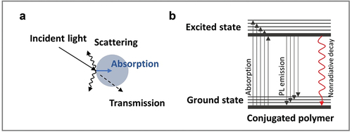
The absorption of the chromophore occurs rapidly after a photon at certain wavelengths of light interacts with the molecules. This typically induces the transfer of absorbed energy to molecules through the activation of the electron in rapid promotion (from the ground state (S0) to the higher excited state (Sn)); the absorption transition is extremely fast (in the order of 10 −15 s). Because of the excess energy under unstable states, when an electron is excited, it tends to quickly jump back to the initial ground state through multiple dissipation mechanisms. The first is vibrational relaxation. In the diagram, this nonradiative relaxation exists in the parallel light lines, which represent the decay of kinetic energy among vibrational levels at the same electronic level (represented by dark lines). The timescale for this process is extremely fast (10−14–10−11 s), facilitating its immediate occurrence following absorbance. When the vibrational energy levels strongly overlap with the electronic energy levels, the excited electron has a high probability of transitioning to a vibrational energy level with a lower electronic level, resulting in an internal conversion with the same mechanism of vibration relaxation. Therefore, internal conversion occurs through the transition of vibrational levels from the vicinity of electronic energy states. At higher energy levels of excitation (Sn), the density distributions of vibrational and electronic eigenstates become similar. This indicates that a considerable overlap of vibrational transitions may result in a higher degree of probability for the occurrence of internal conversion among electronic states, supporting the facile decay of active electrons. However, this process is limited from the first singlet state (S1) to the ground state because of the significant energy difference and the absence of vibrational and electronic energy state overlap between the S1 and S0 states. The timescale of internal conversion to the ground state is overly long, causing the activation of other competing processes to decay the excited electron energy from the first singlet excited state.
First, the direct radiative release of energy from S1 to S0 by photon emission with lower energy and longer wavelength than the incident light is known as fluorescence; this can be used for fluorescence sensing and imaging applications [Citation32,Citation33]. Fluorescence is a slow process (10−9–10−7 s); hence, the probability that this pathway emanates from the transition of the higher electronic states is low. Nevertheless, it frequently competes with the internal conversion from the decay in the S1 state.
Second, the relaxation of molecules can also occur in the form of nonradiative dissipation through intramolecular motions or collisions among molecules and with external solvent molecules. Thus, energy is vibrationally transferred from organic molecules to enclosing neighborhoods and released by thermal energy. Thermal expansion also occurs as a result of the generated heat. Heat expansion generates localized pressure waves, which can be recorded as acoustic waves. In this process, heat generation is widely regarded as the main rule for designing light-based theranostic agents for PTT and PAI [Citation29,Citation34–37]. It is combined with increasing temperature and heat-induced acoustic wave generation.
Instead of the two aforementioned pathways, organic optical molecules are also capable of going through a third transition pathway (from the lowest singlet excited state S1 to the lowest triplet excited state T1). In this path, the molecules happen a transition through intersystem crossing (ISC). This forbidden transition is related to the conversion of the spin multiplicity of a paired electron in the singlet state to an unpaired electron in the triplet state. This can result in radiative photon emission as phosphorescence or the generation of reactive oxygen species (ROSs). The latter is crucial to the implementation of photodynamic therapy [Citation3], which uses chemical processes to specifically target and kill cancer cells and tumors. Because the triplet transition is forbidden, the occurrence of ISC is several orders of magnitude slower than fluorescence (in the order of 10−8–10−3 s) and tends to be limited.
The main method of clarifying the energy transition processes in the photophysical process is achieved using the Jablonski diagram, which explicitly displays the preferable mechanism of energy release following the excitation of a particular molecule. Low fluorescence emitters are complemented by energy decay in the form of heat de-excitation, allowing them to be employed as PA imaging probes and PTT agents for tumor ablation. CPs contain a delocalized electronic structure and long π-conjugation in their backbone; thus, their transited energy is controlled by various intramolecular and intermolecular interactions related to the special features of chemical structure and morphology characteristic. Understanding structure-property relationship is important and must be considered when designing the molecular structure and aggregated state of CPNs, which is an ongoing investigation [Citation38].
2.2. Photothermal conversion efficiency (PCE)
The PCE is vital for evaluating the transformation of absorbed light energy to released heat. Generally, three methods are used to calculate the PCE. The first method is as follows:
In EquationEquation (1)(1)
(1) , the PCE is calculated as the ratio of the thermal energy, Q (produced by the PTAs in a solution), to the total energy, E, of incident light during the irradiation time. Here, cp and m are the specific heat and mass of the solution containing the PTAs, respectively; ∆T represents the increasing temperature of the solution; P represents the power density of the light source; S is the irradiation area; and t is the irradiation period. This approach considers all photons, including those in scattered and transmitted light; however, only the absorbed energy can generate thermal energy. In addition, the concentration of PTAs directly affects the heat generation; however, it is not indicated as an influencing feature in the equation. Therefore, comparing the η value among PTAs under different conditions can be difficult. Some PCE values have been calculated using this method [Citation39–41].
The second method is based on the temperature decay curve of the cooling process after removing the excitation irradiation, as given by EquationEquation (2)(2)
(2) :
where η is the PCE; h represents the heat transfer coefficient of a solution; S represents the surface area of the container; (Τmax − Τsur) is the maximum temperature change in the solution; Qdis denotes the extra energy contributed by the solvent and container; Aλ represents the light absorbance at the excitation wavelength, λ; and I is the power of incident light. This calculation can minimize common errors because only absorbed photons based on light absorbance are considered in the heat generation. Concurrently, the concentration-dependent absorbance of the suspension is introduced to the photothermal process through Aλ value. This enables the objective comparison among various PTAs for photothermal conversion. This method is reported in previous papers [Citation42–44].
A recent method for obtaining the PCE value by theoretically fitting the heating and cooling curves to a macroscopic model of collective particle heating is used [Citation45]. According to EquationEquation (3)(3)
(3) , the temperature T(t) at time point t is expressed in terms of the ambient temperature (T0), arbitrary initial temperature (Ti), and characteristic constants (A and B). Here, A and B represent the rate of energy absorption and heat dissipation, respectively. These were collected by fitting EquationEquation (3)
(3)
(3) to the experimental data of temperature versus time curves during the heating (under laser excitation, A ≠ 0 and Ti = T0) and cooling processes (A = 0 and Ti > T0). The PCE value is then calculated using EquationEquation (4)
(4)
(4) in terms of index A; mass (mi) and specific heat capacity (Ci) of each element (i) in the entire system; incident laser power (P); and absorbance intensity (Aλ) at the excitation wavelength. This approach has been considered in previous studies [Citation46,Citation47]. The equations are
2.3 Strategies for enhancing photothermal conversion
Based on the photothermal conversion mechanism, three main factors can be considered for designing an effective PTA for PTT: the position of maximum absorption, extinction coefficient, and quantum yield of nonradiative decay. Each factor related to the intrinsic molecular engineering of PTA can be considered in terms of the following.
The adjustment of the absorption spectrum of PTAs into the transparent tissue window at longer wavelengths is one of the most important aspects concerning penetration depth. In this range, most of the photoenergy could be transferred to the PTA, and the absorption loss through the tissue components could be significantly minimized. The absorption under NIR irradiation can be attributed to the transition from HOMO to LUMO. A vital approach for shifting the absorbance of conjugated molecules into the NIR window is narrowing the energy bandgap of HOMO–LUMO. Several methods have been used to reduce the energy bandgap of conjugated molecules/polymers. These include tuning the conjugated length of the polymer, controlling the bond length [Citation48], and controlling the D–A molecular orbital interaction [Citation49]. Notably, in relation to intermolecular interactions, the co-crystallized D–A assembly can also reduce the energy bandgap [Citation50]. In addition, the presence of free radicals in conjugated molecules and doped CPs [Citation51] can have the same effect on the redshift in the NIR range, suggesting that the absorbed energy is strongly released in heat form.
The ability of PTAs to absorb light energy is considered a relevant factor in boosting the PCE efficiency (as represented by the extinction coefficient). This ability can also be defined as the molar absorption coefficient [Citation52]. Occasionally, it can be used to account for the mass absorption coefficient [Citation53] or mass extinction coefficient [Citation54]. The molecular structure of CPs and nanocomposites has a strong absorption coefficient in the NIR range of the ‘therapeutic window’ is supposed to increase the amount of generated heat from the absorbed energy. According to quantum mechanics, the extinction coefficient is proportional to the dipole moment of the molecular transition. In this regard, a large spatial overlap of electron clouds was found to influence the transition dipole moment. With the improved coplanarity in the CP molecular structure (as indicated by the considerable overlap of electron clouds), more light energy would be absorbed, thereby, likely for enhancing the PCE [Citation55].
Electrons that are excited to a higher energy level tend to dissipate energy back to a stable ground state through radiative and nonradiative pathways. This decay involves competitive processes, including fluorescence emission, internal conversion, and ISC. Among these, improving the rate of internal conversion is considered a vital factor for enhancing photothermal efficiency. The concepts of temperature and thermal energy are related to molecular vibrations. Hence, the designed molecular structure, which can vibrate between molecules and solvent molecules, can also support vibration relaxation. Some intrinsic molecular modifications (such as the combination of twistable bonding [Citation56] and long alkyl length [Citation57]) in conjugated molecules have been found effective in reducing the fluorescence quantum yield for enhancing vibration relaxation.
3. Improvement of photothermal properties of conjugated polymer
The development of electroactive materials for PTT starts with CPs, which have large π-conjugated backbones and delocalized electronic structures. The hypothesis is that the heat generation mechanism in the CP under sufficient irradiation energy activation can be attributed to the existence of bipolarons (bications), which are created by alternating double bonds within the semiconducting polymer backbone [Citation58]. Because of lattice coupling, the energy released by bipolarons generates polarons in the form of electron–hole pairs [Citation59–61]. These pairs partially support the dissipation of energy to a nonradiative pathway for heat production to ablate a tumor. Recently, the conversion of CPs into nano-dimensionality has also been considerably emphasized because it provides additional features in a biologically aqueous environment. Based on the fundamental molecular structure, CPs contain various multichromophoric units in the main backbone. The π-electron delocalization in each chromophore unit significantly contributes to the entire polymer chain and overall optoelectrical properties of the system. In the nanodimension-preparation of CPs, the chains tend to collapse, which results in a close approach to the π-conjugated multichromophoric state, thereby affecting the excited-state dynamics and intrachain – interchain energy transfer [Citation62–64]. This rearrangement involves various chain conformations and aggregation states in CPNs, resulting in the modification of the photophysical properties of CPNs [Citation62,Citation64,Citation65]. Consequently, the heat energy released (according to the Jablonski diagram) is also affected by various intramolecular–intermolecular interactions affecting from the design of the molecular structure and aggregated state of CPNs; this is currently being investigated [Citation38].
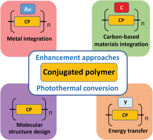
The CPs commonly used in PTT studies include PANI, PPy, PDA, PEDOT:PSS, and D–A CPs. Although considerable efforts have been devoted to investigating the photothermal properties in the NIR range, most organic CPs exhibit nonspecific absorption, preventing them from achieving optimum efficiency. To enhance the PCE in clinical PTT, increasing the concentration of PTAs and/or applying intense laser density is necessary to generate a higher amount of heat. However, these approaches damage the surrounding tissues, causing severe side effects. To resolve this, promising molecules with high absorbance are added to support the enhancement of light-harvesting and light-to-heat conversion reducing the aforementioned drawbacks. Concurrently, multi-components in the as-prepared nano-CP can also provide synergistic functionality, benefiting tumor uptake and treatment efficacy [Citation65,Citation66]. Besides, the common strategy of controlling molecular structures to aid in enhancing electron delocalization in the CP backbone, in relation to enhancing heat generation has also been mentioned. The overall view for recent developed PTA based CP is mentioned in .
Figure 2. Enhanced approaches based on photothermal conversion of CP.
3.1 Metal integration
Recently, the use of metals and metal compounds in CP systems to enhance biological properties has stimulated the interest of researchers [Citation67–70]. Metal nanoparticles are characterized by the well-known surface plasmon oscillation via collective electron oscillation on their surface, resonating with incident light. This greatly enhances the absorption and scattering of metal nanoparticles compared with bulk or other materials [Citation71,Citation72]. Based on the special localized surface plasmon resonance (LSPR), the absorption bands of metal compounds (such as gold and metal oxide nanoparticle) can also be facilely tuned from the visible range to the therapeutic window by modifying their size and morphology. Owing to their distinct features, the combination of metal compounds and CPs has been widely anticipated. In particular, gold nanoparticles (Au NPs) have been identified as an acceptable candidate for assisting the exciton dissociation process in a CP via plasmon–exciton coupling [Citation73–75]. Plasmon coupling provides the exciton more energy to overcome the Coulombic potential that holds the electron–hole pair together, thereby enhancing the probability of dissociation into free polarons [Citation76]. The foregoing may be regarded as a heat-generating process. In 2015, Li et al [Citation77]. successfully coated the surface of gold nanorods (AuNRs) with PPy via facial–interfacial polymerization to improve the temperature increment and photostability compared with pure AuNRs. In addition, the LSPR absorption band of AuNR composites can be fine-tuned by controlling the thickness of the coating of the biocompatible PANI shell [Citation68,Citation78]. Zhang et al [Citation79]. found that adjusting the thickness of the PDA shell on the surface of AuNRs in situ polymerization could robustly enhance the photothermal transduction efficiency (74.4% for AuNRs compared with 35.8% for raw AuNRs). This study provides significant evidence regarding the contribution of the conductive polymer shell to the PCE of the as-prepared metal nanomaterial (). The encapsulation of metal nanoparticles into the functional polymer matrix also promotes biocompatibility, biosecurity, and longer blood circulation; all of these benefit tumor absorption and PTT effectiveness. The half-life of PDA-coated AuNRs, which exhibited prolonged circulation time in the blood, was calculated at 4.5 h. This is six times longer than that of AuNRs without coating. The molecular structure of polymer PDA contains promising functional groups, such as amino and hydroxyl groups [Citation80], supporting the development of multifunctional theranostics through coordinated interactions during polymerization. Copper (II) has an unpaired three-dimensional electron in its molecules, leading to a nonzero nuclear spin magnetic moment. Therefore, the hybrid of Cu(II) and AuNR@CuPDA can reduce the longitudinal relaxation time (T1) of surrounding protons and can potentially function as a magnetic resonance imaging (MRI) contrast agent. Note that Cu(II) released in the PDA shell can behave as a chemotherapeutic agent, effectively inhibiting the tumor growth rate by 31.2%. The tumor could be ablated entirely by irradiating it with a 808-nm laser in PTT; no recurrence was observed. In the hybridization of photothermal polymers, encouraging results have also been found for several types of gold nanomaterials, such as Au NPs [Citation81], Au nanostars [Citation82], Au nanocubes [Citation83], and Au nanoflowers [Citation82]. Other materials (including metal oxides (iron oxide [Citation84,Citation85], titanium oxide [Citation86] and tantalum oxide [Citation87]), copper chalcogenides (CuS) [Citation88,Citation89], metal selenides (Bi2Se3) [Citation90], and metal tellurides (MoSe3) [Citation91], iridium [Citation92]) that are widely used in PTT are anticipated to be combined with photothermal CPs.
Figure 3. PCE-enhancing copper-doped PDA-coated AuNrs for tumor theragnostics. (a,b) TEM images of AuNrs and AuNr@cupda. (c) UV−vis absorption spectra of AuNrs and AuNR@ CuPDA. (d) Real-time temperature (time constants for heat transfer of AuNrs and AuNr@cupda are τ = 404.4 s and τ = 348.5 s corresponding to PCE values 35.8% and 74.4%, respectively). (e) T1-weighted MRI of nude mice bearing KB tumor without AuNr@cupda treatment. (f) T1-weighted MRI of mice 24 h after injection of AuNr@cupda solution. (g) Cone beam CT images of mice treated with saline (left) and AuNr@cupda (right) 24 h after treatment. (h) Infrared thermal images of mice treated with saline (top) and AuNr@cupda (bottom) under 808-nm laser irradiation. (k) Relative growing trend of tumor volume of each group. Reprinted with permission from [Citation79].
![Figure 3. PCE-enhancing copper-doped PDA-coated AuNrs for tumor theragnostics. (a,b) TEM images of AuNrs and AuNr@cupda. (c) UV−vis absorption spectra of AuNrs and AuNR@ CuPDA. (d) Real-time temperature (time constants for heat transfer of AuNrs and AuNr@cupda are τ = 404.4 s and τ = 348.5 s corresponding to PCE values 35.8% and 74.4%, respectively). (e) T1-weighted MRI of nude mice bearing KB tumor without AuNr@cupda treatment. (f) T1-weighted MRI of mice 24 h after injection of AuNr@cupda solution. (g) Cone beam CT images of mice treated with saline (left) and AuNr@cupda (right) 24 h after treatment. (h) Infrared thermal images of mice treated with saline (top) and AuNr@cupda (bottom) under 808-nm laser irradiation. (k) Relative growing trend of tumor volume of each group. Reprinted with permission from [Citation79].](/cms/asset/2b8e2740-d565-425b-a2a6-78e4fd5b04d0/tsta_a_2134976_f0003_oc.jpg)
3.2 Carbon-based nanostructure integration
In addition to metal and metal compounds, carbon materials (such as graphene and carbon nanotube, created by sp2 hybridized carbons) also show promising photothermal effects owing to their high absorption coefficient expanded from the ultraviolet (UV)–visible (vis) to the NIR range [Citation41,Citation93–98]. However, their applications are frequently limited by their low water dispersibility, lack of solution processability, and facile aggregation. The surface modification of carbon nanomaterials with CPs made of large π-conjugated backbones and delocalized electronic structures is a potential method not only for modifying surface properties but also enhancing the PCE of the resulting system. Because of the presence of functional groups in the structure, the surface modification of carbon materials may be alternated by noncovalent and covalent techniques. Non-covalent binding is advantageous in an existing system with π–π stacking, hydrogen bond interactions, or electrostatic interactions [Citation99,Citation100]; this facilitates the efficient intermolecular electron transfer in the composites [Citation41,Citation101,Citation102]. Covalent modification provides stronger connected grafted moieties and leads to more stable composite structures [Citation103]. Instead of using conventional hydrophobic CPs, the development of ionic water-soluble CPs (composed of conjugated aromatic backbones and ionic side chains) on grafted carbon materials for PTT applications is critical owing to their satisfactory solubility in water and the intriguing optoelectronic properties of the system. Geng et al [Citation44]. developed positively and negatively charged reduced graphene oxide (RGO) composites (RGO-g-P3TOPA and RGO-g-P3TOPS, respectively), which were synthesized by covalently grafting poly(3-(3’- thienyloxy) propyltrimethylammonium bromide) (P3TOPA, a cationic CP) and poly(sodium 3-(3’- thienyloxy)propanesulfonate) (P3TOPS, an anionic CP) onto RGO sheets using a grafting-through method [Citation104]. The integration of water-soluble CPs into the RGO sheet enhanced the optical absorption from the UV to the NIR region as well as redshifted the absorption maximum of GO (from approximately 233 to 268 nm) and the absorption peak of the as-prepared CPs [Citation105]. This is due to the increased electron interaction between the RGO basal planes and polymer main chains, leading to reduced bandgaps in both CPs. Concurrently, photoinduced electron transfer (PET) from CPs to RGO effectively occurred [Citation41], especially the quenching of the light emission of P3TOPA in the presence of RGO because of covalent linking and electrostatic attraction among components [Citation106]. The RGO-g-P3TOPS component is linked only by the electrostatic repulsion of negatively charged RGO basal planes and P3TOPS, resulting in lower quenching. Consequently, the photothermal effect of the composites has improved significantly: 88.5% and 88.1% for RGO-g-P3TOPA and RGO-g-P3TOPS, respectively; these values are higher than those of the materials without RGO (i.e. 54.6% and 48.4%, respectively). Notably, the positive charge binding on RGO-g-P3TOPA has attracted the negatively charged Escherichia coli (E. coli) through electrostatic interaction. The RGO-g-P3TOPA composite killed 100% of the E. coli bacteria through photothermal heating. In contrast, for the RGO-g-P3TOPS composite to achieve a similar bactericidal effect, a 16-fold increase in its amount is required ().
Figure 4. Enhanced photothermal conversion based on carbon-based nanostructure integration. (a) Scheme of efficient photothermal bactericidal process by linking positively charged RGO-g- p3topa. (b) TEM images of RGO-g-p3topa and RGO-g-p3tops sheets. (c) Optical-electronic properties of RGO-g-p3topa composite; RGO-g-p3tops composite and their components: enhanced absorption from UV region to NIR range with redshifted absorption peak of GO and CPs in composites (top); photoluminescence quenching of CPs with RGO present (bottom), proving transfer of photoinduced electron from CPs to RGO. (d) Photothermal bactericidal experiment on RGO-g-p3topa (two figures at top) and RGO-g-p3tops (two figures at bottom) composites according to various concentrations indicated by survival rate of E. coli bacteria (left) and related temperature increase (right): result indicates amount of RGO-g-p3tops to be 16-fold to obtain similar bactericidal effect as RGO-g-p3topa. (e) SEM image of treated E. coli in bactericidal experiment using P3TOPA and RGO-g-p3topa with comparable concentrations of P3TOPA component. (f) SEM image of treated E. coli in bactericidal experiment using P3TOPS and RGO-g-p3tops with comparable concentrations of P3TOPA component. Reprinted with permission from [Citation44].
![Figure 4. Enhanced photothermal conversion based on carbon-based nanostructure integration. (a) Scheme of efficient photothermal bactericidal process by linking positively charged RGO-g- p3topa. (b) TEM images of RGO-g-p3topa and RGO-g-p3tops sheets. (c) Optical-electronic properties of RGO-g-p3topa composite; RGO-g-p3tops composite and their components: enhanced absorption from UV region to NIR range with redshifted absorption peak of GO and CPs in composites (top); photoluminescence quenching of CPs with RGO present (bottom), proving transfer of photoinduced electron from CPs to RGO. (d) Photothermal bactericidal experiment on RGO-g-p3topa (two figures at top) and RGO-g-p3tops (two figures at bottom) composites according to various concentrations indicated by survival rate of E. coli bacteria (left) and related temperature increase (right): result indicates amount of RGO-g-p3tops to be 16-fold to obtain similar bactericidal effect as RGO-g-p3topa. (e) SEM image of treated E. coli in bactericidal experiment using P3TOPA and RGO-g-p3topa with comparable concentrations of P3TOPA component. (f) SEM image of treated E. coli in bactericidal experiment using P3TOPS and RGO-g-p3tops with comparable concentrations of P3TOPA component. Reprinted with permission from [Citation44].](/cms/asset/f5d66313-6ed8-4c90-8fcd-ada73c242aad/tsta_a_2134976_f0004_oc.jpg)
3.3 Energy transfer simulation
The use of two sources of absorbed energy based on corresponding CPs in a nanocomposite has been reported as one of the effective means for enhancing the heat generation in PTT [Citation107,Citation108]. In 2019, Xia et al. [Citation109]. We successfully prepared a ‘nanococktail’ of CPNs using a single laser to produce significant dual thermal energy through fluorescence resonance energy transfer (FRET) (). The FRET mechanism describes the non-covalent energy transfer that occurs when the emission of donor fluorogen overlaps with the absorption of an acceptor molecule within a closely related distance [Citation110]. In this study, a (poly(cyclopentadithiophene-alt-diketopyrrolopyrrole) (PDPPP) polymer, which strongly absorbs NIR energy of 600–900 nm, is used as the main source of heat generation and as an energy acceptor. The aggregate-induced emission agent, TPA–BDTO (AIEgen T), has a maximum emission peak at 660 nm, overlapping with the absorption of P, which acts as a donor. The system was then prepared in the form of nanoparticle dispersions of DTP(R) via the encapsulation of amphiphilic polymer (DSPE–PEG2000–Mal (D)), followed by the capping of targeting agents (R). The DTP(R) exhibited two distinct peaks at 530 and 840 nm from donor T and acceptor polymer P, respectively. Interestingly, upon excitation at 530 nm, the DTP CPN displayed the same fluorescence peak at 660 nm; however, its intensity was lower than that of the CPN containing only T. The increase in the amount of quencher (P) decreases the emission intensity in the DTP CPN, suggesting that P receives energy from the emission of T through the FRET mechanism. The DTPR CPN efficiently converted light to heat (reaching 68°C) with a conversion efficiency of 60.3%. This was a 1.9-fold increase compared with that of CPN containing only P (31.5%, 48.6°C). This indicates an effective energy transfer for amplifying the heat released from the synergistic D–A pair (T–P) compared with that generated by pure polymer. Under the same conditions, the DTPR CPN with dual photothermal sources had higher cytotoxicity (93.8%) than the DPR CPN with a single source (70.9%) against SKOV-3 cells. This resulted mechanism also be investigated in group of Xia et al. for enhahnced NIR-II PTT in term of a cell membranced-anchored nanoassembly, indicating a great tumor accumulation and efficient tumor suppression in PTT [Citation111]. On similar version, another report by Pu et al. [Citation112] displayed the charge transfer between p-CP backbones and the anthraquinone part of DOX (an aromatic anticancer drug) to explain fluorescence quenching and enhanced photothermal temperature. Based on the strong hydrophobic and π–π interactions during nanofabrication, DOX can be assembled in the amphiphilic poly(ethylene glycol) (PEG)-grafted poly(cyclopentadithiophene-alt-benzothiadiazole) (PCB) (i.e. PEG–PCB) to form hybrid CPNs. This supports a 75% decrease in fluorescence intensity and is 4.1°C higher than that of a CPN without DOX. Under 808-nm NIR laser exposure, the temperature of the tumor in hybrid CPN-treated mice reached approximately 60°C after 2 min of irradiation. This is approximately 1.5-fold of that of the control mice (39.3°C), leading to the efficient killing of cancer cells and significant inhibition of tumor growth. This study provides an intraparticle molecular engineering approach to enhance photothermal conversion and integrate essential properties into one polymer for multimodal nanotheranostics.
Figure 5. Enhanced photothermal conversion based on energy transfer. (a) Schematic of single (left) and dual (right) PTT strategy under 808-nm laser irradiation with addition of AIEgen T for energy transfer of photothermal CP PDPPP. (b) Preparation of DTPR nanoparticles. (c) Schematic of DTPR nanoparticles for 808-nm-activated image-guided dual PTT. Reprinted with permission from [Citation109].
![Figure 5. Enhanced photothermal conversion based on energy transfer. (a) Schematic of single (left) and dual (right) PTT strategy under 808-nm laser irradiation with addition of AIEgen T for energy transfer of photothermal CP PDPPP. (b) Preparation of DTPR nanoparticles. (c) Schematic of DTPR nanoparticles for 808-nm-activated image-guided dual PTT. Reprinted with permission from [Citation109].](/cms/asset/c7056768-a3ae-4f66-a063-89b1d4783fe3/tsta_a_2134976_f0005_oc.jpg)
3.4 Molecular structural design
By accurately constructing their backbone structures, CPs can be used in PTT by controlling radiative and nonradiative decay pathways to capture more energy from light for heat conversion [Citation113–115]. In 2018, Pu et al [Citation53]. reported the molecular design of optically active polymer nanoparticles incorporated with vinylene bonds, remarkably enhancing the photothermal conversion. These polymer nanoparticles are also promising biodegradable agents for improving precision medicine. Biodegradable CPs with and without vinylene bonds were synthesized poly{2,2′-[(2,5-bis(2-hexyldecyl)-3,6-dioxo-2,3,5,6-tetrahydropyrrolo[3,4-c]pyrrole-1,4-diyl)-dithiophene]-5,5′-diyl-alt-vinylene} (DPPV) and poly{2,2′-[(2,5-bis(2-hexyldecyl)-3,6-dioxo-2,3,5,6-tetrahydropyrrolo[3,4-c]pyrrole-1,4-diyl)-dithiophene]-5,5′-diyl-alt-thiophene-2,5-diyl} (DPPT) (), followed by co-precipitation with amphiphilic polymer PLGA–PEG to provide corresponding SPNV and SPNT nanoparticles, respectively. These spherical nanoparticles are approximately 36 nm in size and exhibit broad absorption in the 600–900-nm NIR energy range, promising for PA imaging and PTT. In particular, the maximum absorption of SPNV is at 819 nm, with mass absorption and molar extinction coefficients of 40 cm−1∙mg−1 ∙mL and 32.1 cm−1∙mol−1 ∙mL, respectively; these are 1.3-fold and 1.2-fold higher than those of the SPNT in its maximum absorption at 828 nm (30 cm−1∙mg−1 ∙mL and 25 cm−1∙mol−1 ∙mL, respectively). Owing to its potential for light absorption, the SPNVs exhibited an extraordinarily increasing temperature at each time point compared with the SPNTs at the same concentration and under the same conditions. The highest temperature reached by SPNV and SPNT were 53 and 43°C, respectively; this resulted in significant improvements in their PCEs of 71% and 29%, respectively. Regarding photothermal conversion, the PA signal of the SPNV achieved by pulsed laser irradiation (680–900 nm) was 2.2-fold stronger than that of the SPNT. This enhancement in light absorption and related improvement of PTT and PAI properties were accompanied by a more planar conformation in the presence of the vinylene group, DPPV. This facilitates electron delocalization within the polymer backbone [Citation116] and more compact-chain packing in nanoparticle preparation [Citation117]. Recently, the biodegradability of combined materials containing the vinylene bond group as a structural unit has been observed as a result of the hydrolysis of this functional group in the existing abundance of the oxidative species environment [Citation118]. During the interaction of hydrogen peroxide (H2O2) and myeloperoxidase (a peroxide enzyme in immune cells), the appearance of a strong oxidant HCLO induced the absorption of SPNV tends to decrease. This demonstrates the breakout of vinylene bonds. In contrast, the absorption of SPNT without a special group in their synthesized molecular structure seems unchanged. The enhanced therapeutic outcome of PTT/PAI and organically benign SPNV compared with SPNT has been demonstrated both in vitro and in vivo as showing considerable potential for medicine. This indicates that the combination of functional groups can efficiently improve the PCE of CPs for PTT applications.
Figure 6. Enhanced photothermal conversion based on vinylene bond combination (DPPV). (a) Chemical structures of SPNV and SPNT. (b) Mass absorption coefficients of SPNV and SPNT. (c) Photothermal images of SPNV, SPNT, and PBS. (d) in vitro biodegradability study of SPNs: schematic of degradation of SPNV with MPO and H2O2 presence. (e) Degradable absorption spectra of SPNV with H2O2 and MPO present. (f) Decreased absorption (Abs/Abs0) of SPNV and SPNT with MPO and H2O2 present in each incubation time period. Reprinted with permission from [Citation53].
![Figure 6. Enhanced photothermal conversion based on vinylene bond combination (DPPV). (a) Chemical structures of SPNV and SPNT. (b) Mass absorption coefficients of SPNV and SPNT. (c) Photothermal images of SPNV, SPNT, and PBS. (d) in vitro biodegradability study of SPNs: schematic of degradation of SPNV with MPO and H2O2 presence. (e) Degradable absorption spectra of SPNV with H2O2 and MPO present. (f) Decreased absorption (Abs/Abs0) of SPNV and SPNT with MPO and H2O2 present in each incubation time period. Reprinted with permission from [Citation53].](/cms/asset/f0ce39de-27cb-42de-a5f7-d832e8c9bdf7/tsta_a_2134976_f0006_oc.jpg)
Recently, the emergence of D–A CP has been highlighted as a new level in the molecular design of CP for PTT applications, owing to the flexibility of D–A units combined. These units induce the activation of intramolecular charge transfer and bandgap control, resulting in virtually complete fluorescence quenching and enhanced heat generation. In the next section, the enhanced approaches to the photothermal properties of D–A CP are further clarified, demonstrating its considerable potential for PTT medicine.
4. Enhancement approaches for photothermal properties of Donor–Acceptor conjugated polymers
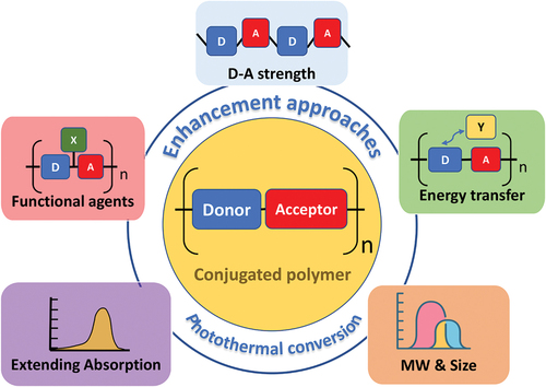
Because of their promising tunable optical properties and excellent photostability, D–A CPs, which have been designed using an alternative array of electron-rich units (donors) and electron-deficient segments (acceptors), have received considerable interest for PTT. The intramolecular charge transfer between the donor and acceptor is created in a unit belonging to the polymer chain. This results in the reduced charge recombination of electrons and holes, thereby leading to fluorescence quenching and enhanced heat generation. With this electronic push–pull structure, the energy bandgap of D–A CPs can be narrowly controlled, enabling efficient NIR absorption. In this review, the regulation of the photophysical properties of D–A CPs for enhancing heat emission has been mainly focused on enhancing the D–A strength, combining functional groups, stimulating energy transfer, controlling the MW and size of the nanostructure, and optimizing wavelength absorption ().
Figure 7. Enhanced approaches based on photothermal conversion of D–A CP.
4.1. D–A strength enhancement
The design of D–A CPs is based on an alternative arrangement of enriching electrons and electron-deficient segments in a repeat unit. The replacement of a donor or an acceptor by those with stronger or weaker features or its combination with different ratios induces an alternative electron density, resulting in an adjustable bandgap and a related optical bandgap [Citation28]. The controlled polymer at the lower bandgap leads to absorption at a longer wavelength, supporting the effective tuning of absorption in the NIR region and heat conversion. In 2018, Huang et al [Citation119]. designed and evaluated two photothermal CPs based on their distinct D–A strengths in combination with the same donor ((E)-1,2-diethoxy-1,2- bis(5-(trimethylstannyl) selenophen-2-yl)ethene (SVS)) with two different acceptors (6,6ʹ-dibromodi(2-octyldodecyl)isoindigo (IIG) and (E)-2,2ʹ-dibromo-4,4ʹ-bis(1-octyl)-[6,6ʹ-bithieno[3,2-b]pyrrolylidene]-5,5ʹ(4 H,4 H)-dione (TIG), denoted as PTIGSVS and PIIGSVS, respectively) (). When PTIGSVS and PIIGSVS are dissolved in a suitable solvent (such as THF), the former exhibits NIR absorption in the range 380–1000 nm with the highest peaks at 472 and 786 nm, and the latter showed a slight blueshift absorption from 380 to 800 nm with λmax at 419 and 715 nm. The highest peaks at longer wavelengths were observed for the intramolecular charge transfer of CPs (786 nm and 715 nm), indicating that the acceptor strength of TIG is greater than that of IIG; this conforms with reported results [Citation120]. To understand the molecular structure-dependent optical absorption, the backbone of CPs was modeled by density functional theory (DFT). A better planar structure with lower dihedral angles of 0.1°, 12.7°, and 0.1° was exhibited by PTIGSVS while the dihedral angles of the planar structure of PIIGSVS were 12.6°, 19.9°, and 0.1°. This resulted in a larger extinction coefficient in the former compared with the latter (249 and 15 Lg−1∙cm−1, respectively). In the nanoparticle form encapsulated by Pluronic PF-127, the optical properties of PIIGSVS CPN and PTIGSVS CPN were defined as broadened spectra in the range 580–1000 nm with the mass extinction coefficient decreasing to 25.5 and 46 Lg−1 cm−1 at 808 nm, respectively. This was attributed to the reduced intramolecular charge transfer and improved intermolecular π–π stacking among the polymer chains in CPN. Meanwhile, the dominant nonradiative decay of PTIGSVS CPNs was observed to be a strongly competitive process in both radiative decay and ISC routes, indicating that PTIGSVS CPNs had strong photothermal effects. As expected, under 808-nm laser irradiation for 5 min, the temperature of PTIGSVS CPN rapidly increases to 45°C, which is significantly higher than the enhanced of PIIGSVS CPN temperature of 13°C corresponding to PCEs of 74% and 11% for PTIGSVS CPN and PIIGSVS CPN, respectively. In vivo PTT, PTIGSVS CPNs were injected on the mice bearing 4T1 tumors, the temperature was raised to 70.5°C after 5 min of irradiation with an 808 nm laser, which was adequate to trigger cancer cell death and totally control tumor development during the therapy time. The tumor virtually disappeared after 15 d of treatment with no recurrence. The survival rate of 4T1 mice with PTIGSVS CPN–PTT treatment was 100% after 60 d; this is in contrast with the result of the control groups in which PBS, laser, or CPN was solely used. These findings indicate that PTIGSVS CPNs have high potential as PTT agents in thermotherapy applications.
Figure 8. Enhanced photothermal conversion based on higher D–A strength in PTIGSVS. (a) Chemical structures of PIIGSVS and PTIGSVS. (b) Optimized conformation revealing more planar structures of PTIGSVS (right) than PIIGSVS (left). (C) Significant increase in UV–vis absorption of PTIGSVS CPNs in NIR range. (d) Quenching photoluminescence spectra of PTIGSVS CPNs. (e) Photothermal thermal effect of PIIGSVS CPNs under 808-nm laser indicating increasing temperature compared with that of PIIGSVS CPNs. (f) in vivo PTT images of mice bearing 4T1 tumors showing results of four treatment groups, including (i) PBS group, (ii) NIR group, (iii) PTIGSVS CPN group, and (IV) PTIGSVS NPs + laser group. Reprinted with permission from [Citation119]. .
![Figure 8. Enhanced photothermal conversion based on higher D–A strength in PTIGSVS. (a) Chemical structures of PIIGSVS and PTIGSVS. (b) Optimized conformation revealing more planar structures of PTIGSVS (right) than PIIGSVS (left). (C) Significant increase in UV–vis absorption of PTIGSVS CPNs in NIR range. (d) Quenching photoluminescence spectra of PTIGSVS CPNs. (e) Photothermal thermal effect of PIIGSVS CPNs under 808-nm laser indicating increasing temperature compared with that of PIIGSVS CPNs. (f) in vivo PTT images of mice bearing 4T1 tumors showing results of four treatment groups, including (i) PBS group, (ii) NIR group, (iii) PTIGSVS CPN group, and (IV) PTIGSVS NPs + laser group. Reprinted with permission from [Citation119]. .](/cms/asset/9b9578fe-9102-472a-9dba-c859e3991fe6/tsta_a_2134976_f0008_oc.jpg)
Another study that supports D–A strength engineering in CPs focused on a controlled adequately planed structure. The as-synthesized polymers, represented by CP1–CP3, were obtained by the copolymerization of a donor, 4 H-dithieno[3,2-b:2,3 -d]pyrrole (DTP), and various acceptors, such as benzothiadiazole (BT), pyridal[2,1,3]thiadiazole (PT), and diketopyrrolopyrrole (DPP). Their D–A attractive force follows a strength order of CP1 < CP2 < CP3 [Citation29]. Evidently, the variation in D–A strength among the CPs leads to different optical and physical properties [Citation121]. In particular, the absorption spectra of CP3 displayed a greater redshift and higher intensity than those of CP2 and CP1. The DFT and time-dependent DFT calculations were conducted to determine the effect of molecular structure on the electron delocalization of CPs. The results showed that although the DPP in CP3 has a bulkier and stiffer molecular structure than the other CPs, the adjacent bisthiophene rings existing in the backbone of CP3 can reduce the torsion angle and hampered space [Citation122]. Consequently, absorption is enhanced, and the intrachain/interchain electronic interaction in the solution/aggreagated state is facilitated owing to the adequately planed structure [Citation123,Citation124]. The calculation also shows that CP3 has the strongest oscillator strength, which is consistent with the high extinction coefficient, promising for phototherapeutic application. The resulted nanoparticles showed the maximum peaks are at 690, 740, and 783 nm, and the mass extinction coefficients are 41.4, 44, and 59.3 L∙g−1∙cm−1 for samples CPN1–CPN3, respectively. Interestingly, CPN3 maintained virtually the same absorption capacity (57.7 L∙g−1∙cm−1) as the CP3 solution at 808 nm, rendering it as a promising PA/PTT agent [Citation125–127]. As for photophysical transition, CPN3 exhibited the lowest emission and negligible ROS, followed by CPN2 and CPN1. Hence, most of the absorbed energy was observed to dissipate by nonradiative decay with the strongly enhanced temperature of CPN3, which approached 68°C under 808-nm laser activation (0.8 W∙cm−2) for 3 min; the temperatures of CPN1 and CPN2 were moderate (i.e. 56 and 61.2°C, respectively). The CPN2 and CPN3 displayed 2.8-fold and 5.8-fold brighter PA signals than that of CPN1 upon excitation at 780 nm. They are also dominantly brighter than that of the indocyanine green (ICG) (as reference) at the same mass concentration, indicating a potential impact on PA images. The in vivo PTT/PA evaluation of CPN3 also shows promising results in treatment when validated in the U87 tumor–mice model. Under laser irradiation, the tumor temperature in mice increased to 52.7°C, and the PA signal 24 h after injection showed a high contrast at a depth of 3.2 nm, which is superior to that of common PA agents. The 100% survival rate of treated mice was 100% after 30 d. No side effects, such as recurrence of tumor growth, body weight loss, or cytotoxicity in normal blood function, were observed, indicating excellent biocompatibility and high tumor ablation efficiency.
The foregoing shows that by controlling the D–A molecular strength in CPs, the optical – physical CPN properties, such as backbone planarity, D–A intensity, emission, and radiative/nonradiative decay route, are efficiently tuned [Citation128]. Hence, this is a critical strategy for designing the molecular structure and aggregated state of CP to achieve optimized efficiency; it is promising for further clinical phototheranostic applications.
4.2. Combination of functional groups
The photothermal conversion of CPNs can be strongly boosted by the introduction of a light-harvesting unit or functional groups in the polymer architecture; this considerably enhances the absorption of the as-prepared polymer [Citation129]. A promising property of a porphyrin–pyrene pendant was observed in early polymeric solar applications [Citation130]. The photoinduced charge transfer in Porphyrin-pyrene faciliates for redshifting and enhancing the Q-band, assisting for broaded absorption and increased absorption coefficient in D-A based molecular architecture [Citation131,Citation132]. In 2017, Lee et al. [Citation133] showed that the integration of the light-harvesting unit (porphyrin–pyrene pendant) into the D–A CP backbone for assembled nanoparticles (PPor–PEG CPNs) () facilitated the highly effective PAI-guided PTT with a calculated PCE value of 62.3%. Because of the enhanced intramolecular charge transfer resulting from the integration of the light-harvesting group, the HOMO and LUMO of the synthesized polymer were modified to −5.59 and −3.31 eV, respectively, thus benefiting the redshift and improving photothermal efficiency. Specifically, under continuous 635-nm laser irradiation (1.4 W∙cm−2), the Ppor–PEG CPNs exhibited an increase in temperature, virtually reaching 55°C for 5 min with excellent photostability compared with ICG. The Ppor–PEG CPNs consistently displayed negligible fluorescence as a result of aggregation-induced quenching while Ppor dissolved in THF displays significant red-light emission; this is promising for the main nonradiative decay in the heat generation of the as-prepared NPs. The Ppor–PEG CPNs also presented a linear correlation between PA effects and related CPN concentrations with the ability to detect the tumor site 24 h after injection. Hence, they have potential use as long-term photoacoustic contrast agents in vivo. To evaluate cytotoxicity, the incubation of PPor–PEG CPNs was prepared using A549 human lung cancer cells and HeLa human cervical cancer cells. The results showed dose-dependent cytotoxicity in the laser exposure. A survival rate of approximately 90% was observed for both cell lines in the absence of laser, indicating the satisfactory biocompatibility of PPor–PEG CPNs. These preliminary results indicate that PPor–PEG CPN is a potential clinically translatable nanomaterial for effective PAI-guided PTT with excellent antitumor properties and biosafety. Recently, the requirement for improving the photothermal conversion of hydrophobic CPN also was strongly responded by the emergence of various hydrophilic functional groups linked into the poorly soluble backbone, enhancing water-soluble properties of CPNs. The addition of the cation quaternary ammonium group in each repeat unit has been proved to be able to modify the soluble properties of the main backbone, inhere, the presence of electron-rich dithieno-pyrrole and electron-deficient benzothiadiazole mobilities serves as the electron donor and acceptor, respectively [Citation134]. Covered by the positively charged surface with the increased water solubility, DPTPBT CPN indicated a strong absorption in the near-infrared region, excellent photostability, promising photothermal performance with a high PCE value (71.1%) and a significantly effective antibacterial effect under 808 nm irradiation. Wu et al. also successfully fabricated D-A CPN (PTPTA), which had a brush-like topological structure with two PEG chains (2000 Da) in each repeating unit [Citation135]. The PTPTA CPN possessed high hydrophilicity, good biocompatibility as well as strong absorption and photothermal conversion in NIR-II, serving as a promising agent for the light-triggered treatment.
Figure 9. Enhanced photothermal conversion based on combination of light-harvesting agent, PPor. (a) Schematic of PPor–PEG NP preparation and photothermal mechanism. (b) Chemical structure of PPor. (c) Absorbed spectrum of free-PPor solution and Ppor–PEG CPN. (d) Photothermal effect of Ppor–PEG CPN under 635–nm laser irradiation (1.4 W cm−2). (e) Linear correlation of PA signal to concentration of PPor–PEG CPN. (f) in vivo infrared thermal images. (g) Temperature increase in tumors of A549 tumor-bearing mice injected with PPor–PEG CPN under treatment condition. (h) Relative tumor volumes. (i) Body weight and (j) H&E staining images of tumor slices of treated mice showing promising results in biosafety evaluation. Reprinted with permission from [Citation133].
![Figure 9. Enhanced photothermal conversion based on combination of light-harvesting agent, PPor. (a) Schematic of PPor–PEG NP preparation and photothermal mechanism. (b) Chemical structure of PPor. (c) Absorbed spectrum of free-PPor solution and Ppor–PEG CPN. (d) Photothermal effect of Ppor–PEG CPN under 635–nm laser irradiation (1.4 W cm−2). (e) Linear correlation of PA signal to concentration of PPor–PEG CPN. (f) in vivo infrared thermal images. (g) Temperature increase in tumors of A549 tumor-bearing mice injected with PPor–PEG CPN under treatment condition. (h) Relative tumor volumes. (i) Body weight and (j) H&E staining images of tumor slices of treated mice showing promising results in biosafety evaluation. Reprinted with permission from [Citation133].](/cms/asset/a2f60d1d-7183-4338-b8fd-03b577af7aef/tsta_a_2134976_f0009_oc.jpg)
The foregoing shows that modifying CP with a light-harvesting unit or functional groups considerably enhances the absorption of the as-prepared CPNs as well as affects their optical– physical and photostability properties. This is positively promoting further clinical phototheranostic applications in the future.
4.3. Energy transfer stimulation
In contrast to molecular engineering, the nanoapproach is based on the intraparticle interaction force in an assembled nanocomposite. The addition of various components not only enhances the absorption spectra of combined materials but also influences their related photophysical properties. This facilitates the light-harvesting application and highly efficient transfer of absorbed energy to bio-applications as useful energy. For instance, electric components with a Fermi level or conduction band that is electrically lower than that of the CP can be utilized in aggregated composites. This can result in the transition of the excited electron to the conduction band instead of the direct relaxation of an activated electron to the ground state, as described in the Jablonski diagram. In this system, the released nonradiative heat is generally accompanied by the transition of electrons due to enhanced electron delocalization. Pu et al [Citation34]. prepared a dual-component nanostructure based on an intraparticle molecular orbital engineering approach. The primary D–A polymer (poly [2,6-(4,4-bis(2-ethylhexyl)-4H-cyclopenta-[2,1-b;3,4-b]dithiophene)-alt-4,7-(2,1,3-benzothiadiazole)] (PCPDTBT)) served as donor, and the secondary electrophilic dopant ((6,6)-phenyl-C71-butyric acid methyl ester (PC70BM)) was presented as acceptor. Based on the careful alignment of molecular orbitals, the HOMO and LUMO energy levels of PCPDTBT (from −4.9 to −3.5 eV) are higher than that of PC70BM (from −6.5 to −4.3 eV). The excited electron in the donor tends to transfer to the conduction band of the acceptor component instead of directly radiatively decaying to the ground state, thereby facilitating fluorescence quenching and enhancing nonradiative heat emission (). This foregoing hybrid is defined as the PET process and observed by optical characterization. In the UV absorption spectra, the CPNs exhibited two distinct peaks: a particularly unchanged peak at 650 nm for PCPDTBT and a remaining peak at 404 nm with various intensities due to the increasing amount of fulleren PC70BM. The figure indicates that the absorbed energy can be dissipated by competing processes: the decrease in photoluminescence intensity and gradual increase in the photoacoustic intensity when more PC70BM was added. This provides a 2.6-fold amplification in the in vitro photoacoustic brightness of CPN-F20 compared with that of CPN-F0. As for the thermal mechanism, the CPN-F20 also displays an increase in the photothermal temperature by 1.3-fold (from 48 to 62°C); this is consistent with the in vivo PTT. This study introduces a new generation of organic optical theranostics centered on CPNs with the amplified PTT/PAI properties based on the presence of hybrid components.
Figure 10. Enhanced photothermal conversion based on PET. (a) Chemical structures of PCPDTBT, PC70BM, and PEG-b – ppg-b – peg. (b) Images of solutions of CPNs with various amounts of PC70BM. (c) Design of PET applied to amplified theranostic SPNs. Reprinted with permission from [Citation34].
![Figure 10. Enhanced photothermal conversion based on PET. (a) Chemical structures of PCPDTBT, PC70BM, and PEG-b – ppg-b – peg. (b) Images of solutions of CPNs with various amounts of PC70BM. (c) Design of PET applied to amplified theranostic SPNs. Reprinted with permission from [Citation34].](/cms/asset/64ecbe8a-84b0-4c6b-9079-022064451541/tsta_a_2134976_f0010_oc.jpg)
The molecular modification of special atoms on the donor and acceptor segments of CP has been reported to play an important role not only in intramolecular interaction but also in intermolecular interaction, influencing their phototherapeutic performance [Citation136]. A series of diketopyrrolopyrrole (DPP) derivatives were synthesized and investigated as DPP-SS, DPP-OF, DPP-SF, and DPP-SeF, with the chalcogen atom changed from O to S and Se and in the presence and absence of fluorine atoms. Their optical absorption was observed to be red-shifted, and the DFT calculation also revealed a more planar backbone due to the strong F⋯H interaction in these polymers. This result not only shows the extent of effective conjugation between the electron-rich and electron-deficient moieties, but it also promotes–stacking of the polymer backbone. This improves both intramolecular and intermolecular charge transport, yielding promising photothermal conversion [Citation137]. CPNs formulated by strong π–π and F⋯H interactions showed strong NIR absorption, high photostability and quenching fluorescence, and excellent photothermal effect in both in vitro and in vivo for tumor elimination when combined with amphiphilic polymer for nanofabrication. These findings suggest that the synergistic effect of fluorine and chalcogen atoms can be accessed, promoting intra- and intermolecular charge transport and benefiting antitumor efficiency through photothermal conversion.
4.4. Control of molecular weight and particle size
One of the features of inorganic gold nanostructures or metal nanomaterials is their size-dependent optical properties. This indicates that by tuning their size, the absorption spectra can be systemically relocated. Thus, they are potential PTAs with intense absorption in the NIR biological window. In contrast, the absorption spectra of organic conjugated molecules and polymers are dominated by their molecular structure, especially on their molecular weight [Citation138,Citation139]. Hwang et al. demonstrated the effect of repeating unit numbers on improved photothermal performance and fluorescent brightness upon excitation at 1064 nm. D-A CP PBQx was synthesized in the hybridization of (4,8-bis((2-octyldodecyl)oxy)benzo-[1,2,b:4,5-b′]dithiophene-2,6-diyl)bis(trimethylstannane) (BDT) as the donor and 4,9-dibromo-6,7-bis(4-(hexyloxy)phenyl)-[1,2,5]- thiadiazole[3,4-g]quinoxaline (TQ) as the acceptor, varying on their repeating unit with x = 3, 5, 8, 11, 45, respectively. Among the PBQx CPN, PBQ45 CPN not only had a red-shifted absorption spectrum but also had the highest mass extinction coefficients. This agrees with the DFT calculation, attributing to the improved ICT with the higher repeating unit. Under irradiation, the PBQ 45 had 5 times the brightness (QYs extinction coefficient) of the PBQ5 CPN and 6.7 times the photothermal performance (PCE mass), indicating a promisingly in regulate-based phototheragnostic performance by a simple process [Citation139]. Wu et al. reported that tuning the optical absorption of polymer nanoparticles could be based on the particle morphology and persistence length of D–A polymers () [Citation140]. The molecular weight (MW) is defined as one of the main material factors with a substantial impact on the charge transport of polymeric semiconductors [Citation141,Citation142]. Specifically, the low-MW polymer creates a loosely interconnected crystal domain. In contrast, the high-MW polymer generates defective crystals with numerous chains linking the crystal domains, enhancing the charge-carrier mobility in the heterojunction and resulting in an effective electronic system. A strong correlation between MW and mobility has also been observed in D–A polymers in organic field-effect transistors [Citation142–145]. Based on this, Wu et al [Citation140], in a therapeutic study, developed D–A CPNs of diketopyrrolopyrrole-dithiophene (DPP–DT) prepared with different MW levels. These include DPP–DT–H (MW = 153 kDa, PDI = 1.73), DPP–DT–M (MW = 104 kDa, PDI = 1.9), and DPP–DT–L (MW = 15 kDa, PDI = 4.19). Because of the impact of persistence length and large MW, DPP–DT–H displayed the highest peak mass extinction coefficient (81.7 Lg−1∙cm−1 at 772 nm). This was significantly higher than those of DPP–DT–L (73.8 Lg−1∙cm−1 at 758 nm) and DPP–DT–M (66.3 Lg−1∙cm−1 at 756 nm) as well as some other control PTT agents, such as AuNRs (20 Lg−1∙cm−1) [Citation126], graphene oxide (5.94 Lg−1∙cm−1) [Citation127], and RGO (21.1 Lg−1∙cm−1) [Citation146]. The dependent absorption based on MW is consistent with Nelson’s result [Citation116]. The investigation of CPN dimensions by changing the concentration was observed to have a significant impact on optical properties. When the dimensions of DPP–DT–H Pdots varied from 13 to 255 nm, the optical absorption changed from 691 to 811 nm in a dependent manner. The control of MW and fine-tuning of CPN sizes could result in delocalized NIR absorption (with λmax changing from 630 to 811 nm and the main band ranging from 559 to 934 nm), suggesting promising size-dependent properties with tunable absorption of organic CPNs. With the highest mass extinction coefficient, the DPP–DT–H CPN solution (0.1 mg∙mL−1) could reach the highest temperature of approximately 50°C after 10 min of continuous 808-nm laser irradiation (0.5 W∙cm−1) and an effective PCE value of 55%. The in vivo PAI experiment showed a 2.6-fold increase in the maximum PA signal intensity of tumors treated with CPN compared with the signal intensity of tumors treated with saline. After treatment with CPN, mice with H22 tumors survived for over 60 d without tumor recurrence, which contrasts with the result of the control group. These results indicate that CPNs are promising for clinical applications in cancer treatment.
Figure 11. Effect of controlling MW and particles size of CPN on photothermal conversion. (a) Synthetically processed D–A CP DPP–DT with various MWs. (b) Scheme of PEGylated DPP–DT Pdots used for PAI and PTT agents. (c) Mass absorption of DPP–DT CPs with various MWs compared with AuNR. (d) Absorption spectra of DPP–DT–H CPNs with corresponding hydrodynamic particle sizes; dotted lines represent absorption spectra of DPP–DT–H solution in chloroform. (e) Absorption spectra of DPP–DT nanoparticles dependent on Mn values and particle sizes. Reprinted with permission from [Citation140].
![Figure 11. Effect of controlling MW and particles size of CPN on photothermal conversion. (a) Synthetically processed D–A CP DPP–DT with various MWs. (b) Scheme of PEGylated DPP–DT Pdots used for PAI and PTT agents. (c) Mass absorption of DPP–DT CPs with various MWs compared with AuNR. (d) Absorption spectra of DPP–DT–H CPNs with corresponding hydrodynamic particle sizes; dotted lines represent absorption spectra of DPP–DT–H solution in chloroform. (e) Absorption spectra of DPP–DT nanoparticles dependent on Mn values and particle sizes. Reprinted with permission from [Citation140].](/cms/asset/eb11cc53-db94-4c39-8972-b24952c9cdcf/tsta_a_2134976_f0011_oc.jpg)
4.5. Extending NIR wavelength regions
Although CPNs have been admitted as a class of promising agents in photomedicine PTT/PAI because of their excellent light-harvesting ability, high photostability, high photothermal conversion ability, and biocompatibility, their absorption is mostly in the NIR I region (700–1000 nm); moreover, it is still limited by low tissue penetration depth. In contrast, the second NIR optical window (NIR-II, 1000–1700 nm) is more promising for deep tissue penetration because it is considerably less scattered and has a lower tissue background [Citation147,Citation148]. More importantly, according to the American National Standard for Safe Use of Lasers (ANSI Z136.1–2014), the standard maximum permissible laser power (MPE) in practical PTT applications is 1064 nm (1 W∙cm−2) to protect the skin; this is higher than 808 nm (0.33 W∙cm−2) and 980 nm (0.72 W∙cm−2) [Citation149]. Consequently, the development of organic NIR-II photothermal materials for effective tissue penetration and biosafety for biomedical applications is highly desirable. Generally, narrowing the energy bandgap between the HOMO and LUMO in D–A CPs is a typical strategy to relocate the light absorption into the NIR-II window [Citation150]. Based on the relationship between bandgap and wavelength, the suitable incorporation of D–A molecular orbital interactions can lead to the transit of LUMO and HOMO of the as-prepared CP. These orbitals can be lower and higher than those of the acceptor and donor segments, respectively, inducing a narrow bandgap for enhancing light absorption at longer wavelengths [Citation150].
Recently, Liu et al [Citation151]. successfully designed a thieno-isoindigo derivative-based D–A CP (BTPBF–DTS) with a narrow bandgap of 1.41 eV; The BTPBF-DTS CP was fabricated by copolymerization of the electron-rich donor unit (DTS) and the electron-deficient acceptor thieno-isoindigo (BTPBF), exhibiting intense absorption from 700 to 1100 nm. As expected, the CPN NPSBTBFDTS@HA displayed a promising property for photothermal conversion (45.8% PCE at 1064-nm excitation wavelength). Similarly, another D–A CP was developed by the copolymerization of the donor DTS and acceptor unit benzobisthiadiazole (BBT) [Citation147]. The obtained nanoparticle dispersion has a broad absorption in the NIR range with a maximum peak located at 1090 nm. This resulted in the calculated PCE reaching as high as 65%, which was considerably greater than most reported PCEs [Citation152]. In 2019, Huang et al [Citation153]. highlighted the electron-withdrawing capabilities of acceptor segments on the absorption spectrum under NIR-II laser irradiation. Three D–A semiconducting polymers (CP1–CP3) were synthesized as the consistent frame (D–A1–D–A2) with thiophene as a donor unit, diketopyrrolopyrrole as a constant acceptor (A1), and three A2 acceptors (i.e. thienothiophene (TT), thienoisoindigo (TIIG), and BBT, listed in the order of increasing electron-withdrawing abilities) [Citation15,Citation154]. All the polymer solutions, CP1–CP3, have a broad light absorption range from visible to NIR-II with maximum peaks at 946, 1046, and 1333 nm, respectively (). The DFT calculations were performed to clarify the relationship between molecular structure and bandgap, suggesting that the increased D–A strength induces a reduced bandgap, thereby assisting in the redshifted absorption of CPs from CP1 to CP3. The CPNs were then prepared using water-soluble nanodispersions. Notably, CPN3 displayed a maximum absorption peak at 1300 nm, which was more redshifted than those of CPN2 (1000 nm) and CPN (899 nm). According to the absorption spectra, the photothermal measurements of CPN2 and CPN3 can probably reach 69.5 and 57.8°C, respectively, under 1064-nm laser irradiation (1 W∙cm−2). These values are higher than the increased temperature of CPN1 at 915-nm laser irradiation (1 W∙cm−2) of 42°C, followed by the MPE limit for clinical applications. The corresponding PCE values for CPN1 to CPN3 are calculated as 60%, 35% and 28.5%, respectively, suggesting that the NIR-II window is more appropriate for deep tissues and photothermal conversion. As for the thermal expansion process, the CPNs were further used as NIR-II PA agents, and the strong PA signal of CPN3 at 1280 nm was applied to commercial small-animal NIR-II PAI systems. Under 1064-nm NIR laser exposure, the temperature of the tumor in CPN3-treated mice increased from 30.2 to 75.2°C within 5 min of irradiation, leading to the efficient killing of cancer cells and significant inhibition of tumor growth. This study indicates that optimizing the NIR region of CPNs is a promising method for precise diagnosis and effective treatment. This is because it enables deep tissue penetration and low scattering, supporting the transformation of PTA from fundamental science to clinical applications.
Figure 12. Enhanced photothermal conversion based on extension of absorption region to NIR-II biological window. (a) Chemical structures of CP1–CP3. (b) Absorption spectra of CPN1–CPN3. (c) Photothermal heating and cooling curves of CPN solutions under 915-nm and 1064-nm laser irradiation based on MPE limiting laser conditions. (d) Photoacoustic spectra of CPN2–CPN3. (e) Image of deep-tissue NIR-II PTT experimental sample setup: tube was placed under chicken-breast tissue layers. (f) Fitted temperature change curves of SPN solutions under 915-nm or 1064-nm irradiation according to different depths of chicken breast tissue. (g) in vivo deep-tissue photothermal heating effects of SPNs under different laser irradiation wavelengths. Reprinted with permission from [Citation153].
![Figure 12. Enhanced photothermal conversion based on extension of absorption region to NIR-II biological window. (a) Chemical structures of CP1–CP3. (b) Absorption spectra of CPN1–CPN3. (c) Photothermal heating and cooling curves of CPN solutions under 915-nm and 1064-nm laser irradiation based on MPE limiting laser conditions. (d) Photoacoustic spectra of CPN2–CPN3. (e) Image of deep-tissue NIR-II PTT experimental sample setup: tube was placed under chicken-breast tissue layers. (f) Fitted temperature change curves of SPN solutions under 915-nm or 1064-nm irradiation according to different depths of chicken breast tissue. (g) in vivo deep-tissue photothermal heating effects of SPNs under different laser irradiation wavelengths. Reprinted with permission from [Citation153].](/cms/asset/8e1ff5bb-e6df-4012-befe-e89170314826/tsta_a_2134976_f0012_oc.jpg)
5. Discussions and perspectives
Phototherapeutic is becoming increasingly significant in current biomedical science. PTT and PDT have recently been considered the two popular phototherapy procedures for treating malignancies due to their effectiveness, noninvasiveness, low side effects, fast recovery, and excellent specificity [Citation155]. In PDT, the ROS generation is tightly linked by chemical processes under the effects of oxygen and activated light via the triplet-state process. The PS is often activated by the high energy in the visible light range, which affect tissue penetration [Citation156]. In addition, the PDT process would not function in the absence of oxygen in tissue, and as a result, it might be influenced by a variety of circumstances. Tumor tissues have been discovered to be hypoxic due to their rapid growth and insufficient vasculature [Citation157]. The provision of oxygen as the main source continually during photodynamic reactions is becoming a hot issue. Many strategies to control the physicochemical and pharmacokinetic properties of organic PS have been designed to enhance the effective PDT including molecular engineering structure with a heavy atom or spin convertor and hybrid with other therapeutic functions [Citation158–162]. However, the accomplishment of D–A CP-based PDT is still limited and needs more exploration [Citation163–166].
On the other hand, organic thermal agents based on CPs have been recognized as promising for theragnostic with the relatively simple principle. They are widely applied to in vivo PTT/PAI because of their outstanding optical properties, satisfactory biocompatibility, simple processability, and variable surface functionalization. The efficiency of the light-based thermal conversion of CPN is closely related to the wavelength absorption peak, extinction coefficient, and nonradiative decay efficiency. This review summarizes the recent advances in the design of nanoarchitectures of CPs for PTT applications, especially to clarify the development of D–A CPs. A molecular engineering method based on modified intrachain interactions in the CP backbone is an important proposition for guiding the photothermal conversion in D–A CP. It includes the suitable selection of donor and acceptor for promoting D–A strength and/or controlling the coplanar structure, the addition of functional groups, and the increased persistence length of CPs. These can simultaneously enhance the electron delocalization along the backbone and strengthen their extinction coefficient and nonradiative decay yield. Currently, the nanoengineering approach is widely investigated. As a result, related problems regarding complex synthesis and cost-consuming processes in molecular engineering methods are resolved, leading to the development of a simple and effective means to improve the PTT effect. The addition of noncovalent substitutes with intrinsic electronic alignment not only enhances absorption and additional functions but also affects the photophysical properties of the entire D–A CPN. Consequently, synergy, which is better than the individual functions of components, is achieved.
Metal and carbon materials have exhibited many promising results in the role of hybrid CPN-based PTA. However, to the best of our knowledge, the combination of these materials, with their flexible electron ability and unique properties of D–A CP, has not been extensively investigated. Concurrently, the fabrication of a hybrid system contain D–A CP and these promising material with in-depth understanding of various intermolecular interactions/photophysical processes between them (such as PET, FRET, and exciton coupling [Citation167], facilitating the conversion of energy in the nonradiative decay of the system) is necessary for the design process. These types of combined materials not only enable exceptional performance through synergistic effects, but also assemble multifunctional in a single system with minimal side effects [Citation168–173]. Soker et al. created a targeted theragnostic D–A CPN with both photothermal and fluorescence CPs in an assembled unit that can conduct heat ablation and enable visualized capabilities on colorectal cancer [Citation174]. The use of chemotherapeutic medicines has also been emphasized in order to improve the outcome of treatment. Aside from molecular engineering in D–A CP, co-delivery of DOX using pH-sensitive nanoparticles is a feasible photothermal therapy approach with potentially synergistic pharmacological effects [Citation138]. Additionally, the design and optimization of multifunctional nanomaterials under the use of a single component have also been strongly noticed recently. Chen et al. fabricated D–A CPN in the presence of isoindigo derivative as acceptor moiety displaying a remarkable NIR absorption peak around 782 nm [Citation166]. Under single-wavelength NIR light irradiation, the energy D–A CPN was released through both singlet-to-triplet transitions triggered by charge transfer and nonradiative decay caused by high electron-deficiency of electron acceptor isoindigo derivative, producing distinct singlet oxygen and simultaneous thermal effect for causing favorable ROS and potent hyperthermia at the tumor. This promotes effective treatment through a highly efficient photoconversion for dually photoactive PDT/PTT treatment. Another study of NIR-II fluorescence/NIR-II photoacoustic imaging-guided photothermal therapy based on D–A CPN for orthotopic osteosarcoma has been reported. These nanoplatforms could be used to improve the efficacy of targeted hyperthermia and incorporate the benefits of NIRII fluorescence imaging and NIR-II PAI for tracking guidance by modifying molecular structure in a way that reduces nonradiative decay pathways and balances the released energy source [Citation175–177]. Therefore, these features must be clarified to enable the design of a potential class of PTAs with multiple functions and optimum heat efficiency [Citation178,Citation179].
In 2013, Polyachenko et al. first reported D–A CPNs for medicinal purposes [Citation61]. The D–A structure introduces intramolecular/intermolecular charge transfer along the backbone to redshift and enhance the Q-band absorption in the NIR range [Citation53,Citation61,Citation180,Citation181]. However, the low water solubility of D–A CP limits its use in bio-applications. Nanoprecipitation or emulsion is a popular method to solve this problem. It is based on dissolving CP in a suitable solvent, followed by immediately changing the environment to one that is anti-solvent under an external force to fabricate nanoscale CP. Although CPN has been proven capable of enhancing solubility, an immediate change in the environment could result in breakdown effects in the conformation of CP. These include bending and twisting as well as shortening of the conjugated length in the system, leading to the blueshifted absorption of CPN and prevention of achieving the optimum of the original characteristics. Therefore, limiting photothermal property as well as other photo functions of CPN. It is important to explore the method for understanding and maintaining the conformation of D–A CP in confined dimensions with the same original properties. On the way, its solution should also be formulated and demonstrated through computational simulations to support the rational prediction, research, and design of CPNs with the optimized photothermal conversion.
Finally, issues concerning metabolism, biosafety, and clinical practice efficiency must be addressed. The ability of NIR-II absorption is viewed as an optimal region for providing a more exact diagnostic and effective therapy with minimum side effects, assuring long-term safety for the biomedical use of PAI-guided PTT. The developed CPNs, which have a strong capacity to absorb the longer wavelengths of NIR-II as well as outstanding biocompatibility and biometabolism [Citation182, Citation183–186], are promising solutions for all requirements. The production of NIR-II CPNs, which are potentially metabolizable due to their ultrasmall size, has recently been investigated [Citation187]. Furthermore, rational molecular design has gradually shifted toward the development of diverse multifunctional photoresponsive materials. Their growth, however, is in its early stages and requires careful monitoring. Thus, more effort must be devoted to the thorough exploration of these potential properties of deep tissue-penetrating CPs. After resolving these problems, D–A CPs are anticipated to provide an effective therapeutic modality against cancer. Their use has the potential of virtually transforming PTT from fundamental science to clinical applications.
6. Conclusion
This review has summarized the recent findings of studies for enhancing the photothermal conversion of D-A CPNs, a promising material class of CP for PTT application. Because of their flexible optical properties and biocompatibility, D–A CPs have been mainly exploited through molecular engineering and nanoengineering approaches, resulting in promising contributions to in vitro and in vivo PTT. This study also emphasizes the relevance of the Jablonski diagram in directing the design and manufacture of photoinduced agents, offering new insights and leading to more beneficial work for the enhancement approaches of PTT in the field of cancer therapy.
Acknowledgement
The research was supported by the National Research Foundation of Korea [NRF-2020M3A9E4104385; NRF-2022R1A2C1013016], GRRC program of Gyeonggi province (GRRC-Gachon2020(B01), AI-based Medical Image Analysis) and the Korea Institute of Energy Technology Evaluation and Planning (KETEP) and the Ministry of Trade, Industry & Energy (MOTIE) (Grant No. 20214000000280).
Disclosure statement
There is no known competing financial interest.
Additional information
Notes on contributors

Thi-Thuy Duong Pham
Thi-Thuy Duong Pham is received her Ph.D. degree in Chemical Engineering and Materials Science at Chung-Ang University, Korea in 2022. Her current research interest is developing assembly structures in polymeric nanomaterials for diagnostic and therapeutic applications.

Le Minh Tu Phan
Le Minh Tu Phan is currently an Assistant Professor of Biochemistry at Gachon University, Republic of Korea. He received his Master of Medicine in Functional Medicine at Hue University of Medicine and Pharmacy, Vietnam in 2014, and obtained his Ph.D. degree in Biochemistry at Chung-Ang University, Korea in 2020. He is presently working in Clinical Biochemistry and interested in developing superior biosensing and phototherapy platforms based on advanced nanobiomaterials that can be utilized in biomedical applications.

Sungbo Cho
Sungbo Cho is a Professor of the Electronic Engineering at Gachon University, Seongnam, Korea. He received his B.S. and M.S. in Electrical engineering at Korea University in 2001, Republic of Korea and Dr.-Ing. degree in Physics and Mechatronics at Saarland University/Fraunhofer Institute for Biomedical Engineering, Germany in 2008. His interest has been focused on the Biomicroelectromechanical systems manipulating DNA, protein, cells, or tissues & Bioimpedance characterizing non-destructively and real-time electrical properties. His research topics are from basic understanding of biology to fabrication of in vitro/vivo sensors and electronic devices for clinical applications.

Juhyun Park
Juhyun Park is Full Professor in Department of Intelligent Energy and Industry, School of Chemical Engineering and Materials Science, Chung-Ang University. He received his Ph.D. degree in Polymer Science and Technology/Materials Science and Engineering at Massachusetts Institute of Technology (MIT) in 2006. His current research work concerns assembly structures in polymeric nanomaterials of particles, rod, wires and 3D porous networks, and their relationship with mechanical, photocatalytic, photothermal, piezoelectric, and antimicrobial properties.
References
- Kalbasi A, Komar C, Tooker GM, et al. Tumor-Derived CCL2 mediates resistance to radiotherapy in pancreatic ductal adenocarcinoma. Clin Cancer Res. 2017;23(1):137. DOI:10.1158/1078-0432.CCR-16-0870
- Miller KD, Siegel RL, Lin CC, et al., Cancer treatment and survivorship statistics, 2016. Cancer J Clin. 2016; 66 (4): 271–289.
- Liu Y, Bhattarai P, Dai Z, et al. Photothermal therapy and photoacoustic imaging via nanotheranostics in fighting cancer. Chem Soc Rev. 2019;48(7):2053–2108. DOI:10.1039/C8CS00618K
- Zhao L, Liu Y, Chang R, et al. Supramolecular photothermal nanomaterials as an emerging paradigm toward precision cancer therapy. Adv Funct Mater. 2019;29(4):1806877. DOI:10.1002/adfm.201806877
- Shanmugam V, Selvakumar S, Yeh C-S. Near-infrared light-responsive nanomaterials in cancer therapeutics. Chem Soc Rev. 2014;43(17):6254–6287.
- Nomura S, Morimoto Y, Tsujimoto H, et al. Highly reliable, targeted photothermal cancer therapy combined with thermal dosimetry using a near-infrared absorbent. Sci Rep. 2020;10(1):9765. DOI:10.1038/s41598-020-66646-x
- Phan TTV, Bui NQ, Cho S-W, et al. Photoacoustic imaging-guided photothermal therapy with tumor-targeting HA-FeOoh@ppy nanorods. Sci Rep. 2018;8(1):8809. DOI:10.1038/s41598-018-27204-8
- Li Y, Miao Z, Shang Z, et al. A visible- and NIR-light responsive photothermal therapy agent by chirality-dependent MoO3−x nanoparticles. Adv Funct Mater. 2020;30(4):1906311. DOI:10.1002/adfm.201906311
- Tsai W-K, Wang C-I, Liao C-H, et al. Molecular design of near-infrared fluorescent Pdots for tumor targeting: aggregation-induced emission versus anti-aggregation-caused quenching. Chem Sci. 2019;10(1):198–207. DOI:10.1039/C8SC03510E
- Antaris AL, Chen H, Cheng K, et al. A small-molecule dye for NIR-II imaging. Nat Mater. 2016;15(2):235–242. DOI:10.1038/nmat4476
- Zhou B, Li Y, Niu G, et al. Near-infrared organic dye-based nanoagent for the photothermal therapy of cancer. ACS Appl Mater Interfaces. 2016;8(44):29899–29905. DOI:10.1021/acsami.6b07838
- Jung HS, Verwilst P, Sharma A, et al. Organic molecule-based photothermal agents: an expanding photothermal therapy universe. Chem Soc Rev. 2018;47(7):2280–2297. DOI:10.1039/C7CS00522A
- Li S, Deng Q, Zhang Y, et al. Rational design of conjugated small molecules for superior photothermal theranostics in the NIR-II biowindow. Adv Mater. 2020;32(33):2001146. DOI:10.1002/adma.202001146
- Facchetti A. π-conjugated polymers for organic electronics and photovoltaic cell applications. Chem Mater. 2011;23(3):733–758.
- Dou L, Liu Y, Hong Z, et al. Low-bandgap near-IR conjugated polymers/molecules for organic electronics. Chem Rev. 2015;115(23):12633–12665. DOI:10.1021/acs.chemrev.5b00165
- Ohshita J, Hwang Y-M, Mizumo T, et al. Synthesis of dithienogermole-containing π-conjugated polymers and applications to photovoltaic cells. Organometallics. 2011;30(12):3233–3236. DOI:10.1021/om200081b
- Traina CA, Bakus RC, Bazan GC. Design and synthesis of monofunctionalized, water-soluble conjugated polymers for biosensing and imaging applications. J Am Chem Soc. 2011;133(32):12600–12607.
- Feng X, Lv F, Liu L, et al. Conjugated polymer nanoparticles for drug delivery and imaging. ACS Appl Mater Interfaces. 2010;2(8):2429–2435. DOI:10.1021/am100435k
- Wang Y, Feng L, Wang S. Conjugated polymer nanoparticles for imaging, cell activity regulation, and therapy. Adv Funct Mater. 2019;29(5):1806818.
- Lyu Y, Tian J, Li J, et al. Semiconducting polymer nanobiocatalysts for photoactivation of intracellular redox reactions. Angew Chem Int Ed Engl. 2018;57(41):13484–13488. DOI:10.1002/anie.201806973
- Sun H, Lv F, Liu L, et al. Conjugated polymer materials for photothermal therapy. Adv Ther. 2018;1(6):1800057. DOI:10.1002/adtp.201800057
- Nguyen HT, Phung CD, Thapa RK, et al. Multifunctional nanoparticles as somatostatin receptor-targeting delivery system of polyaniline and methotrexate for combined chemo–photothermal therapy. Acta Biomater. 2018;68:154–167.
- Wang X, Ma Y, Sheng X, et al. Ultrathin polypyrrole nanosheets via space-confined synthesis for efficient photothermal therapy in the second near-infrared window. Nano Lett. 2018;18(4):2217–2225. DOI:10.1021/acs.nanolett.7b04675
- Poinard B, Neo SZY, Yeo ELL, et al. Polydopamine nanoparticles enhance drug release for combined photodynamic and photothermal therapy. ACS Appl Mater Interfaces. 2018;10(25):21125–21136. DOI:10.1021/acsami.8b04799
- Cao S, Tong X, Dai K, et al. A super-stretchable and tough functionalized boron nitride/pedot:pss/poly(n-isopropylacrylamide) hydrogel with self-healing, adhesion, conductive and photothermal activity. J Mater Chem A. 2019;7(14):8204–8209. DOI:10.1039/C9TA00618D
- Cui L, Rao J. Semiconducting polymer nanoparticles as photoacoustic molecular imaging probes. Wires Nanomed Nanobi. 2017;9(2):e1418.
- Gibson GL, McCormick TM, Seferos DS. Atomistic band gap engineering in donor–acceptor polymers. J Am Chem Soc. 2012;134(1):539–547.
- Chen C-H, Hsieh C-H, Dubosc M, et al. Synthesis and characterization of bridged bithiophene-based conjugated polymers for photovoltaic applications: acceptor strength and ternary blends. Macromolecules. 2010;43(2):697–708. DOI:10.1021/ma902206u
- Guo B, Sheng Z, Hu D, et al. Molecular engineering of conjugated polymers for biocompatible organic nanoparticles with highly efficient photoacoustic and photothermal performance in cancer theranostics. ACS Nano. 2017;11(10):10124–10134. DOI:10.1021/acsnano.7b04685
- Jeon B, Kim T, Lee D, et al. Photothermal polymer nanocomposites of tungsten bronze nanorods with enhanced tensile elongation at low filler contents. Polymers. 2019;11(11):1740. DOI:10.3390/polym11111740
- Lu L, Zheng T, Wu Q, et al. Recent advances in bulk heterojunction polymer solar cells. Chem Rev. 2015;115(23):12666–12731. DOI:10.1021/acs.chemrev.5b00098
- Li J, Rao J, Pu K. Recent progress on semiconducting polymer nanoparticles for molecular imaging and cancer phototherapy. Biomaterials. 2018;155:217–235.
- Wolfbeis OS. An overview of nanoparticles commonly used in fluorescent bioimaging. Chem Soc Rev. 2015;44(14):4743–4768.
- Lyu Y, Fang Y, Miao Q, et al. Intraparticle molecular orbital engineering of semiconducting polymer nanoparticles as amplified theranostics for in vivo photoacoustic imaging and photothermal therapy. ACS Nano. 2016;10(4):4472–4481.
- Ou H, Li J, Chen C, et al. Organic/polymer photothermal nanoagents for photoacoustic imaging and photothermal therapy in vivo. Sci China Mater. 2019;62(11):1740–1758.
- Xie C, Upputuri PK, Zhen X, et al. Self-quenched semiconducting polymer nanoparticles for amplified in vivo photoacoustic imaging. Biomaterials. 2017;119:1–8.
- Pu K, Mei J, Jokerst JV, et al. Diketopyrrolopyrrole-based semiconducting polymer nanoparticles for in vivo photoacoustic imaging. Advanced Materials. 2015. (1521-4095 (Electronic)).
- Yoon J, Kwag J, Shin TJ, et al. Nanoparticles of conjugated polymers prepared from phase-separated films of phospholipids and polymers for biomedical applications. Adv Mater. 2014;26(26):4559–4564. DOI:10.1002/adma.201400906
- Zeng J, Goldfeld D, Xia Y. A plasmon-assisted optofluidic (PAOF) system for measuring the photothermal conversion efficiencies of gold nanostructures and controlling an electrical switch. Angew Chem Int Ed. 2013;52(15):4169–4173.
- Chen J, Wen K, Chen H, et al. Achieving high-performance photothermal and photodynamic effects upon combining D–A structure and nonplanar conformation. Small. 2020;16(17):2000909. DOI:10.1002/smll.202000909
- Meng D, Yang S, Guo L, et al. The enhanced photothermal effect of graphene/conjugated polymer composites: photoinduced energy transfer and applications in photocontrolled switches. Chem Commun. 2014;50(92):14345–14348. DOI:10.1039/C4CC06849A
- Singh RK, Kurian AG, Patel KD, et al. Label-free fluorescent mesoporous bioglass for drug delivery, optical triple-mode imaging, and photothermal/photodynamic synergistic cancer therapy. ACS Appl Bio Mater. 2020;3(4):2218–2229. DOI:10.1021/acsabm.0c00050
- Tian Q, Jiang F, Zou R, et al. Hydrophilic Cu9S5 nanocrystals: a photothermal agent with a 25.7% heat conversion efficiency for photothermal ablation of cancer cells in vivo. ACS Nano. 2011;5(12):9761–9771. DOI:10.1021/nn203293t
- Xiao L, Sun J, Liu L, et al. Enhanced photothermal bactericidal activity of the reduced graphene oxide modified by cationic water-soluble conjugated polymer. ACS Appl Mater Interfaces. 2017;9(6):5382–5391. DOI:10.1021/acsami.6b14473
- Breitenborn H, Dong J, Piccoli R, et al. Quantifying the photothermal conversion efficiency of plasmonic nanoparticles by means of terahertz radiation. APL Photonics. 2019;4(12):126106. DOI:10.1063/1.5128524
- Merkl P, Zhou S, Zaganiaris A, et al. Plasmonic coupling in silver nanoparticle aggregates and their polymer composite films for near-infrared photothermal biofilm eradication. ACS Appl Nano Mater. 2021;4(5):5330–5339. DOI:10.1021/acsanm.1c00668
- Urie R, McBride M, Ghosh D, et al. Antimicrobial laser-activated sealants for combating surgical site infections. Biomater Sci. 2021;9(10):3791–3803. DOI:10.1039/D0BM01438A
- Kertesz M, Lee YS. Energy gap and bond length alternation in heterosubstituted narrow gap semiconducting polymers. J Phys Chem. 1987;91(11):2690–2692.
- Kawabata K, Saito M, Osaka I, et al. Very small bandgap π-conjugated polymers with extended thienoquinoids. J Am Chem Soc. 2016;138(24):7725–7732. DOI:10.1021/jacs.6b03688
- Wang Y, Zhu W, Du W, et al. Cocrystals strategy towards materials for near-infrared photothermal conversion and imaging. Angew Chem Int Ed Engl. 2018;57(15):3963–3967. DOI:10.1002/anie.201712949
- Bubnova O, Khan ZU, Wang H, et al. Semi-metallic polymers. Nat Mater. 2014;13(2):190–194. DOI:10.1038/nmat3824
- Jain PK, Lee KS, El-Sayed IH, et al. calculated absorption and scattering properties of gold nanoparticles of different size, shape, and composition: applications in biological imaging and biomedicine. J Phys Chem B. 2006;110(14):7238–7248. DOI:10.1021/jp057170o
- Lyu Y, Zeng J, Jiang Y, et al. Enhancing both biodegradability and efficacy of semiconducting polymer nanoparticles for photoacoustic imaging and photothermal therapy. ACS Nano. 2018;12(2):1801–1810. DOI:10.1021/acsnano.7b08616
- Li D, Zhang G, Xu W, et al. Investigating the effect of chemical structure of semiconducting polymer nanoparticle on photothermal therapy and photoacoustic imaging. Theranostics. 2017;7(16):4029–4040. DOI:10.7150/thno.19538
- Yuan L, Lu K, Xia B, et al. Acceptor end-capped oligomeric conjugated molecules with broadened absorption and enhanced extinction coefficients for high-efficiency organic solar cells. Adv Mater. 2016;28(28):5980–5985. DOI:10.1002/adma.201600512
- Saltiel J, Zafiriou OC, Megarity ED, et al. Tests of the singlet mechanism for cis-trans photoisomerization of the stilbenes. J Am Chem Soc. 1968;90(17):4759–4760. DOI:10.1021/ja01019a063
- Xue S, Qiu X, Sun Q, et al. Alkyl length effects on solid-state fluorescence and mechanochromic behavior of small organic luminophores. J Mater Chem C. 2016;4(8):1568–1578. DOI:10.1039/C5TC04358A
- Chen X, Inganäs O. Three-step redox in polythiophenes: evidence from electrochemistry at an ultramicroele-ctrode. J Phys Chem. 1996;100(37):15202–15206.
- Zhou J, Lu Z, Zhu X, et al. NIR photothermal therapy using polyaniline nanoparticles. Biomaterials. 2013;34(37):9584–9592. DOI:10.1016/j.biomaterials.2013.08.075
- Di B, Meng Y, Wang YD, et al. Formation and evolution dynamics of bipolarons in conjugated polymers. J Phys Chem B. 2011;115(5):964–971. DOI:10.1021/jp110875b
- MacNeill CM, Coffin RC, Carroll DL, et al. Low band gap donor-acceptor conjugated polymer nanoparticles and their NIR-mediated thermal ablation of cancer cells. Macromol Biosci. 2013;13(1):1. DOI:10.1002/mabi.201200241
- Hedley GJ, Ruseckas A, Samuel IDW. Light harvesting for organic photovoltaics. Chem Rev. 2017;117(2):796–837.
- Jana B, Ghosh A, Patra A. Photon harvesting in conjugated polymer-based functional nanoparticles. J Phys Chem Lett. 2017;8(18):4608–4620.
- Jiang Y, McNeill J. Light-harvesting and amplified energy transfer in conjugated polymer nanoparticles. Chem Rev. 2017;117(2):838–859.
- Kundu S, Patra A. Nanoscale strategies for light harvesting. Chem Rev. 2017;117(2):712–757.
- Xie J, Gu P, Zhang Q. Nanostructured conjugated polymers: toward high-performance organic electrodes for rechargeable batteries. ACS Energy Lett. 2017;2(9):1985–1996.
- Phillips RL, Miranda OR, You C-C, et al. Rapid and efficient identification of bacteria using gold-nanoparticle–poly(para-phenyleneethynylene) constructs. Angew Chem Int Ed Engl. 2008;47(14):2590–2594. DOI:10.1002/anie.200703369
- Yin A, He Q, Lin Z, et al. Plasmonic/nonlinear optical material core/shell nanorods as nanoscale plasmon modulators and optical voltage sensors. Angew Chem Int Ed Engl. 2016;55(2):583–587. DOI:10.1002/anie.201508586
- Wu C, Szymanski C, McNeill J. Preparation and encapsulation of highly fluorescent conjugated polymer nanoparticles. Langmuir. 2006;22(7):2956–2960.
- Tan G, Kantner K, Zhang Q, et al. Conjugation of polymer-coated gold nanoparticles with antibodies-synthesis and characterization. Nanomaterials (Basel). 2015;5(3):1297–1316. DOI:10.3390/nano5031297
- Marignier JL, Belloni J, Delcourt MO, et al. Microaggregates of non-noble metals and bimetallic alloys prepared by radiation-induced reduction. Nature. 1985;317(6035):344–345. DOI:10.1038/317344a0
- Belloni J, Mostafavi M, Remita H, et al. Radiation-induced synthesis of mono- and multi-metallic clusters and nanocolloids. New J Chem. 1998;22(11):1239–1255. DOI:10.1039/a801445k
- Fofang NT, Park T-H, Neumann O, et al. Plexcitonic nanoparticles: plasmon−exciton coupling in nanoshell−j-aggregate complexes. Nano Lett. 2008;8(10):3481–3487. DOI:10.1021/nl8024278
- Park JH, Lim YT, Park OO, et al. Enhancement of photostability in blue-light-emitting polymers doped with gold nanoparticles. Macromol Rapid Commun. 2003;24(4):331–334. DOI:10.1002/marc.200390051
- Sih BC, Wolf MO. Metal nanoparticle—conjugated polymer nanocomposites. ChemComm. 2005;27(27):3375–3384.
- Vijayakumar C, Balan B, Saeki A, et al. Gold nanoparticle assisted self-assembly and enhancement of charge carrier mobilities of a conjugated polymer. J Phys Chem C. 2012;116(33):17343–17350. DOI:10.1021/jp3039253
- Du C, Wang A, Fei J, et al. Polypyrrole-stabilized gold nanorods with enhanced photothermal effect towards two-photon photothermal therapy. J Mater Chem B. 2015;3(22):4539–4545. DOI:10.1039/C5TB00560D
- Jiang N, Shao L, Wang J. (Gold nanorod core)/(polyaniline shell) plasmonic switches with large plasmon shifts and modulation depths. Adv Mater. 2014;26(20):3282–3289.
- Liu S, Wang L, Lin M, et al. Cu(ii)-doped polydopamine-coated gold nanorods for tumor theranostics. ACS Appl Mater Interfaces. 2017;9(51):44293–44306. DOI:10.1021/acsami.7b13643
- Ge R, Lin M, Li X, et al. Cu2±loaded polydopamine nanoparticles for magnetic resonance imaging-guided PH- and near-infrared-light-stimulated thermochemotherapy. ACS Appl Mater Interfaces. 2017;9(23):19706–19716. DOI:10.1021/acsami.7b05583
- Li J, Han J, Xu T, et al. Coating urchin like gold nanoparticles with polypyrrole thin shells to produce photothermal agents with high stability and photothermal transduction efficiency. Langmuir. 2013;29(23):7102–7110. DOI:10.1021/la401366c
- Wang L, Meng D, Hao Y, et al. A gold nanostar based multi-functional tumor-targeting nanoplatform for tumor theranostic applications. J Mater Chem B. 2016;4(35):5895–5906. DOI:10.1039/C6TB01304J
- Jeon J-W, Ledin PA, Geldmeier JA, et al. Electrically controlled plasmonic behavior of gold nanocube@polyaniline nanostructures: transparent plasmonic aggregates. Chem Mater. 2016;28(8):2868–2881. DOI:10.1021/acs.chemmater.6b00882
- Zhang X, Xu X, Li T, et al. Composite photothermal platform of polypyrrole-enveloped Fe(3)O(4) nanoparticle self-assembled superstructures. ACS Appl Mater Interfaces. 2014;6(16):14552–14561. DOI:10.1021/am503831m
- Ge R, Li X, Lin M, et al. Fe3o4@polydopamine composite theranostic superparticles employing preassembled Fe3O4 nanoparticles as the core. ACS Appl Mater Interfaces. 2016;8(35):22942–22952. DOI:10.1021/acsami.6b07997
- Guo W, Wang F, Ding D, et al. TiO2–x based nanoplatform for bimodal cancer imaging and NIR-triggered chem/photodynamic/photothermal combination therapy. Chem Mater. 2017;29(21):9262–9274. DOI:10.1021/acs.chemmater.7b03241
- Jin Y, Li Y, Ma X, et al. Encapsulating tantalum oxide into polypyrrole nanoparticles for X-ray CT/photoacoustic bimodal imaging-guided photothermal ablation of cancer. Biomaterials. 2014;35(22):5795–5804. DOI:10.1016/j.biomaterials.2014.03.086
- Liu X, Swihart MT. Heavily-doped colloidal semiconductor and metal oxide nanocrystals: an emerging new class of plasmonic nanomaterials. Chem Soc Rev. 2014;43(11):3908–3920.
- Wei T, Liu Y, Dong W, et al. Surface-dependent localized surface plasmon resonances in CuS nanodisks. ACS Appl Mater Interfaces. 2013;5(21):10473–10477. DOI:10.1021/am4039568
- Li Z, Hu Y, Howard KA, et al. Multifunctional bismuth selenide nanocomposites for antitumor thermo-chemotherapy and imaging. ACS Nano. 2016;10(1):984–997. DOI:10.1021/acsnano.5b06259
- Wang C, Bai J, Liu Y, et al. Polydopamine coated selenide molybdenum: a new photothermal nanocarrier for highly effective chemo-photothermal synergistic therapy. ACS Biomater Sci Eng. 2016;2(11):2011–2017. DOI:10.1021/acsbiomaterials.6b00416
- Zhang H, Chen X, Li S, et al. An enhanced photothermal therapeutic iridium hybrid platform reversing the tumor hypoxic microenvironment. Molecules. 2022;27(9):2629. DOI:10.3390/molecules27092629
- Qin XC, Guo ZY, Liu ZM, et al. Folic acid-conjugated graphene oxide for cancer targeted chemo-photothermal therapy. J Photochem Photobiol B. 2013;120:156–162.
- Chen Y-W, Su Y-L, Hu S-H, et al. Functionalized graphene nanocomposites for enhancing photothermal therapy in tumor treatment. Adv Drug Deliv Rev. 2016;105:190–204.
- Kim SH, Lee JE, Sharker SM, et al. In vitro and in vivo tumor targeted photothermal cancer therapy using functionalized graphene nanoparticles. Biomacromolecules. 2015;16(11):3519–3529. DOI:10.1021/acs.biomac.5b00944
- Qian W, Yan C, He D, et al. pH-triggered charge-reversible of glycol chitosan conjugated carboxyl graphene for enhancing photothermal ablation of focal infection. Acta Biomater. 2018;69:256–264.
- Xu J-W, Yao K, Xu Z-K. Nanomaterials with a photothermal effect for antibacterial activities: an overview. Nanoscale. 2019;11(18):8680–8691.
- Li D, Gao D, Qi J, et al. Conjugated polymer/graphene oxide complexes for photothermal activation of DNA unzipping and binding to protein. ACS Appl Bio Mater. 2018;1(1):146–152. DOI:10.1021/acsabm.8b00047
- Cai X, Ozawa TC, Funatsu A, et al. Tuning the surface charge of 2D oxide nanosheets and the bulk-scale production of superlatticelike composites. J Am Chem Soc. 2015;137(8):2844–2847. DOI:10.1021/jacs.5b00317
- Singh M, Holzinger M, Tabrizian M, et al. Noncovalently functionalized monolayer graphene for sensitivity enhancement of surface plasmon resonance immunosensors. J Am Chem Soc. 2015;137(8):2800–2803. DOI:10.1021/ja511512m
- Meng D, Yang S, Sun D, et al. A dual-fluorescent composite of graphene oxide and poly(3-hexylthiophene) enables the ratiometric detection of amines. Chem Sci. 2014;5(8):3130–3134. DOI:10.1039/c4sc00598h
- Hou W, Zhao N-J, Meng D, et al. Controlled growth of well-defined conjugated polymers from the surfaces of multiwalled carbon nanotubes: photoresponse enhancement via charge separation. ACS Nano. 2016;10(5):5189–5198. DOI:10.1021/acsnano.6b00673
- McGrail BT, Rodier BJ, Pentzer E. Rapid functionalization of graphene oxide in water. Chem Mater. 2014;26(19):5806–5811.
- Mohammadi Sejoubsari R, Martinez AP, Kutes Y, et al. “Grafting-through”: growing polymer brushes by supplying monomers through the surface. Macromolecules. 2016;49(7):2477–2483. DOI:10.1021/acs.macromol.6b00183
- Yu D, Yang Y, Durstock M, et al. Soluble P3HT-grafted graphene for efficient bilayer−heterojunction photovoltaic devices. ACS Nano. 2010;4(10):5633–5640. DOI:10.1021/nn101671t
- Sun J, Xiao L, Meng D, et al. Enhanced photoresponse of large-sized photoactive graphene composite films based on water-soluble conjugated polymers. Chem Commun. 2013;49(49):5538–5540. DOI:10.1039/c3cc40563j
- Kelkar SS, McCabe-Lankford E, Albright R, et al. Dual wavelength stimulation of polymeric nanoparticles for photothermal therapy. Lasers Surg Med. 2016;48(9):893–902. DOI:10.1002/lsm.22583
- Jiang Y, Li J, Zhen X, et al. Dual-peak absorbing semiconducting copolymer nanoparticles for first and second near-infrared window photothermal therapy: a comparative study. Adv Mater. 2018;30(14):1705980. DOI:10.1002/adma.201705980
- Long Z, Dai J, Hu Q, et al. Nanococktail based on aiegens and semiconducting polymers: a single laser excited image-guided dual photothermal therapy. Theranostics. 2020;10(5):2260–2272. DOI:10.7150/thno.41317
- Hu D, Sheng Z, Zhu M, et al. Förster resonance energy transfer-based dual-modal theranostic nanoprobe for in situ visualization of cancer photothermal therapy. Theranostics. 2018;8(2):410–422. DOI:10.7150/thno.22226
- Long Z, Hu J-J, Yuan L, et al. A cell membrane-anchored nanoassembly with self-reporting property for enhanced second near-infrared photothermal therapy. Nano Today. 2021:41. DOI:10.1016/j.nantod.2021.101312.
- Jiang Y, Cui D, Fang Y, et al. Amphiphilic semiconducting polymer as multifunctional nanocarrier for fluorescence/photoacoustic imaging guided chemo-photothermal therapy. Biomaterials. 2017;145:168–177.
- Chen P, Ma Y, Zheng Z, et al. Facile syntheses of conjugated polymers for photothermal tumour therapy. Nat Commun. 2019;10(1):1192. DOI:10.1038/s41467-019-09226-6
- Jin X, Xing X, Deng Q, et al. Molecular engineering of diketopyrrolopyrrole-conjugated polymer nanoparticles by chalcogenide variation for photoacoustic imaging guided photothermal therapy. J Mater Chem B. 2021;9(14):3153–3160. DOI:10.1039/D1TB00193K
- Jiang Y, Duan X, Bai J, et al. Polymerization-induced photothermy: a non-donor-acceptor approach to highly effective near-infrared photothermal conversion nanoparticles. Biomaterials. 2020;255:120179.
- Vezie MS, Few S, Meager I, et al. Exploring the origin of high optical absorption in conjugated polymers. Nat Mater. 2016;15(7):746–753. DOI:10.1038/nmat4645
- Schwartz BJ. Conjugated polymers as molecular materials: how chain conformation and film morphology influence energy transfer and interchain interactions. Annu Rev Phys Chem. 2003;54(1):141–172.
- Miao Q, Xie C, Zhen X, et al. Molecular afterglow imaging with bright, biodegradable polymer nanoparticles. Nature Biotechnology. 2017;35(11):1102–1110. DOI:10.1038/nbt.3987
- Dong T, Wen K, Chen J, et al. Significant enhancement of photothermal and photoacoustic efficiencies for semiconducting polymer nanoparticles through simply molecular engineering. Adv Funct Mater. 2018;28(23):23. DOI:10.1002/adfm.201800135
- Lu C, Chen H-C, Chuang W-T, et al. Interplay of molecular orientation, film formation, and optoelectronic properties on isoindigo- and thienoisoindigo-based copolymers for organic field effect transistor and organic photovoltaic applications. Chem Mater. 2015;27(19):6837–6847. DOI:10.1021/acs.chemmater.5b03197
- Hodgkiss JM, Tu G, Albert-Seifried S, et al. Ion-induced formation of charge-transfer states in conjugated polyelectrolytes. J Am Chem Soc. 2009;131(25):8913–8921. DOI:10.1021/ja902167a
- Pandey L, Risko C, Norton JE, et al. Donor–acceptor copolymers of relevance for organic photovoltaics: a theoretical investigation of the impact of chemical structure modifications on the electronic and optical properties. Macromolecules. 2012;45(16):6405–6414. DOI:10.1021/ma301164e
- Xu Y-X, Chueh C-C, Yip H-L, et al. Improved Charge Transport and Absorption Coefficient in Indacenodithieno[3,2-b]thiophene-based Ladder-Type Polymer Leading to Highly Efficient Polymer Solar Cells. Adv Mater. 2012;24(47):6356–6361. DOI:10.1002/adma.201203246
- Mondal R, Ko S, Norton JE, et al. Molecular design for improved photovoltaic efficiency: band gap and absorption coefficient engineering. J Mater Chem. 2009;19(39):7195–7197. DOI:10.1039/b915222a
- Zhang S, Sun C, Zeng J, et al. Ambient aqueous synthesis of ultrasmall PEGylated Cu2−xSe nanoparticles as a multifunctional theranostic agent for multimodal imaging guided photothermal therapy of cancer. Adv Mater. 2016;28(40):8927–8936. DOI:10.1002/adma.201602193
- Lyu Y, Xie C, Chechetka SA, et al. Semiconducting polymer nanobioconjugates for targeted photothermal activation of neurons. J Am Chem Soc. 2016;138(29):9049–9052. DOI:10.1021/jacs.6b05192
- Yang K, Zhang S, Zhang G, et al. Graphene in mice: ultrahigh in vivo tumor uptake and efficient photothermal therapy. Nano Lett. 2010;10(9):3318–3323. DOI:10.1021/nl100996u
- Duan X, Zhang Q, Jiang Y, et al. Semiconducting polymer nanoparticles with intramolecular motion-induced photothermy for tumor phototheranostics and tooth root canal therapy. Adv Mater. 2022;34(17):e2200179. DOI:10.1002/adma.202200179
- Li C, Luo Z, Yang L, et al. Self-assembled porphyrin polymer nanoparticles with NIR-II emission and highly efficient photothermal performance in cancer therapy. Mater Today Bio. 2022;13:100198.
- Chao YH, Jheng JF, Wu JS, et al. Porphyrin-incorporated 2D D-A polymers with over 8.5% polymer solar cell efficiency. Adv Mater. 2014;26(30):5205–5210. DOI:10.1002/adma.201401345
- Li D-D, Wang J-X, Ma Y, et al. A donor–acceptor conjugated polymer with alternating isoindigo derivative and bithiophene units for near-infrared modulated cancer thermo-chemotherapy. ACS Appl Mater Interfaces. 2016;8(30):19312–19320. DOI:10.1021/acsami.6b05495
- Zhang J, Chen W, Kalytchuk S, et al. Self-assembly of electron donor–acceptor-based carbazole derivatives: novel fluorescent organic nanoprobes for both one- and two-photon cellular imaging. ACS Appl Mater Interfaces. 2016;8(18):11355–11365. DOI:10.1021/acsami.6b03259
- Zhang J, Yang C, Zhang R, et al. Biocompatible D-A semiconducting polymer nanoparticle with light-harvesting unit for highly effective photoacoustic imaging guided photothermal therapy. Adv Funct Mater. 2017;27(13):13. DOI:10.1002/adfm.201605094
- Zhang C, Wang K, Guo X, et al. A cationic conjugated polymer with high 808 nm NIR-triggered photothermal conversion for antibacterial treatment. J Mater Chem C. 2022;10(7):2600–2607. DOI:10.1039/D1TC03128G
- Li J, Li S, Yang S, et al. Semiconductor polymer with strong nir-ii absorption for photoacoustic imaging and photothermal therapy. ACS Appl Bio Mater. 2022;5(5):2224–2231. DOI:10.1021/acsabm.2c00080
- Liu F, Ma F, Chen Q, et al. Synergistic non-bonding interactions based on diketopyrrolo-pyrrole for elevated photoacoustic imaging-guided photothermal therapy. Biomater Sci. 2021;9(3):908–916. DOI:10.1039/D0BM01569E
- Li J, Pu K. Development of organic semiconducting materials for deep-tissue optical imaging, phototherapy and photoactivation. Chem Soc Rev. 2019;48(1):38–71.
- Yu D, Wang Y, Chen J, et al. Co-delivery of NIR-II semiconducting polymer and pH-sensitive doxorubicin-conjugated prodrug for photothermal/chemotherapy. Acta Biomater. 2022;137:238–251.
- Zhou H, Lu Z, Zhang Y, et al. Simultaneous enhancement of the long-wavelength NIR-II brightness and photothermal performance of semiconducting polymer nanoparticles. ACS Appl Mater Interfaces. 2022;14(7):8705–8717. DOI:10.1021/acsami.1c20722
- Chen H, Zhang J, Chang K, et al. Highly absorbing multispectral near-infrared polymer nanoparticles from one conjugated backbone for photoacoustic imaging and photothermal therapy. Biomaterials. 2017;144:42–52.
- Kline RJ, McGehee MD, Kadnikova EN, et al. Controlling the field-effect mobility of regioregular polythiophene by changing the molecular weight. Adv Mater. 2003;15(18):1519–1522. DOI:10.1002/adma.200305275
- Koch FPV, Rivnay J, Foster S, et al. The impact of molecular weight on microstructure and charge transport in semicrystalline polymer semiconductors–poly(3-hexylthiophene), a model study. Prog Polym Sci. 2013;38(12):1978–1989. DOI:10.1016/j.progpolymsci.2013.07.009
- Tsao HN, Cho DM, Park I, et al. Ultrahigh mobility in polymer field-effect transistors by design. J Am Chem Soc. 2011;133(8):2605–2612. DOI:10.1021/ja108861q
- Zhang W, Smith J, Watkins SE, et al. Indacenodithiophene semiconducting polymers for high-performance, air-stable transistors. J Am Chem Soc. 2010;132(33):11437– 11439. DOI:10.1021/ja1049324
- Kim M, Ryu SU, Park SA, et al. Donor–acceptor‐conjugated polymer for high‐performance organic field‐effect transistors: a progress report. Adv Funct Mater. 2019;30:20.
- Robinson JT, Tabakman SM, Liang Y, et al. Ultrasmall reduced graphene oxide with high near-infrared absorbance for photothermal therapy. J Am Chem Soc. 2011;133(17):6825–6831. DOI:10.1021/ja2010175
- Wei Z, Xin F, Zhang J, et al. Donor-acceptor conjugated polymer-based nanoparticles for highly effective photoacoustic imaging and photothermal therapy in the NIR-II window. Chem Commun (Camb). 2020;56(7):1093–1096. DOI:10.1039/C9CC07821E
- Cai Y, Wei Z, Song C, et al. Optical nano-agents in the second near-infrared window for biomedical applications. Chem Soc Rev. 2019;48(1):22–37. DOI:10.1039/C8CS00494C
- Yang Q, Hu Z, Zhu S, et al. Donor engineering for NIR-II molecular fluorophores with enhanced fluorescent performance. J Am Chem Soc. 2018;140(5):1715–1724. DOI:10.1021/jacs.7b10334
- Liu X, Shen W, He R, et al. Strategy to modulate the electron-rich units in donor–acceptor copolymers for improvements of organic photovoltaics. J Phys Chem C. 2014;118(31):17266–17278. DOI:10.1021/jp503248a
- Wei Z, Xue F, Xin F, et al. A thieno-isoindigo derivative-based conjugated polymer nanoparticle for photothermal therapy in the NIR-II bio-window. Nanoscale. 2020;12(38):19665–19672. DOI:10.1039/D0NR03771K
- Zou Q-O, Abbas M, Zhao L, et al. Biological photothermal nanodots based on self-assembly of peptide-porphyrin conjugates for antitumor therapy. J Am Chem Soc. 2017;139(5):1921–1927. DOI:10.1021/jacs.6b11382
- Zhang W, Sun X, Huang T, et al. 1300 nm absorption two-acceptor semiconducting polymer nanoparticles for NIR-II photoacoustic imaging system guided NIR-II photothermal therapy. Chem Commun (Camb). 2019;55(64):9487–9490. DOI:10.1039/C9CC04196F
- Liu C, Wang K, Gong X, et al. Low bandgap semiconducting polymers for polymeric photovoltaics. Chem Soc Rev. 2016;45(17):4825–4846. DOI:10.1039/C5CS00650C
- Shibu ES, Hamada M, Murase N, et al. Nanomaterials formulations for photothermal and photodynamic therapy of cancer. J Photochem Photobiol C Photochem Rev. 2013;15:53–72.
- Lal S, Clare SE, Halas NJ. Nanoshell-enabled photothermal cancer therapy: impending clinical impact. Acc Chem Res. 2008;41(12):1842–1851.
- Li X-F, O’Donoghue JA. Hypoxia in microscopic tumors. Cancer Lett. 2008;264(2):172–180.
- Wu W, Feng G, Xu S, et al. A photostable far-red/near-infrared conjugated polymer photosensitizer with aggregation-induced emission for image-guided cancer cell ablation. Macromolecules. 2016;49(14):5017–5025. DOI:10.1021/acs.macromol.6b00958
- Wu W. High-performance conjugated polymer photosensitizers. Chem. 2018;4(8):1762–1764.
- Wu W, Mao D, Xu S, et al. Polymerization-enhanced photosensitization. Chem. 2018;4(8):1937–1951. DOI:10.1016/j.chempr.2018.06.003
- Feng G, Wu W, Xu S, et al. Far red/near-Infrared AIE dots for image-guided photodynamic cancer cell ablation. ACS Appl Mater Interfaces. 2016;8(33):21193–21200. DOI:10.1021/acsami.6b06136
- Liu S, Zhang H, Li Y, et al. Strategies to enhance the photosensitization: polymerization and the donor–acceptor even–odd effect. Angew Chem Int Ed. 2018;57(46):15189–15193. DOI:10.1002/anie.201810326
- Zhou W, Chen Y, Zhang Y, et al. Iodine-rich semiconducting polymer nanoparticles for ct/fluorescence dual-modal imaging-guided enhanced photodynamic therapy. Small. 2020;16(5):e1905641. DOI:10.1002/smll.201905641
- Zhu H, Li J, Qi X, et al. Oxygenic hybrid semiconducting nanoparticles for enhanced photodynamic therapy. Nano Lett. 2018;18(1):586–594. DOI:10.1021/acs.nanolett.7b04759
- Zhu H, Fang Y, Miao Q, et al. Regulating near-infrared photodynamic properties of semiconducting polymer nanotheranostics for optimized cancer therapy. ACS Nano. 2017;11(9):8998–9009. DOI:10.1021/acsnano.7b03507
- Yang T, Liu L, Deng Y, et al. Ultrastable near-infrared conjugated-polymer nanoparticles for dually photoactive tumor inhibition. Adv Mater. 2017;29:31.
- Ng KK, Zheng G. Molecular interactions in organic nanoparticles for phototheranostic applications. Chem Rev. 2015;115(19):11012–11042.
- Yang Y, Fan X, Li L, et al. Semiconducting polymer nanoparticles as theranostic system for near-infrared-II fluorescence imaging and photothermal therapy under safe laser fluence. ACS Nano. 2020;14(2):2509–2521. DOI:10.1021/acsnano.0c00043
- Song X, Lu X, Sun B, et al. Conjugated polymer nanoparticles with absorption beyond 1000 nm for NIR-II fluorescence imaging system guided nir-ii photothermal therapy. ACS Appl Polym. 2020;2(10):4171–4179. DOI:10.1021/acsapm.0c00637
- Lu X, Chen J, Li J, et al. Single nanoparticles as versatile phototheranostics for tri-modal imaging-guided photothermal therapy. Biomater Sci. 2019;7(9):3609–3613. DOI:10.1039/C9BM00997C
- Li J, Jiang R, Wang Q, et al. Semiconducting polymer nanotheranostics for NIR-II/Photoacoustic imaging-guided photothermal initiated nitric oxide/photothermal therapy. Biomaterials. 2019;217:119304.
- Hu X, Tang Y, Hu Y, et al. Gadolinium-chelated conjugated polymer-based nanotheranostics for photoacoustic/magnetic resonance/NIR-II fluorescence imaging-guided cancer photothermal therapy. Theranostics. 2019;9(14):4168–4181. DOI:10.7150/thno.34390
- Ruan C, Liu C, Hu H, et al. NIR-II light-modulated thermosensitive hydrogel for light-triggered cisplatin release and repeatable chemo-photothermal therapy. Chem Sci. 2019;10(17):4699–4706. DOI:10.1039/C9SC00375D
- McCarthy B, Cudykier A, Singh R, et al. Semiconducting polymer nanoparticles for photothermal ablation of colorectal cancer organoids. Sci Rep. 2021;11(1):1532. DOI:10.1038/s41598-021-81122-w
- Li D, Zhang C, Tai X, et al. 1064 nm activatable semiconducting polymer-based nanoplatform for NIR-II fluorescence/nir-II photoacoustic imaging guided photothermal therapy of orthotopic osteosarcoma. Chem Eng J. 2022;445:136836.
- Yin C, Tai X, Li X, et al. Side chain engineering of semiconducting polymers for improved NIR-II fluorescence imaging and photothermal therapy. Chem Eng J. 2022;428:428. DOI:10.1016/j.cej.2021.132098
- Chen Y, Sun B, Jiang X, et al. Double-acceptor conjugated polymers for NIR-II fluorescence imaging and NIR-II photothermal therapy applications. J Mater Chem B. 2021;9(4):1002–1008. DOI:10.1039/D0TB02499F
- Natali M, Campagna S, Scandola F. Photoinduced electron transfer across molecular bridges: electron- and hole-transfer superexchange pathways. Chem Soc Rev. 2014;43(12):4005–4018.
- Stennett EMS, Ciuba MA, Levitus M. Photophysical processes in single molecule organic fluorescent probes. Chem Soc Rev. 2014;43(4):1057–1075.
- Guo L, Liu W, Niu G, et al. Polymer nanoparticles with high photothermal conversion efficiency as robust photoacoustic and thermal theranostics. J Mater Chem B. 2017;5(15):2832–2839. DOI:10.1039/C7TB00498B
- Geng J, Sun C, Liu J, et al. Biocompatible conjugated polymer nanoparticles for efficient photothermal tumor therapy. Small. 2015;11(13):1603–1610. DOI:10.1002/smll.201402092
- Chu Y, Liao S, Liao H, et al. Second near-infrared photothermal therapy with superior penetrability through skin tissues. CCS Chem. 2022;4(9):3002–3013. DOI:10.31635/ccschem.021.202101539
- Long Q, Xi W, Chen Y, et al. Biometabolizable conjugated polymers for photothermal therapy under the guidance of dual-modal imaging of prostate cancer. ACS Appl Polym Mater. 2022;4(6):4355–4363. DOI:10.1021/acsapm.2c00326
- Wang F, Men X, Chen H, et al. Second near-infrared photoactivatable biocompatible polymer nanoparticles for effective in vitro and in vivo cancer theranostics. Nanoscale. 2021;13(31):13410–13420. DOI:10.1039/D1NR03156B
- Zhang H, Tian S, Li M, et al. Novel donor-acceptor conjugated polymer-based nanomicelles for photothermal therapy in the NIR window. Biomacromolecules. 2022;23(8):3243–3256. DOI:10.1021/acs.biomac.2c00330
- Li X, Liu L, Li S-O, et al., Biodegradable π-conjugated oligomer nanoparticles with high photothermal conversion efficiency for cancer theranostics. ACS Nano (1936-086X (Electronic)).
- Men X, Wang F, Chen H, et al. Ultrasmall semiconducting polymer dots with rapid clearance for second near‐infrared photoacoustic imaging and photothermal cancer therapy. Adv Funct Mater. 2020;30:24.