Abstract
Inhibitory effects of some drugs were investigated on human erythrocyte 6-phosphogluconate dehydrogenase obtained with a 6552-fold purification in a yield of 78% using 2′, 5′-ADP Separose 4B affinity gel. Which on SDS polyacrylamide gel electrophoresis showed a single band. Larnoxicam, metronidazole, imipenem, ornidazole, vancomycin, clindamycin, and amoxicillin exhibited inhibitory effects on the enzyme in vitro with IC50 values of 0.17, 0.23, 0.43, 21.79, 46.39, 117.43 and 287.35 mM, and the Ki constants 0.40 ± 0.04, 0.57 ± 0.06, 0.77 ± 0.11, 42.40 ± 2.89, 65.60 ± 4.03, 130.22 ± 9.21, and 287.58 ± 10.56 mM, respectively. While vancomycin, clindamycin and amoxicillin showed competitive inhibition the other drugs displayed noncompetitive inhibition.
Introduction
6-Phosphogluconate dehydrogenase (E.C.1.1.1.44; 6PGD), the third enzyme in the pentose phosphate metabolic pathway, catalyzes the conversion of 6-phospogluconate (6-PGA) and NADP+, to D-ribulose-5-phosphate and NADPH which protects the cell against oxidizing agents by producing reduced glutathione (GSH) [Citation1,Citation2]. NADPH is also a coenzyme participating in the synthesis of a number of biomolecules such as fatty acids, steroids, and some amino acids [Citation3,Citation4]. In the absence of NADPH, the concentration of GSH in living cells declines, resulting in cell death. Since GSH is indirectly produced by 6PGD, therefore, 6PGD can be considered as an indirect antioxidant enzyme [Citation4,Citation5]. Many agents are known to activate or inhibit enzymes in vitro and in vivo Citation6-9 so affecting metabolic pathways. Inhibition of 6PGD leads to decreased NADPH and GSH which will cause cell damage especially in older erythrocytes, resulting in some problems in living cells Citation6-8.
Few reports could be found in the literature on the in vitro effects of drugs on human erythrocyte 6PGD although some antibiotics [Citation9,Citation10] and vitamin C has been reported to inhibit or stimulate 6PGD [Citation11]. This study was aimed at purifying human erythrocyte 6PGD, and determining the effects of some commonly used drugs on human red blood cell 6PGD activity.
Materials and methods
Materials
6-PGA, NADP+, Tris and the other chemicals were from Sigma and the drugs were purchased from Hoechst Marian Roussel (Turkey).
Activity determination
6PGD activity was measured spectrophotometrically at 25°C as described by Beutler [Citation12]. Briefly, the enzyme sample was added to a 1 mL (final volume) incubation mixture containing 0.1 M Tris-HCl +0.5 mM EDTA (pH 8.0), 10 mM MgCl2, 0.2 mM NADP+ and 0.6 mM 6-phosphogluconate and the increase in absorption at 340 nm measured due to the reduction of NADP+ at 25°C. One enzyme unit represents the reduction of 1 μmol of NADP+ min− 1 at 25°C, pH 8.0.
Preparation of the hemolysate
Fresh human blood samples were collected in tubes containing EDTA, then centrifuged (15 min, 2,500 × g) and the plasma and buffy coat (leucocytes) removed. The packed red cells were washed three times with KCl (0.16 M), hemolyzed with 5 times the volume of ice-cold water and then centrifuged (4°C, 10,000 × g, for 30 min) to remove the ghosts and intact cells [Citation10].
Ammonium sulphate precipitation
The hemolyzate was subjected to precipitation with ammonium sulphate (gradient 35%–65%). Enzyme activity was determined both in the supernatant and in the precipitate for each respective precipitation. The precipitated enzyme was dissolved in phosphate buffer (50 mM, pH = 7.0) and gave a clear solution which contained partially purified enzyme [Citation10].
Purification of the 6PGD
The ammonium sulphate fraction (35–65%) of the hemolysate obtained above was loaded onto a 2′,5′ -ADP Sepharose 4B affinity column and the flow rate was adjusted to 20 mL/h. The column was then sequentially washed with a 25 mL buffer of 0.1M K-acetate and 0.1M K-phosphate (pH 6.0) and 25 mL of a buffer of 0.1M K-acetate and 0.1M K-phosphate (pH 7.85). Washing continued until an absorbance of 0.05 at 280 nm was obtained. Elution of the enzyme was carried out with a mixture containing 80 mM K-phosphate, 80 mM KCl, 5 mM NADP+ and 10 mM EDTA (pH 7.85). Enzyme activity was measured in all fractions, and the activity-containing fractions were pooled, then dialyzed in 50 mM K-acetate +50 mM K-phosphate buffer (pH 7.0) for 2 h with two changes of buffer. All procedures were performed at 4°C [Citation10].
Protein determination
The protein content in all samples was quantified spectrophotometrically at 595 nm according to Bradford's method [Citation13], using bovine serum albumin as standard.
Sds Polyacrylamide Gel Electrophoresis (Sds-page)
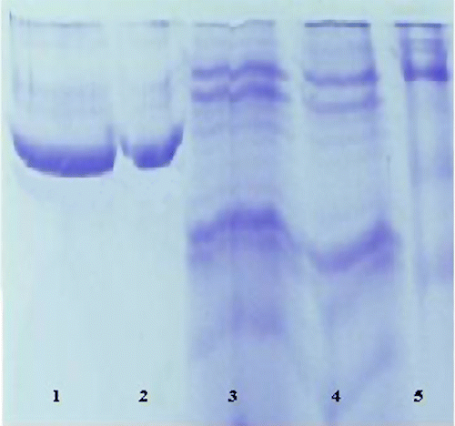
Enzyme purity was examined using Laemmli's procedure [Citation14] with 3% and 8% acrylamide concentrations for running and stacking gel, respectively. Escherichia coli β-galactosidase (116,000), rabbit phosphorylase B (97,400), bovine albumin (66,000), chicken ovalbumin (45,000), and bovine carbonic anhydrase (29,000) were used as standards (Sigma: MW-SDS-200) (See ).
Figure 1 SDS-PAGE bands of 6PGD (Lane 1and 2: Human erythrocytes 6PGD. Lane 3: Ammonium sulfate precipitation. Lane 4: Hemolysate. Lane 5: Standards: Rabbit myosin (205,000), E. coli β-galactosidase (116,000), rabbit phosphorylase B (97,400), bovine albumin (66,000), chicken ovalbumin (45,000), and bovine carbonic anhydrase (29,000).

In vitro effect of drugs
In order to determine the effects of some drugs on human 6PGD, concentrations of larnoxicam (0.0538–0.269 mM), metronidazole (0.0292–0.292 mM), imipenem (0.197–0.591 mM), ornidazole (7.588–37.94 mM), vancomycin (17.25–86.25 mM), clindamycin (55.88 – 159.03 mM), amoxicillin (87.87–332.90 mM), rifamycin (8.23 – 164.59 mM), sulfanylamide (1.111–11.11 mM), sulfanylacetamide (9.00–50.40 mM) and lincomycin (0.43–6.24 mM), were added to the reaction mixture and the enzyme activity was measured. An experiment in the absence of drug was used as control (100% activity). The IC50 values were obtained from activity (%) vs. drug concentration plots (for example ).
Figure 2 Activity % vs [Larnoxicam] regression analysis graphs for human erythrocytes 6PGD in the presence of 5 different larnoxicam concentrations.
![Figure 2 Activity % vs [Larnoxicam] regression analysis graphs for human erythrocytes 6PGD in the presence of 5 different larnoxicam concentrations.](/cms/asset/5ed8b733-239e-4abc-954d-5b8580060562/ienz_a_222823_f0002_b.gif)
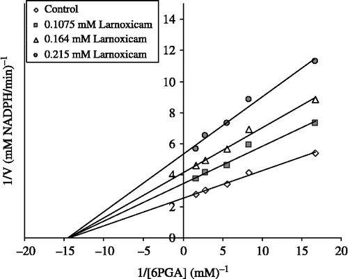
In order to determine the Ki values, the substrate (6-PGA) concentrations were 0.20, 0.24, 0.30, 0.33, and 0.35 mM. Inhibitors (drugs) solutions were added to the reaction mixture at 3 different fixed concentrations. Lineweaver-Burk graphs [Citation15] were drawn using 1/V vs. 1/[S] values and the Ki values were calculated from these graphs (see ). Regression analysis graphs were drawn using % inhibition values vs drug concentration by a statistical package (SPSS-for windows; version 10.0) on a computer (student t-test; n = 3).
Results
Purification of the enzyme led to a specific activity of 25.75 EU/mg protein, a yield of 78% and 6552-fold purification (). SDS polyacrylamide gel electrophoresis was performed after the purification of the enzyme, and the electrophoretic pattern is shown in Figure 1.
Table I. Purification scheme for 6-phosphogluconate dehydrogenase from human erythrocytes.
IC50 values of larnoxicam, metronidazole, imipenem, ornidazole, vancomycin, clindamycin and amoxicillin were 0.17, 0.23, 0.43, 21.79, 46.39, 117.43 and 287.35 mM, and the Ki constants were 0.40 ± 0.045 (non-competitive), 0.57 ± 0.069 (non-competitive), 0.77 ± 0.11 (non-competitive), 42.40 ± 2.89 (non-competitive), 65.60 ± 4.03 (competitive), 130.22 ± 9.21 (competitive) and 287.58 ± 10.56 (competitive) mM, respectively. Representative graphs are shown for larnoxicam (Figures. 2 and 3).
Discussion
It is known that many drugs have adverse effects on the body when used for therapeutic or other purposes [Citation16] which may be dramatic and systematic [Citation17]. A good example of this is that of pamaquine used for malaria treatment which caused severe adverse effects in patients within a few days, resulting in black urination, hyperbilirubinemia, a dramatic decrease in blood Hb levels and finally death, which occurred in cases of severe G6PD deficiency [Citation18]. Therefore, investigation of the effects of some drugs on the enzyme activity of human erythrocyte G6PD is very important.
Here, human erythrocyte 6PGD enzyme was purified with a 78% yield and 6552-fold purification in 5 - 6 hours using 2′,5′-ADP Sepharose 4B affinity gel chromatography (). SDS-PAGE showed that high purity for the enzyme had been obtained (Figure 1).
Inhibitory effects of some drugs on 6PGD activity in humans have been reported in many investigations [Citation9,Citation10]. For example, it has been reported that netilmicin sulphate, cefepime, amikacin, ısepamycin, chloramphenicol, ceftazidim, teicoplanin, ampicillin, ofloxacin, levofloxacin, cefotaxime, penicillin G, gentamicin sulphate, ciprofloxacin inhibit human erythrocyte 6PGD [Citation10] and ampicillin and amikacin inhibit rat red cells 6PGA [Citation9]. However, to the best of our knowledge, the inhibitory effects of the drugs examined here on 6PGD in human erythrocyte 6PGD have not been studied.
In order to show inhibitory effects, while the most suitable parameter is the Ki constant, some researchers use the IC50 value. Therefore, in this study, both the Ki and IC50 parameters of these drugs for 6PGD were determined.
IC50 values of larnoxicam, metronidazole, imipenem, ornidazole, vancomycin, clindamycin and amoxicillin were 0.17, 0.23, 0.43, 21.79, 46.39, 117.43 and 287.35 mM, and the Ki constants were 0.40 ± 0.04, 0.57 ± 0.06, 0.77 ± 0.11, 42.40 ± 2.89, 65.60 ± 4.03, 130.22 ± 9.21, and 287.58 ± 10.56 mM, respectively. Ki values show that larnoxicam had the highest inhibitory effect, followed by metronidazole, imipenem, ornidazole, vancomycin, clindamycin and amoxicillin, respectively. IC50 values showed the same trend.
In this investigation, by using the obtained Ki and IC50 values, undesirable side effects of these drugs on 6PGD activity and body metabolism and fatty acid synthesis can be reduced.
The dosages of the examined drugs (iv) used clinically give blood drug concentrations as follows; larnoxicam ∼4, metronidazole ∼58, imipenem ∼315, ornidazole ∼455, vancomycin 345, clindamycin ∼237 and amoxicillin ∼532 μM [Citation19]. By taking into account these concentrations, the inhibition data calculated from plots were found to be 2%, 13%, 23%, 1%, 0.4%, 0% and 0.1%, respectively. According to these data, if it is required to give metronidazole and imipenem to patients, their dosage should be very well controlled to decrease hemolytic and other side effects due to possible inhibition of 6PGD.
References
- Bianchi D, Bertrant O, Haupt K, Coello N. Effect of gluconic acid as a secondary carbon source on non-growing L-lysine producers cells of Corynebacterium glutamicum. Purification and properties of 6-phosphogluconate dehydrogenase. Enz Microb Technol 2001; 28: 754–759
- Lehninger AL, Nelson DL, Cox MM. Principles of biochemistry2nd edn. 2000; 558–560
- Kahler SG, Kirkman HN. Intracellular glucose-6-phosphate dehydrogenase does not monomerize in human erythrocytes. J Biol Chem 1983; 258: 717–718
- Srivastava SK, Beutler E. Glutathione metabolism of the erythrocyte. The enzymic cleavage of glutathione-haemoglobin preparations by glutathione reductase. Biochem J 1989; 119: 353
- Kozar RA, Weibel CJ, Cipolla J, Klein AJ, Haber MM, Abedin MZ, Trooskin SZ. Antioxidant enzymes are induced during recovery from acute lung injury. Crit Care Med 2000; 28: 2486–2491
- Beutler E. Blood 1994; 84: 3613–3636
- Edward E, Morse MD. Toxic effects of drugs on erythrocytes. Ann Clin Lab Sci 1988; 18: 13–18
- Jacobasch G, Rapoport SM. Hemolytic anemias due to erythrocyte enzyme deficiencies. Mol Asp Med 1996; 17: 143–170
- Ciftci M, Beydemir S, Yılmaz H, Bakan E. Effects of some drugs on rat erythrocyte 6-phosphogluconate dehydrogenase: An in vitro and in vivo study. J Pharmacol 2002; 54: 275–280
- Akyuz M, Erat M, Ciftci M, Gumustekin K, Bakan N. Effects of some antibiotics on human erythrocyte 6-phosphogluconate dehydrogenase: An in vitro and in vivo study. J Enzyme Inhibit Med Chem 2004, 19: 361–365
- Puskas F, Gergely PJ, Banki K, Perl A. Stimulation of the pentose phosphate pathway and glutathione levels by dehydroascorbate, the oxidized form of vitamin C. FASEB J 2000; 14: 1352–1361
- Beutler E. 1971; 12, Red Cell Metabolism Manual of Biochemical Methods (Academic Press, London)
- Bradford MM. A rapid and sensitive method for the quantization of microgram quantities of protein utilizing the principle of protein-dye binding. Anal Biochem 1976; 72: 248–251
- Laemmli DK. Cleavage of structural proteins during assembly of the head of bacteriophage T4. Nature 1970; 227: 680–683
- Segel IE. Enzyme kinetics. John Wiley and Sons, Toronto 1975
- Hochster R, Kates MM, Quastel JH. Metabolic inhibitors. Academic Press, New York 1972; 71–89
- Ciftci M, Kufrevioglu OI, Gundogdu M, Ozmen I. Effects of some antibiotics on enzyme activity of glucose-6-phosphate dehydrogenase from human erythrocytes Pharmacol. Res 2000; 41(1)109–113
- Keha EE, Kufrevioglu OI. Biyokimya (Turkish) 2nd. Aktif Yayinevi, Istanbul 2000; 356–366
- Kayaalp SO. Rasyonal tedavi yönünden tıbbi farmakoloji. Hacettepe-Tas Yayıncılık (Turkish), Ankara 2002
