
 ?Mathematical formulae have been encoded as MathML and are displayed in this HTML version using MathJax in order to improve their display. Uncheck the box to turn MathJax off. This feature requires Javascript. Click on a formula to zoom.
Abstract
The activation of the β-class carbonic anhydrases (CAs, EC 4.2.1.1) from the bacteria Brucella suis and Francisella tularensis with amine and amino acids was investigated. BsuCA 1 was sensitive to activation with amino acids and amines, whereas FtuCA was not. The most effective BsuCA 1 activators were L-adrenaline and D-Tyr (KAs of 0.70–0.95 µM). L-His, L-/D-Phe, L-/D-DOPA, L-Trp, L-Tyr, 4-amino-L-Phe, dopamine, 2-pyridyl-methylamine, D-Glu and L-Gln showed activation constants in the range of 0.70–3.21 µM. FtuCA was sensitive to activation with L-Glu (KA of 9.13 µM). Most of the investigated compounds showed a weak activating effect against FtuCA (KAs of 30.5–78.3 µM). Many of the investigated amino acid and amines are present in high concentrations in many tissues in vertebrates, and their role in the pathogenicity of the two bacteria is poorly understood. Our study may bring insights in processes connected with invasion and pathogenic effects of intracellular bacteria.
1. Introduction
The pathogenic bacteria Brucella suisCitation1,Citation2 and Francisella tularensisCitation3,Citation4 provoke serious diseases both in human and livestock, are difficult to treat by antibiotics, and have the potential to induce widespread infections. In fact, both Brucella and Francisella might be used as bioterrorism organisms due to the fact that quite low doses of pathogens (as few as 10–100 bacteria) are highly infectious, leading to ravaging epidemicsCitation1. Furthermore, they persist in the environment and are rapidly transmitted via different routes, including by aerosols and from human to humanCitation1. Brucellosis, like tularemia (the infection produced by F. tularensis) are neglected diseases, although their prevalence in humans and domestic/wild animals is not at all lowCitation1–4. These pathogens also became resistant to many currently used antibioticsCitation1–4, with the danger that the treatment of infected patients/animals will become increasingly difficult. Thus, searching for new drug targets addressing these complex issues is of stringent relevance.
Carbonic anhydrases (CAs, EC 4.2.1.1) are ubiquitous metalloenzymes in all kingdomsCitation5–7. They catalyze the reversible hydration of CO2 with formation of bicarbonate and protons, converting thus efficiently two neutral molecules (CO2 and H2O) in a weak base (bicarbonate) and a very strong acid (H+ ions)Citation8–10. For this reason, in most organisms investigated so far, from simple (such as bacteria and archaea) to complex ones (plants, animals etc.) these enzymes are involved in pH regulation as well as several crucial metabolic pathwaysCitation5–7,Citation11–17. At least seven distinct CA genetic families are known to date (α-, β-, γ-, ζ-, η- and θ-CAs)Citation5–7, and their diffusion and physiological roles have been investigated in details mainly in vertebrates, including humans, who posses only α-CAs, but with quite a large number of isoforms (15 CA isoforms are known in humans, human carbonic anhydrase (hCA) I-XIV, with two V-type ones, CA VA and VB)Citation5. The investigation of CAs belonging to other classes, such as those found in bacteria, is on the other hand a rather recent field, although notable advances were registered in the last yearsCitation5–7,Citation13,Citation18,Citation19. CA inhibitors (CAIs) belonging to many diverse chemotypes and possessing a wealth of inhibition mechanismsCitation5–7 are clinically used for the management of a variety of disorders, including edema, epilepsy, glaucoma, obesity, hypoxic tumors, neuropathic pain and arthritisCitation5–7,Citation11–17. On the other hand, CA activators (CAAs) started to be investigated in detail only in the last two decades, after the CA activation mechanism has been explained by one of our groupsCitation8–10.
Indeed, CAAs have been demonstrated to participate in the CA catalytic cycleCitation8, which is shown schematically in EquationEquations (1)(1)
(1) and Equation(2)
(2)
(2) below:
(1)
(1)
(2)
(2)
In the first step, a zinc-bound hydroxide species of the enzyme with a strong nucleophilicity, attacks the CO2 substrate, which is weakly bound in a hydrophobic pocket nearby, being optimally orientated for the hydration reaction to occur by the attack of the zinc hydroxide nucleophile (EquationEquation 1(1)
(1) )Citation8. In the next step of the process, the formed bicarbonate in the hydration reaction is replaced by an incoming water molecule, leading to the formation of an acidic enzymatic species, EZn2+—OH2 (EquationEquation 1
(1)
(1) ). In order to regenerate the zinc hydroxide species, a proton must be transferred from the Zn(II)-bound water molecule to the external medium (EquationEquation 2
(2)
(2) ). This is also the rate-determining step of the entire catalytic cycleCitation8–10. In the presence of activators (A in EquationEquation 3
(3)
(3) ), this rate-determining step is facilitated by an additional proton release pathway, which involves the activator A bound within the enzyme active site. It should be noted that all CAAs known to date possess in their molecule protonatable moieties of the amine, carboxylate or imidazole type, with pKa values in the range of 5–8Citation8–10.
(3)
(3)
By the formation of enzyme-activator complex (EquationEquation 3(3)
(3) ), the proton transfer reaction becomes intramolecular, and is thus more rapid compared with the intermolecular process in which for example buffer molecules participateCitation8–10,Citation20. The enzyme-activator complexes were thoroughly characterized for α-CAs of human origin, such as hCA I and II, by means of kinetic and X-ray crystallographic techniques, which allowed the identification of the activator-binding site within the CA cavityCitation8,Citation9,Citation21–29. However, CAA research involving various activators has been relatively neglected compared with that of CAIs, with only a relatively limited number of studies focusing on activators of bacterial CAsCitation29. Recently, it has been demonstrated that such activators may have pharmacological applications for enhancing cognition, in the management of CA deficiencies, for therapy memory and for obtaining artificial tissues a well as for investigating the effects of endogenous amino acids and amines on CA activityCitation30.
Here we report the first activation study of the CAs investigated so far in the genome of the two pathogenic bacteria mentioned earlier. Indeed, B. suis encodes for two β-CAs, BsuCA1 and BsuCA 2Citation18, whereas in the genome of F. tularensis only one such enzyme is present (again belonging to the β-CA class, FtuCA)Citation18 which were cloned and characterized by some of us earlierCitation18. The inhibition of these enzymes with sulfonamide, anions and other derivatives was reported earlierCitation31–33, but no activation studies of the two enzymes are available in the literature. Here we report the first activation study of BsuCA 1 and FtuCA with a panel of amino acids and amines, known to act as CAAs for enzymes belonging to various other genetic familiesCitation29–34.
2. Materials and methods
2.1. Materials
Amino acids and amines 1–24 were commercially available, highest purity reagents from Sigma-Aldrich, Milan, Italy. BsuCA 1 and FtuCA were recombinant proteins produced as reported earlier by our groupsCitation18.
2.2. CA enzyme activation assay
An Sx.18Mv-R Applied Photophysics (Oxford, UK) stopped-flow instrument has been used to assay the catalytic activity of various CA isozymes for CO2 hydration reactionCitation20. Phenol red (at a concentration of 0.2 mM) was used as indicator, working at the absorbance maximum of 557 nm, with 10 mM Tris (pH 8.4) as buffer, and 0.1 M Na2SO4 (for maintaining constant ionic strength, which is not inhibitory against these enzymesCitation18), following the CA-catalyzed CO2 hydration reaction for a period of 10 s at 25 °C. The CO2 concentrations ranged from 1.7 to 17 mM for the determination of the kinetic parameters and activation constants. For each activator at least six traces of the initial 5–10% of the reaction have been used for determining the initial velocity. The uncatalyzed rates were determined in the same manner and subtracted from the total observed rates. Stock solutions of activators (10 mM) were prepared in distilled-deionized water and dilutions up to 1 nM were done thereafter with the assay buffer. Activator and enzyme solutions were pre-incubated together for 15 min (standard assay at room temperature) prior to assay, in order to allow for the formation of the E–A complex. The activation constant (KA), defined similarly with the inhibition constant KI, can be obtained by considering the classical Michaelis–Menten equation (EquationEquation 4(4)
(4) ), which has been fitted by non-linear least squares by using PRISM 3:
(4)
(4)
where [A]f is the free concentration of activator.
Working at substrate concentrations considerably lower than KM ([S] ≪KM), and considering that [A]f can be represented in the form of the total concentration of the enzyme ([E]t)and activator ([A]t), the obtained competitive steady-state equation for determining the activation constant is given by EquationEquation (5)(5)
(5) :
(5)
(5)
where v0 represents the initial velocity of the enzyme-catalyzed reaction in the absence of activatorCitation21–29. The results obtained with this type of kinetic assay are in excellent agreement with those obtained by use of other independent methods, including native mass spectrometry and fluorescence spectroscopyCitation35.
3. Results and discussion
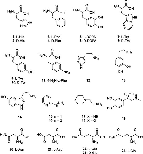
Natural and non-natural amino acids and amines 1–24 were included among the investigated compounds as activators of the two bacterial β-CAs investigated here (. These compounds were employed for investigations as CAAs against many classes of CAs, including the bacterial, archaeal and mammalian ones, as mentioned earlierCitation8–10,Citation21–29. The salient feature of these compounds is the presence of protonatable moieties of the amine, carboxylate or imidazole type, which makes them appropriate for participating in the proton shuttling processes between the active site and the reaction medium, as described by EquationEquation (3)(3)
(3) .
Figure 1. Amino acids and amines 1–24 investigated as activators.
Data of demonstrate that the two bacterial β-CAs investigated here shows a more effective CO2 hydrase activity compared with hCA I, a widely spread isoform in humansCitation8. Considering the kcat, BsuCA 1 is 3.2-times more effective as a catalyst for CO2 hydration compared with hCA I, whereas FtuCA is even more active, being 4.9-times more effective compared with the human enzyme. On the other hand, the Michaelis constants (KM) of the bacterial enzymes are higher compared with the two human enzymes showed in for comparison reasons. In fact the Michaelis constant of hCA I and II is in the range of 4.0–9.3 mM, whereas for the bacterial enzymes the KM values are in the range of 11.0–16.4 mM, denoting that CO2 has a lower affinity for the bacterial enzymes than for the human ones hCA I/II. We have chosen L-Trp for performing detailed kinetic measurements because this amino acid was a rather effective activator for both enzymes investigated here (as well as the human isoforms hCA I and II used as standard enzymes in such experiments – see Section 3). In the presence of 10 µM L-Trp as activator, the KM of BsuCA 1/FtuCA remained unchanged (data not shown) but the kcat was 4.60 times higher than in the absence of the activator for BsuCA 1, and 1.60-times higher for FtuCA, respectively (). This situation, in which the KM does not depend on the presence of an activator and kcat is strongly affected by the activator, has been observed for all CAs investigated to date, belonging to all known CA genetic families. This strongly indicates that the CA activation mechanism is similar for all enzyme classes, involving facilitation of the proton transfer process by the activator molecule bound within the enzyme active site in the enzyme-activator complexCitation8–10. It should be also noted that L-Trp is a much more effective activator for BsuCA 1 than for FtuCA (see Section 3).
Table 1. Activation of hCA isozymes I, II and BsuCA 1/FtuCA, with L-Trp, at 25 °C, for the CO2 hydration reactionCitation20.
In the activation constants (KAs) of the bacterial β-CAs investigated here (BsuCA 1 and FtuCA) and of the human isoforms hCA I and II, with the amino acid and amine derivatives 1–24 () are shown. The human CA activation data are reported for comparison reasons.
Table 2. Activation constants of hCA I, hCA II and the bacterial β-CAs investigated here with amino acids and amines 1–24, by a stopped-flow CO2hydrase assayCitation20.
Data of show the following interesting features for the activation of BsuCA 1 and FtuCA with amino acid and amine derivatives 1–24:
The enzyme from B. suis, BsuCA 1 was generally rather sensitive to activation with amino acids and amines of type 1–24, with activation constants in the range of 0.70–43.1 µM. On the contrary, 13 out of the 24 compounds investigated here were inactive as activators of FtuCA (KA > 100 µM), only one compounds had a KA < 10 µM (L-Glu, 22), and most of the investigated active compounds showed a weak activating effect, with KAs ranging between 30.5 and 78.3 µM. Thus, only L-Glu may be considered a an effective FtuCA activator. Small changes in its scaffold, such as the transformation of the COOH moiety in CONH2 lead to a drastic loss of activity (compound 24), whereas the D-enantiomer 23 was devoid of activity (). Thus, the structure-activity relationship will be discussed for the activation of BsuCA 1, as the profile for activating FtuCA with these compounds is quite flat and the activation effects are modest or absent.
The most effective BsuCA 1 activators were L-His, L-/D-Phe, L-/D-DOPA, L-Trp, L-/D-Tyr, 4-amino-L-Phe, dopamine, 2-pyridyl-methylamine, L-adrenaline, D-Glu and L-Gln. These compounds showed activation constants in the range of 0.70–3.21 µM. Generally the L-amino acid derivatives were more efficient BsuCA 1 activators compared with the corresponding D-enantiomers, but some exceptions from this rule were also encountered, with D-Tyr and D-Glu being better activators than the corresponding L-amino acids (). Rather unexpectedly, the most efficient activator was L-adrenaline, with a KA of 0.70 µM.
Slightly less effective BsuCA 1 activators were D-His, D-Trp, histamine, heterocyclic amines 16–18, L-Asn, L-Asp and L-Glu, which had activation constants in the range of 3.71–43.1 µM. Only compound 17 can be considered a weak activator against this isoform (KA of 43.1 µM), with all the other ones being moderate activators.
The activation profile of the two bacterial enzymes investigated here is very different from the ones of the two human isoform hCA I and II showed in as reference enzymes. Furthermore, as mentioned above, the two bacterial enzymes differ considerably in their interaction with these activators.
4. Conclusions
The first activation study of the β-class CA enzymes from the pathogenic bacteria B. suis and F. tularensis is reported here. A panel of 24 amino acid and amine derivative were included in the study. BsuCA 1 was sensitive to activation with amino acids and amines, which showed activation constants in the range of 0.70–43.1 µM. The most effective BsuCA 1 activators were L-adrenaline and D-Tyr (KAs of 0.70–0.95 µM). L-His, L-/D-Phe, L-/D-DOPA, L-Trp, L-Tyr, 4-amino-L-Phe, dopamine, 2-pyridyl-methylamine, D-Glu and L-Gln showed activation constants in the range of 0.70–3.21 µM. FtuCA was not sensitive to activation with many of the investigated compounds (KA > 100 µM), and only L-Glu had a KA < 10 µM. Most of the investigated active compounds showed a weak activating effect against FtuCA, with KAs ranging between 30.5 and 78.3 µM, such a L-/D-His, L-/D-Phe, L-/D-Trp, 2-pyridyl-methylamine. It should be noted that many of the investigated amino acid and amines are present in rather high concentrations in many tissues in vertebrates, and their role in the pathogenicity of the two bacteria is poorly understood. Our study may thus bring some insights in the intricate processes connected with the invasion and pathogenic effects of intracellular bacteria when attacking their hosts.
Acknowledgements
The Australian Research Council is also acknowledged for support.
Disclosure statement
No potential conflict of interest was reported by the authors.
Additional information
Funding
Related Research Data
References
- a) El-Sayed A, Awad W. Brucellosis: evolution and expected comeback. Int J Vet Sci Med 2018;6(Suppl):S31–S5; b) Dadar M, Shahali Y, Whatmore AM. Human brucellosis caused by raw dairy products: a review on the occurrence, major risk factors and prevention. Int J Food Microbiol 2019;292:39–47; c) Tuon FF, Gondolfo RB, Cerchiari N. Human-to-human transmission of Brucella - a systematic review. Trop Med Int Health 2017;22:539–46.
- a) Hull NC, Schumaker BA. Comparisons of brucellosis between human and veterinary medicine. Infect Ecol Epidemiol 2018;8:1500846; b) Cross AR, Baldwin VM, Roy S, et al. Zoonoses under our noses. Microbes Infect 2019;21:10–9; c) Köhler S, Ouahrani-Bettache S, Winum JY. Brucella suis carbonic anhydrases and their inhibitors: towards alternative antibiotics? J Enzyme Inhib Med Chem 2017;32:683–7.
- a) Hirschmann JV. From squirrels to biological weapons: the early history of tularemia. Am J Med Sci 2018;356:319–28; b) Pilo P. Phylogenetic lineages of Francisella tularensis in animals. Front Cell Infect Microbiol 2018;8:258.
- a) Faber M, Heuner K, Jacob D, Grunow R. Tularemia in Germany-a re-emerging zoonosis. Front Cell Infect Microbiol 2018;8:40; b) Caspar Y, Maurin M. Francisella tularensis susceptibility to antibiotics: a comprehensive review of the data obtained in vitro and in animal models. Front Cell Infect Microbiol 2017;7:122.
- a) Supuran CT. Structure and function of carbonic anhydrases. Biochem J 2016;473:2023–32; b) Supuran CT. Advances in structure-based drug discovery of carbonic anhydrase inhibitors. Expert Opin Drug Discov 2017;12:61–88; c) Supuran CT. Carbonic anhydrases: novel therapeutic applications for inhibitors and activators. Nat Rev Drug Discov 2008;7:168–81; d) Neri D, Supuran CT. Interfering with pH regulation in tumours as a therapeutic strategy. Nat Rev Drug Discov 2011;10:767–77; e) Supuran CT, Vullo D, Manole G, et al. Designing of novel carbonic anhydrase inhibitors and activators. Curr Med Chem Cardiovasc Hematol Agents 2004;2:49–68.
- a) Capasso C, Supuran CT. An overview of the alpha-, beta-and gamma-carbonic anhydrases from Bacteria: can bacterial carbonic anhydrases shed new light on evolution of bacteria? J Enzyme Inhib Med Chem 2015;30:325–32; b) Del Prete S, Vullo D, Fisher GM, et al. Discovery of a new family of carbonic anhydrases in the malaria pathogen Plasmodium falciparum–the η-carbonic anhydrases. Bioorg Med Chem Lett 2014;24:4389–96; c) Supuran CT. Structure-based drug discovery of carbonic anhydrase inhibitors. J Enzyme Inhib Med Chem 2012;27:759–72; d) Ozensoy Guler O, Capasso C, Supuran CT. A magnificent enzyme superfamily: carbonic anhydrases, their purification and characterization. J Enzyme Inhib Med Chem 2016;31:689–94.
- a) Supuran CT. Carbonic anhydrases: from biomedical applications of the inhibitors and activators to biotechnological use for CO2 capture. J Enzyme Inhib Med Chem 2013;28:229–30; b) Supuran CT. How many carbonic anhydrase inhibition mechanisms exist? J Enzyme Inhib Med Chem 2016;31:345–60; c) Alterio V, Di Fiore A, D'Ambrosio K, et al. Multiple binding modes of inhibitors to carbonic anhydrases: how to design specific drugs targeting 15 different isoforms? Chem Rev 2012;112:4421–68; d) Abbate F, Winum JY, Potter BV, et al. Carbonic anhydrase inhibitors: X-ray crystallographic structure of the adduct of human isozyme II with EMATE, a dual inhibitor of carbonic anhydrases and steroid sulfatase. Bioorg Med Chem Lett 2004;14:231–4.
- a) Supuran CT. Carbonic anhydrase activators. Future Med Chem 2018;10:561–73; b) Tanini D, Capperucci A, Supuran CT, Angeli A. Sulfur, selenium and tellurium containing amines act as effective carbonic anhydrase activators. Bioorg Chem 2019;87:516–22; c) Rami M, Winum JY, Supuran CT, et al. (Hetero)aryl substituted thiazol-2,4-yl scaffold as human carbonic anhydrase I, II, VII and XIV activators. J Enzyme Inhib Med Chem 2019;34:224–9.
- a) Briganti F., Mangani S., Orioli P., et al., Carbonic anhydrase activators: x-ray crystallographic and spectroscopic investigations for the interaction of isozymes I and II with histamine. Biochemistry 1997;36:10384–92; b) Clare B.W., Supuran C.T., Carbonic anhydrase activators. 3: structure-activity correlations for a series of isozyme II activators. J Pharm Sci 1994;83:768–73; c) Ilies M, Scozzafava A, Supuran CT. Carbonic anhydrase activators. In: Supuran CT, Scozzafava A, Conway J, eds. Carbonic anhydrase – its inhibitors and activators. Boca Raton: CRC Press; 2004:317–52; d) Bhatt A, Mondal UK, Supuran CT, et al. Crystal structure of carbonic anhydrase II in complex with an activating ligand: implications in neuronal function. Mol Neurobiol 2018;55:7431–7.
- a) Akocak S, Lolak N, Vullo D, et al. Synthesis and biological evaluation of histamine Schiff bases as carbonic anhydrase I, II, IV, VII, and IX activators. J Enzyme Inhib Med Chem 2017;32:1305–12; b) Angeli A, Vaiano F, Mari F, et al. Psychoactive substances belonging to the amphetamine class potently activate brain carbonic anhydrase isoforms VA, VB, VII, and XII. J Enzyme Inhib Med Chem 2017;32:1253–9; c) Licsandru E, Tanc M, Kocsis I, Barboiu M, Supuran CT. A class of carbonic anhydrase I - selective activators. J Enzyme Inhib Med Chem 2017;32:37–46; d) Akocak S, Lolak N, Bua S, et al. α-Carbonic anhydrases are strongly activated by spinaceamine derivatives. Bioorg Med Chem 2019;27:800–4.
- a) Scozzafava A, Menabuoni L, Mincione F, Supuran CT. Carbonic anhydrase inhibitors. a general approach for the preparation of water-soluble sulfonamides incorporating polyamino − polycarboxylate tails and of their metal complexes possessing long-lasting, topical intraocular pressure-lowering properties. J Med Chem 2002;45:1466–76; b) Pacchiano F, Aggarwal M, Avvaru BS, et al. Selective hydrophobic pocket binding observed within the carbonic anhydrase II active site accommodate different 4-substituted-ureido-benzenesulfonamides and correlate to inhibitor potency. Chem Commun (Camb) 2010;46:8371–3; c) Carta F, Scozzafava A, Supuran CT. Sulfonamides: a patent review (2008–2012). Expert OpinTher Pat 2012;22:747–58; d) Briganti F, Pierattelli R, Scozzafava A, Supuran CT. Carbonic anhydrase inhibitors. Part 37. Novel classes of carbonic anhydrase inhibitors and their interaction with the native and cobalt-substituted enzyme: kinetic and spectroscopic investigations. Eur J Med Chem 1996;31:1001–10; e) Supuran CT, Clare BW. Carbonic anhydrase inhibitors. Part 57. Quantum chemical QSAR of a group of 1,3,4-thiadiazole and 1,3,4-thiadiazoline disulfonamides with carbonic anhydrase inhibitory properties. Eur J Med Chem 1999;34:41–50.
- a) Perfetto R, Del Prete S, Vullo D, et al. Cloning, expression and purification of the α-carbonic anhydrase from the mantle of the Mediterranean mussel, Mytilusg alloprovincialis. J Enzyme Inhib Med Chem 2017;32:1029–35; b) Abdoli M, Angeli A, Bozdag M, et al. Synthesis and carbonic anhydrase I, II, VII, and IX inhibition studies with a series of benzo[d]thiazole-5- and 6-sulfonamides. J Enzyme Inhib Med Chem 2017;32:1071–8; c) De Simone G, Langella E, Esposito D, et al. Insights into the binding mode of sulphamates and sulphamides to hCA II: crystallographic studies and binding free energy calculations. J Enzyme Inhib Med Chem 2017;32:1002–11; d) Scozzafava A, Menabuoni L, Mincione F, et al. Carbonic anhydrase inhibitors: perfluoroalkyl/aryl-substituted derivatives of aromatic/heterocyclic sulfonamides as topical intraocular pressure-lowering agents with prolonged duration of action. J Med Chem 2000;43:4542–51.
- a) Supuran CT, Capasso C. Carbonic anhydrase from Porphyromonas gingivalis as a drug target. Pathogens 2017;6:E30; b) Capasso C, Supuran CT. Inhibition of bacterial carbonic anhydrases as a novel approach to escape drug resistance. Curr Top Med Chem 2017;17:1237–48; c) Supuran CT, Capasso C. New light on bacterial carbonic anhydrases phylogeny based on the analysis of signal peptide sequences. J Enzyme Inhib Med Chem 2016;31:1254–60; d) Vullo D, Del Prete S, Di Fonzo P, et al. Comparison of the sulfonamide inhibition profiles of the β- and γ-carbonic anhydrases from the pathogenic bacterium Burkholderia pseudomallei. Molecules 2017;22:421–435; e) Prete SD, Vullo D, di Fonzo P, et al. Sulfonamide inhibition profile of the γ-carbonic anhydrase identified in the genome of the pathogenic bacterium Burkholderia pseudomallei the etiological agent responsible of melioidosis. Bioorg Med Chem Lett 2017;27:490–5.
- a) Supuran CT. Applications of carbonic anhydrases inhibitors in renal and central nervous system diseases. Expert Opin Ther Pat 2018;28:713–21; b) Supuran CT. Carbonic anhydrase inhibitors and their potential in a range of therapeutic areas. Expert Opin Ther Pat 2018;28:709–12; c) Monti SM, Supuran CT, De Simone G. Anticancer carbonic anhydrase inhibitors: a patent review (2008 - 2013). Expert Opin Ther Pat 2013;23:737–49; d) Pastorekova S, Casini A, Scozzafava A, et al. Carbonic anhydrase inhibitors: the first selective, membrane-impermeant inhibitors targeting the tumor-associated isozyme IX. Bioorg Med Chem Lett 2004;14:869–73.
- a) Scozzafava A, Supuran CT, Carta F. Antiobesity carbonic anhydrase inhibitors: a literature and patent review. Expert Opin Ther Pat 2013;23:725–35; b) Supuran CT. Carbonic anhydrase inhibition and the management of hypoxic tumors. Metabolites 2017;7:E48; c) Supuran CT. Carbonic anhydrase inhibitors in the treatment and prophylaxis of obesity. Expert Opin Ther Pat 2003;13:1545–50; d) Winum JY, Temperini C, El Cheikh K, et al. Carbonic anhydrase inhibitors: clash with Ala65 as a means for designing inhibitors with low affinity for the ubiquitous isozyme II, exemplified by the crystal structure of the topiramate sulfamide analogue. J Med Chem 2006;49:7024–31; e) Rehman SU, Chohan ZH, Gulnaz F, Supuran CT. In-vitro antibacterial, antifungal and cytotoxic activities of some coumarins and their metal complexes. J Enzyme Inhib Med Chem 2005;20:333–40.
- a) Supuran CT. Carbonic anhydrase inhibition and the management of neuropathic pain. Expert Rev Neurother 2016;16:961–8; b) Di Cesare Mannelli L, Micheli L, Carta F, et al. Carbonic anhydrase inhibition for the management of cerebral ischemia: in vivo evaluation of sulfonamide and coumarin inhibitors.J Enzyme Inhib Med Chem 2016;31:894–9.
- a) Margheri F, Ceruso M, Carta F, et al. Overexpression of the transmembrane carbonic anhydrase isoforms IX and XII in the inflamed synovium. J Enzyme Inhib Med Chem 2016;31(suppl 4):60–3; b) Bua S, Di Cesare Mannelli L, Vullo D, et al. Design and synthesis of novel nonsteroidal anti-inflammatory drugs and carbonic anhydrase inhibitors hybrids (NSAIDs-CAIs) for the treatment of rheumatoid arthritis. J Med Chem 2017;60:1159–70.
- a) Del Prete S, Vullo D, Osman SM, et al. Anion inhibitors of the β-carbonic anhydrase from the pathogenic bacterium responsible of tularemia, Francisella tularensis. Bioorg Med Chem 2017;25:4800–4; b) Del Prete S, Vullo D, Osman SM, et al. Sulfonamide inhibition profiles of the β-carbonic anhydrase from the pathogenic bacterium Francisella tularensis responsible of the febrile illness tularemia. Bioorg Med Chem 2017;25:3555–61; c) Joseph P, Turtaut F, Ouahrani-Bettache S, et al. Cloning, characterization, and inhibition studies of a beta-carbonic anhydrase from Brucella suis. J Med Chem 2010;53:2277–85; d) Joseph P, Ouahrani-Bettache S, Montero JL, et al. A new β-carbonic anhydrase from Brucella suis, its cloning, characterization, and inhibition with sulfonamides and sulfamates, leading to impaired pathogen growth. Bioorg Med Chem 2011;19:1172–8.
- a) Angeli A, Del Prete S, Donald WA, et al. The γ-carbonic anhydrase from the pathogenic bacterium Vibrio cholerae is potently activated by amines and amino acids. Bioorg Chem 2018;77:1–5; b) Angeli A, Del Prete S, Osman SM, et al. Activation studies with amines and amino acids of the β-carbonic anhydrase encoded by the Rv3273 gene from the pathogenic bacterium Mycobacterium tuberculosis. J Enzyme Inhib Med Chem 2018;33:364–9; c) Angeli A, Del Prete S, Osman SM, et al. Activation studies of the α- and β-carbonic anhydrases from the pathogenic bacterium Vibrio cholerae with amines and amino acids. J Enzyme Inhib Med Chem 2018;33:227–33; d) Vullo D, Del Prete S, Osman SM, et al. Comparison of the amine/amino acid activation profiles of the β- and γ-carbonic anhydrases from the pathogenic bacterium Burkholderia pseudomallei. J Enzyme Inhib Med Chem 2018;33:25–30; e) Vullo D, Del Prete S, Osman SM, et al. Burkholderia pseudomallei γ-carbonic anhydrase is strongly activated by amino acids and amines. Bioorg Med Chem Lett 2016;27:77–80.
- Khalifah RG. The carbon dioxide hydration activity of carbonic anhydrase. I. Stop-flow kinetic studies on the native human isoenzymes B and C. J Biol Chem 1971;246:2561–73.
- a) Temperini C, Scozzafava A, Supuran CT. Carbonic anhydrase activation and the drug design. Curr Pharm Des 2008;14:708–715; b) Isik S, Guler OO, Kockar F, et al. Saccharomyces cerevisiae β-carbonic anhydrase: inhibition and activation studies. Curr Pharm Des 2010;16:3327–36.
- a) Temperini C, Scozzafava A, Vullo D, Supuran CT. Carbonic anhydrase activators. Activation of isozymes I, II, IV, VA, VII, and XIV with l- and d-histidine and crystallographic analysis of their adducts with isoform II: engineering proton-transfer processes within the active site of an enzyme. Chemistry 2006;12:7057–66; b) Temperini C, Scozzafava A, Vullo D, Supuran CT. Carbonic anhydrase activators. Activation of isoforms I, II, IV, VA, VII, and XIV with L- and D-phenylalanine and crystallographic analysis of their adducts with isozyme II: stereospecific recognition within the active site of an enzyme and its consequences for the drug design. J Med Chem 2006;49:3019–27; c) Temperini C, Innocenti A, Scozzafava A, Supuran CT. Carbonic anhydrase activators: kinetic and X-ray crystallographic study for the interaction of D- and L-tryptophan with the mammalian isoforms I-XIV. Bioorg Med Chem 2008;16:8373–8.
- a) Temperini C, Innocenti A, Scozzafava A, et al. Carbonic anhydrase activators: L-Adrenaline plugs the active site entrance of isozyme II, activating better isoforms I, IV, VA, VII, and XIV. Bioorg Med Chem Lett 2007;17:628–35; b) Temperini C, Scozzafava A, Puccetti L, Supuran CT. Carbonic anhydrase activators: X-ray crystal structure of the adduct of human isozyme II with L-histidine as a platform for the design of stronger activators. Bioorg Med Chem Lett 2005;15:5136–41; c) Temperini C, Scozzafava A, Supuran CT. Carbonic anhydrase activators: the first X-ray crystallographic study of an adduct of isoform I. Bioorg Med Chem Lett 2006;16:5152–6.
- a) Vullo D, Nishimori I, Innocenti A, et al. Carbonic anhydrase activators: an activation study of the human mitochondrial isoforms VA and VB with amino acids and amines. Bioorg Med Chem Lett 2007;17:1336–40; b) Pastorekova S, Vullo D, Nishimori I, et al. Carbonic anhydrase activators: activation of the human tumor-associated isozymes IX and XII with amino acids and amines. Bioorg Med Chem 2008;16:3530–6; c) Nishimori I, Onishi S, Vullo D, et al. Carbonic anhydrase activators. The first activation study of the human secretory isoform VI. Bioorg Med Chem 2007;15:5351–7.
- a) Parkkila S, Vullo D, Puccetti L, et al. Carbonic anhydrase activators: activation of isozyme XIII with amino acids and amines. Bioorg Med Chem Lett 2006;16:3955–9; b) Vullo D, Innocenti A, Nishimori I, et al. Carbonic anhydrase activators: activation of the human isoforms VII (cytosolic) and XIV (transmembrane) with amino acids and amines. Bioorg Med Chem Lett 2007;17:4107–12; c) Vullo D, Nishimori I, Scozzafava A, Supuran CT. Carbonic anhydrase activators: activation of the human cytosolic isozyme III and membrane-associated isoform IV with amino acids and amines. Bioorg Med Chem Lett 2008;18:4303–7.
- a) Innocenti A, Hilvo M, Parkkila S, et al. Carbonic anhydrase activators. Activation of the membrane-associated isoform XV with amino acids and amines. Bioorg Med Chem Lett 2009;19:3430–3; b) Supuran CT, Dinculescu A, Balaban AT. Carbonic anhydrase activators. Part 5. CA II activation by 2,4,6-trisubstituted pyridinium cations with 1-(ω-aminoalkyl) side chains. Rev Roum Chim 1993;38:343–9; c) Supuran CT, Barboiu M, Luca C, et al. Carbonic anhydrase activators. Part 14. Synthesis of mono- and bis- pyridinium salt derivatives of 2-amino-5-(2-aminoethyl)- and 2-amino-5-(3-aminopropyl)-1,3,4-thiadiazole, and their interaction with isozyme II. Eur J Med Chem 1996;31:597–606; d) Ilies MA, Banciu MD, Ilies M, et al. Carbonic anhydrase activators. Part 17. Synthesis and activation study of a series of 1-(1,2,4-triazole-(1H)-3-yl)-2,4,6-trisubstituted-pyridinium salts against isozymes I, II and IV. Eur J Med Chem 1997;32:911–8.
- a) Ilies M, Banciu MD, Ilies MA, et al. Carbonic anhydrase activators: design of high affinity isozymes I, II, and IV activators, incorporating tri-/tetrasubstituted-pyridinium-azole moieties. J Med Chem 2002;45:504–10; b) Dave K, Scozzafava A, Vullo D, et al. Pyridinium derivatives of histamine are potent activators of cytosolic carbonic anhydrase isoforms I, II and VII. Org Biomol Chem 2011;9:2790–800; c) Dave K, Ilies MA, Scozzafava A, et al. An inhibitor-like binding mode of a carbonic anhydrase activator within the active site of isoform II. Bioorg Med Chem Lett 2011;21:2764–8.
- a) Scozzafava A, Supuran CT. Carbonic anhydrase activators: human isozyme II is strongly activated by oligopeptides incorporating the carboxyterminal sequence of the bicarbonate anion exchanger AE1. Bioorg Med Chem Lett 2002;12:1177–80; b) Scozzafava A, Supuran CT. Carbonic anhydrase activators: high affinity isozymes I, II, and IV activators, incorporating a beta-alanyl-histidine scaffold. J Med Chem 2002;45:284–91; c) Abdo MR, Vullo D, Saada MC, et al. Carbonic anhydrase activators: activation of human isozymes I, II and IX with phenylsulfonylhydrazido l-histidine derivatives. Bioorg Med Chem Lett 2009;19:2440–3; d) Saada MC, Montero JL, Vullo D, et al. Carbonic anhydrase activators: gold nanoparticles coated with derivatized histamine, histidine, and carnosine show enhanced activatory effects on several mammalian isoforms. J Med Chem 2011;54:1170–7; e) Zhang Y, Legrand YM, Petit E, et al. Dynamic encapsulation and activation of carbonic anhydrase in multivalent dynameric host matrices. Chem Commun (Camb) 2016;52:4053–5.
- a) Vullo D, De Luca V, Scozzafava A, Carginale V, Rossi M, Supuran CT, Capasso C. The first activation study of a bacterial carbonic anhydrase (CA). The thermostable α-CA from Sulfurihydrogenibium yellowstonense YO3AOP1 is highly activated by amino acids and amines. Bioorg Med Chem Lett 2012;22:6324–7; b) Innocenti A, Zimmerman SA, Scozzafava A, Ferry JG, Supuran CT. Carbonic anhydrase activators: activation of the archaeal beta-class (Cab) and gamma-class (Cam) carbonic anhydrases with amino acids and amines. Bioorg Med Chem Lett 2008;18:6194–8; c) Vullo D, Del Prete S, Osman SM, et al. Comparison of the amine/amino acid activation profiles of the β- and γ-carbonic anhydrases from the pathogenic bacterium Burkholderia pseudomallei. J Enzyme Inhib Med Chem 2018;33:25–30; d) Vullo D, Del Prete S, Osman SM, et al. Burkholderia pseudomallei γ-carbonic anhydrase is strongly activated by amino acids and amines. Bioorg Med Chem Lett 2017;27:77–80; e) Angeli A, Del Prete S, Osman SM, et al. Activation studies of the γ-carbonic anhydrases from the antartic marine bacteria Pseudoalteromonas haloplanktis and Colwellia psychrerythraea with amino acids and amines. Marine Drugs 2019;17:238–47; f) Angeli A, Del Prete S, Alasmary FAS, et al. The first activation studies of the η-carbonic anhydrase from the malaria parasite Plasmodium falciparum with amines and amino acids. Bioorg Chem 2018;80:94–8.
- a) Canto de Souza L, Provensi G, Vullo D, et al. Carbonic anhydrase activation enhances object recognition memory in mice through phosphorylation of the extracellular signal-regulated kinase in the cortex and the hippocampus. Neuropharmacology 2017;118:148–56; b) Wang X, Schröder HC, Schlossmacher U, et al. Modulation of the initial mineralization process of SaOS-2 cells by carbonic anhydrase activators and polyphosphate. Calcif Tissue Int 2014;94:495–509; c) Sanku RKK, John JS, Ilies MA, Walker EA. Potential learning and memory disruptors and enhancers in a simple, 1-day operant task in mice. Behav Pharmacol 2018;29:482–92.
- a) Monti SM, Meccariello A, Ceruso M, et al. Inhibition studies of Brucella suis β-carbonic anhydrases with a series of 4-substituted pyridine-3-sulphonamides. J Enzyme Inhib Med Chem 2018;33:255–9; b) Ombouma J, Vullo D, Köhler S, et al. N-glycosyl-N-hydroxysulfamides as potent inhibitors of Brucella suis carbonic anhydrases. J Enzyme Inhib Med Chem 2015;30:1010–2.
- a) Riafrecha LE, Vullo D, Ouahrani-Bettache S, et al. Inhibition of β-carbonic anhydrases from Brucella suis with C-cinnamoyl glycosides incorporating the phenol moiety. J Enzyme Inhib Med Chem 2015;30:1017–20; b) Riafrecha LE, Vullo D, Supuran CT, Colinas PA. C-glycosides incorporating the 6-methoxy-2-naphthyl moiety are selective inhibitors of fungal and bacterial carbonic anhydrases. J Enzyme Inhib Med Chem 2015;30:857–61; c) Maresca A, Scozzafava A, Köhler S, et al. Inhibition of beta-carbonic anhydrases from the bacterial pathogen Brucella suis with inorganic anions. J Inorg Biochem 2012;110:36–9.
- a) Supuran CT. Bacterial carbonic anhydrases as drug targets: toward novel antibiotics? Front Pharmacol 2011;2:34; b) Supuran CT. Inhibition of bacterial carbonic anhydrases and zinc proteases: from orphan targets to innovative new antibiotic drugs. Curr Med Chem 2012;19:831–44.
- a) Stefanucci A, Angeli A, Dimmito MP, et al. Activation of β- and γ-carbonic anhydrases from pathogenic bacteria with tripeptides. J Enzyme Inhib Med Chem 2018;33:945–50; b) Angeli A, Buonanno M, Donald WA, et al. The zinc - but not cadmium - containing ζ-carbonic from the diatom Thalassiosira weissflogii is potently activated by amines and amino acids. Bioorg Chem 2018;80:261–5; c) Angeli A, Donald WA, Parkkila S, Supuran CT. Activation studies with amines and amino acids of the β-carbonic anhydrase from the pathogenic protozoan Leishmania donovani chagasi. Bioorg Chem 2018;78:406–10; d) Angeli A, Del Prete S, Donald WA, et al. The γ-carbonic anhydrase from the pathogenic bacterium Vibrio cholerae is potently activated by amines and amino acids. Bioorg Chem 2018;77:1–5.
- Nguyen GTH, Tran TN, Podgorski MN, et al. Nanoscale ion emitters in native mass spectrometry for measuring ligand-protein binding affinities. ACS Cent Sci 2019;5:308–18.