ABSTRACT
Retroperitoneal liposarcoma (RPLS) is a rare tumor, especailly those over 20 kg that are called “giant liposarcoma," whose characteristics and treatments remain relatively unknown. Herein, we report a giant RPLS measuring 65 × 45 × 30 cm in diameter and 31 kg in weight, which we successfully performed complete excision through interdisciplinary cooperation. The patient had an uneventful postoperative course and was discharged without complications. Afterwards he underwent radiotherapy and had no evidence of tumor recurrence or symptoms of metastasis at 3-month CT scan and 8-month follow-up. We also first review the 13 cases reported in literature published in PubMed regarding giant RPLS. Giant RPLS commonly occurs in adults aged 40–60 y and presents atypical clinical manifestations. CT scan is the most useful examination and preoperative biopsy is controversial. Complete surgical resection still remains the principal treatment. Giant RPLS can also be removed, even reach to R0 excision, by a multidisciplinary team in a specialized center after meticulous planning even though its gigantic tumor size. Local radiotherapy following surgery may improve the rate of recurrence. Besides, closely follow-up and routine examinations are required.
Introduction
Liposarcoma, which appears to originate from primitive mesenchymal cells rather than from mature adipose tissue, is one of the most common soft tissue sarcomas, accounting 10–12% of all soft tissue sarcomas.Citation1-3 Furthermore, liposarcoma originating within the retroperitoneum is the most popular histologic type, which constitutes 41% of these tumors.Citation2 Retroperitoneal liposarcoma (RPLS) commonly occurs in adults aged 40–60 years, with a slight male predominance.Citation3
Most tumors that grow to excessively large are benign (for example, ovarian mucinous cystadenomas).Citation4 Unfortunately, RPLS is exceptional. These tumors are usually late to be detected owing to their absence of symptoms in retroperitoneum and reach a large size (> 15 cm) by time of diagnosis. However, resected tumors weighing over 20 kg are extremely rare and considered to be “giant liposarcoma.”Citation5 Due to the rarity of giant RPLS, few surgeons know its clinicopathological features and gain enough experience in the treatment, thus resulting in delayed diagnosis and inadequated therapy. Herein, we report our experience in the treatment of a giant RPLS weighing 31 kg that we managed marginal resection. Meanwhile, we also provide a review of 13 cases of giant RPLS through PubMed search of English language articles. To the best of our knowledge, this review is the first document regarding its clinicopathological characteristics of giant RPLS and communicating the experience in treatment.
Case report
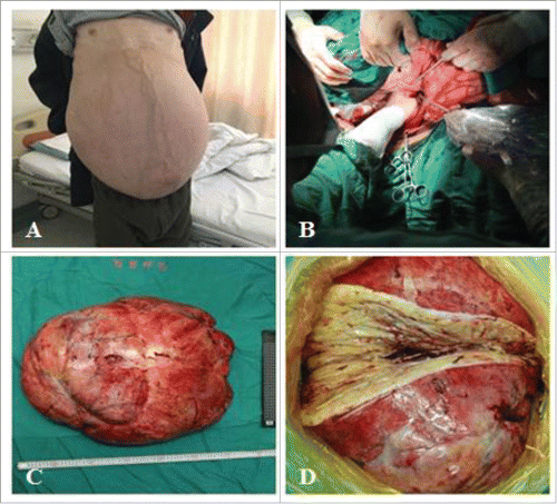
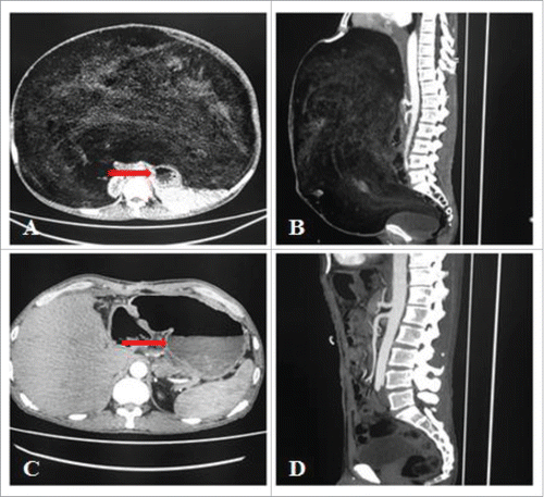
A 45-year-old male reported gradual increase in abdominal girth, without significant abdominal pain, nausea, vomiting, constipation or dyspepsia. On examination, a diffuse hard, non-mobile mass with ill-defined margins that occupied the entire abdomen was palpated and there was edema in both lower extremities (), with a ECOG score of 2 and ASA score of 4. As for routine investigations, haemograms showed mild anemia (hemoglobin 80 g/L), renal and liver functions test were within normal limits. Computed tomography (CT) showed a bulky mass, with mixed content, mainly of fat density, filling the entire abdominal cavity ( and ). The scan also revealed the lump originating from the retroperitoneum probably indicative of RPLS. The intestinal loops and other abdominal organs were pushed aside but without signs of infiltration or distant metastasis.
Figure 1. A: Appearance of the retroperitoneal liposarcoma as a giant mass. B: At surgery, well-encapsulated tumor and its exact supplying vessels were found. C: Gross appearance of resected tumor, measuring 65 × 45 × 30 cm and weighting 31 kg. D: The cut section of the specimen, showing a homogenous, yellow look.

Figure 2. A and B: Abdominal computed tomography (CT) shows a heterogeneous retroperitoneal mass occupying the entire abdominal cavity, with stomach being pushed aside (red arrow). C and D: The 3-month follow-up CT scan, with the normal stomach (red arrow).

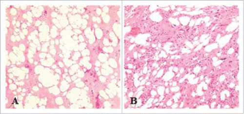
For the curative resection intention, we arranged CT aortography of abdominal aortic for him and performed bilateral intraureteral catheterization. Following this, an interdisciplinary team worked out a meticulous plan and then successfully performed en bloc resection of the tumor through a midline incision. In the operation, we found that the tumor originated from the omentum majus in front of caput pancreatis and was supplied by a branch of arteriae gastroduodenalis (). The operation took 4 hours, and the estimated blood loss was only 400 mL. The specimen measured 65 × 45 × 30 cm and weighted 31 kg (). Grossly, the tumor was well encapsulated and was consistent with R0 resection. The cut section showed a homogenous, yellow appearance (). Histologic studies revealed a well-differentiated liposarcoma ( and ). According to the grading system of the French Federation of Cancer Centers (FNCLCC), the tumor was classified as grade I.
Figure 3. Histopathologic findings. A (original magnification, × 40; hematoxylin and eosin staining) and B (original magnification, × 100; hematoxylin and eosin staining) views demonstrate a well-differentiated liposarcoma.

In the beginning postoperative period, the patient required respiratory support. Spontaneous respiration reached a sufficient level 2 d later. Afterwards he had an uneventful postoperative course and was discharged without complications 10 d after the operation, and thereafter he underwent radiotherapy 3 weeks after surgery. In addition, the patient underwent follow-up CT scans at 3 months after surgery, there has been no evidence of tumor recurrence ( and ). To date, at 8-month follow-up, he had neither symptoms nor signs of recurrence or metastasis.
Review
In English literature, from 1975 to Octobor 2016, only 13 cases of giant RPLS have been reported ().Citation6-18 Of the 14 patients (including ours), there were 9 male (64.3%) and 5 female (35.7%) with a median age of 53 (39–72) years and they all presented atypical clinical manifestations such as abdominal distention or increasing in abdominal girth. All the 14 giant RPLS were diagnosed by CT scan. Besides, 4 cases (28.6%) took preoperative biopsy examination and made exact preoperative diagnosis of liposarcoma. In terms of histologic subtypes, 7 cases (50%) were well-differentiated, 4 cases (28.6%) were dedifferentiated and the rest of 3 cases (28%) were myxoid/round cell. All the 14 cases of giant RPLS underwent surgical operations and 12 of them (85.7%) achieved R0 excision, with half of combined resections, including 5 cases of nephrectomy and 2 cases of bowel resection. Surprisingly, the tumor weight of 6 cases (42.9%), which was nearly half of all, exceeded 40 kg, with the heaviest of 47 kg. Nevertheless, 3 cases (21.4%) had postoperative complications and recovered smoothly without a secondary operation. The follow-up ranged from 6–63 (median, 24) months, and 4 cases recurred at 9, 12, 16 and 40 months respectively.
Table 1. Clinical features, preoperative evaluation, type of therapeutic approach, histopathologic characteristics at diagnosis, follow-up and clinical outcomes of 14 patients with giant RPLS.
Discussion
Retroperitoneal liposarcoma (RPLS) is a rare tumor accounting for less than 0.2% of all malignancies, but it is the most common variant of retroperitoneal tumor.Citation2 Giant RPLS that exceed 20 kg are extremely rare even though RPLS sometimes weigh as much as 10 kg or more. Similar to RPLS, giant RPLS occurs with a peak incidence between 40 and 60 y and affect male more frequently. Besides, giant RPLS are usually asymptomatic. Their most typical manifestations are discomfort or a palpable abdominal mass rather than emergences such as hematochezia, haematemesis or obstruction, resulting in their prolonged natural history and bulk.Citation19 Given this, few surgeons gain enough experience in this field, with the diagnosis often being made late, and therapy is inadequate.
For giant RPLS, the most appropriate diagnostic tests is abdominal CT, which not only defines their size, consistency, and relation between the tumor and the adjacent tissue but also allows us to detect residual tumor and recurrences.Citation20 Meanwhile, selective angiography can help us realize the tumor blood supply so that we can work out a more detailed security plan. In our case, due to the preoperative CT aortography of abdominal aortic, we could ligate the supplying vessels of tumor exactly thereby only 400 mL blood loss in operation. Moreover, intravenous pyelography is necessary because unilateral nephrectomy is often required in complete resection.Citation21 In great majority of RPLS, the CT imaging appearance (location, density, displacement, invasion and metastasis) is nearly diagnostic and preoperative biopsy seems unnecessary.Citation22 Otherwise, image-guided percutaneous coaxial core needle biopsy is strongly suggested for those imaging are not pathognomonic.Citation23,Citation24
RPLS can be classified into 4 histologic types (well-differentiated, myxoid/round cell, pleomorphic, and dedifferentiated) and 3 grades (grade I, II and III) according to the grading system of French Federation Cancer Centers (FNCLCC).Citation25 Among the 4 subtypes, well-differentiated (46%) were the most common form, followed by myxoid/round cell (28%), dedifferentiated (18%) and pleomorphic (8%), as DalalCitation26 showed. Both the histologic subtype and grade were prognostic variables. In a large cohort of 72 patients with primary RPLS, NeuhausCitation27 found that R0 excision and histologic grade I were the only variables significantly associated with a decrease in the rate of local recurrence. In a recent study, GronchiCitation28 reported that overall 5-year survival for well-differentiated subtypes was 90%, contrasting to 30–50% of pleomorphic subtypes. The rate of de-differentiated and myxoid/round cell subtypes was 75% and 60–90%, respectively.
Complete surgical resection is the cornerstone of therapy, and it is the most consistent prognostic factor.Citation29 Size alone does not contraindicate complete resection of giant RPLS, which is currently the only potentially curative treatment. Even huge, those bulks can be removed as well by a multidisciplinary team in a specialized center, which is consistent with the consensus management of RPS in the adult.Citation30 It has been demonstratedCitation31 that they can be successfully resected obtaining negative microscopic margins with combined resection of involved organs such as kidney, ureter and large bowel. Long-term prognosis without complete resection is poor with average 5 and 10-year survival rates of 16.7% and 8.0% respectively.Citation32 LewisCitation33 reported their single-institutional data of 500 retroperitoneal sarcoma cases, a median survival of 103 months for those who underwent complete resection with grossly negative margins in contrast to 18 months for those who underwent incomplete resection.
Retroperitoneal location is itself a negative prognostic factor for the reason that the retroperitoneal provides a sizeable potential space allowing significant growth before appearance of clinical signs and symptoms. Meanwhile, local recurrence and distant metastasis closely related to the tumor size. The 5-year rate of metastasis is approximately 3% when the size of the tumor is less than 2.5 cm. Nevertheless, the rate is multiplied to 55–60% in case that tumors are larger than 20 cm.Citation17 To obtain clear surgical margins and considering the highly aggressive nature of retroperitoneal liposarcomas and their propensity for local recurrence and distant metastasis, complementary adjuvant/neoadjuvant therapy may be required.Citation34 However, adjuvant therapy including chemotherapy and radiotherapy are of few study-proven value and just benefit a minority of patients.Citation35-37 Furthermore, after 2 small randomized trials, BeaneCitation38 and YangCitation39 et al. argued that postoperative radiotherapy improved local control only instead of overall survival.
Large tumor size should not be regarded as a contraindication to surgical resection. Even gigantic, complete surgical cure can also be achieved in giant RPLS with a thorough program. Giant RPLS patients are usually malnourished, hypoproteinemia or anemia as a result of tumor consumption. Therefore, much longer preoperative interval for enteral or parenteral nutrition support to improve patients’ condition is indispensable. Multidisciplinary cooperation is particularly important in the treatment of gaint RPLS. Furthermore, prudent surgical program and closely monitoring vital sign intra-operation play significant role in the whole course of the patients’ hospitalization period. Moreover, meticulous management of postoperation is crucial component of the entire process. Finally, long-term follow-up CT scan is also considered to be important, especially within 2 y after initial resection, which is peak recurrence period of giant RPLS.Citation40
Conclusions
In summary, giant RPLS is aggressive tumor and particularly rare, with no typical presentation. CT scan is the most valuable examination for diagnosis. It can be completely resected, oftentimes with combined resection of unilateral kidney and/or intestines, by an intradisciplinary team in a specialized center even though its huge tumor size. Of the whole process, multidisciplinary cooperation and meticulous preoperative preparation are particularly important. Postoperative radiotherapy may improve local control. R0 excision, histologic subtype of well-differentiated and grade I are the most significant prognostic risk factors. Long-term follow-up examination is absolutely necessary as well.
Disclosure of potential conflicts of interest
No potential conflicts of interest were disclosed.
Funding
This study was suppoted by National Natural Science Foundation of China (No. 81572413) and Scientific and Technological Application Foundation Project of Wuhan (No. 2015060101010044).
References
- Dong M, Bi J, Liu X, Wang B, Wang J. Significant partial response of metastatic intra-abdominal and pelvic round cell liposarcoma to a small-molecule VEGFR-2 tyrosine kinase inhibitor apatinib: A case report. Medicine (Baltimore) 2016; 95:e4368; PMID:27495042; https://doi.org/10.1097/MD.0000000000004368
- Taguchi S, Kume H, Fukuhara H, Morikawa T, Kakutani S, Takeshima Y, Miyazaki H, Suzuki M, Fujimura T, Nakagawa T, et al. Symptoms at diagnosis as independent prognostic factors in retroperitoneal liposarcoma. Mol Clin Oncol 2016; 4:255-60; PMID:26893871; https://doi.org/10.3892/mco.2015.701
- Molina G, Hull MA, Chen YL, DeLaney TF, De Amorim Bernstein K, Choy E, Cote G, Harmon DC, Mullen JT, Haynes AB. Preoperative radiation therapy combined with radical surgical resection is associated with a lower rate of local recurrence when treating unifocal, primary retroperitoneal liposarcoma. J Surg Oncol 2016; 114:814-20; PMID:27634478; https://doi.org/10.1002/jso.24427
- Leão P, Vilaça S, Oliveira M, Falcão J. Giant recurrent retroperitoneal liposarcoma initially presenting as inguinal hernia: Review of literature. Int J Surg Case Rep 2012; 3:103-6; PMID:22288059; https://doi.org/10.1016/j.ijscr.2011.03.009
- Makni A, Triki A, Fetirich F, Ksantini R, Chebbi F, Jouini M, Kacem M, Ben Safta Z. Giant retroperitoneal liposarcoma. Report of 5 cases. Ann Ital Chir 2012; 83:161-6: PMID:22462339
- Hazen B, Cocieru A. Giant Retroperitoneal Sarcoma. J Gastrointest Surg 2017; 21:602-3; PMID:27613734; https://doi.org/10.1007/s11605-016-3258-0
- Oh SD, Oh SJ, Suh BJ, Shin JY, Oh CK, Park JK, Kim YM, Kim BM. A Giant Retroperitoneal liposarcoma encasing the entire left kidney and adherent to adjacent structures: A case report. Case Rep Oncol 2016; 9:368-72; PMID:27462239; https://doi.org/10.1159/000447488
- Sharma M, Mannan R, Bhasin TS, Manjari M, Punj R. Giant inflammatory variant of well differentiated liposarcoma: a case report of a rare entity. J Clin Diagn Res 2013; 7:1720-1; PMID:24086890; https://doi.org/10.7860/JCDR/2013/5998.3267
- Bansal VK, Misra MC, Sharma A, Chabbra A, Murmu LR. Giant retroperitoneal liposarcoma- renal salvage by autotransplantation. Indian J Surg 2013; 75:159-61; PMID:24426418; https://doi.org/10.1007/s12262-012-0474-z
- Amir M, Akhtar S, Pervaiz M, Asad-ur-Rahman, Khawaja A, Ahmad I. Giant dedifferentiated retroperitoneal liposarcoma. J Coll Physicians Surg Pak 2011; 21:569-71; PMID:21914419; https://doi.org/09.2011/JCPSP.569571
- Akhoondinasab MR, Omranifard M. Huge retroperitoneal liposarcoma. J Res Med Sci 2011; 16:565-7; PMID:22091275
- De Nardi P, Bissolati M, Cristallo M, Staudacher C. Recurrent giant liposarcoma of the spermatic cord. Urology 2012; 79:113-4; PMID:21492916; https://doi.org/10.1016/j.urology.2011.02.004
- Hashimoto Y, Hatakeyama S, Tachiwada T, Yoneyama T, Koie T, Kamimura N, Yanagisawa T, Hakamada K, Ohyama C. Surgical treatment of a giant liposarcoma in a Japanese man. Adv Urol 2010; 2010:943073; PMID:21197426; https://doi.org/10.1155/2010/943073
- Clar H, Leithner A, Gruber G, Werkgartner G, Beham A, Windhager R. Interdisciplinary resection of a giant retroperitoneal liposarcoma of 25 kg. ANZ J Surg 2009; 79:957; PMID:20003012; https://doi.org/10.1111/j.1445-2197.2009.05160.x
- Benseler V, Obed A, Schubert T, Schlitt HJ, Bolder U. Case report–surgical therapy of a retroperitoneal liposarcoma weighing 45 kg. Zentralbl Chir 2009; 134:174-7; PMID:19294618; https://doi.org/10.1055/s-2008-1076878
- Morandeira A, Prieto J, Poves I, Sánchez Cano JJ, Díaz C, Baeta E. Giant retroperitoneal sarcoma. Can J Surg 2008; 51:E79-80; PMID:18815633
- McCallum OJ, Burke JJ 2nd, Childs AJ, Ferro A, Gallup DG. Retroperitoneal liposarcoma weighing over one hundred pounds with review of the literature. Gynecol Oncol 2006; 103:1152-4; PMID:17007913; https://doi.org/10.1016/j.ygyno.2006.08.005
- Yol S, Tavli S, Tavli L, Belviranli M, Yosunkaya A. Retroperitoneal and scrotal giant liposarcoma: report of a case. Surg Today 1998; 28:339-42; PMID:9548324; https://doi.org/10.1007/s005950050136
- Selmani R, Begovic G, Janevski V, Rushiti Q, Karpuzi A. Giant retroperitoneal liposarcoma: А case report. Prilozi 2011; 32:323-32; PMID:2182219
- Lahat G, Madewell JE, Anaya DA, Qiao W, Tuvin D, Benjamin RS, Lev DC, Pollock RE. Computed tomography scan-driven selection of treatment for retroperitoneal liposarcoma histologic subtypes. Cancer 2009; 115:1081-90; PMID:19156920; https://doi.org/10.1002/cncr.24045
- Neuhaus SJ, Barry P, Clark MA, Hayes AJ, Fisher C, Thomas JM. Surgical management of primary and recurrent retroperitoneal liposarcoma. Br J Surg 2005; 92:246-52; PMID:15505870; https://doi.org/10.1002/bjs.4802
- Wang Q, Juan YH, Li Y, Xie JJ, Liu H, Huang H, Liu Z, Zheng J, Saboo US, Saboo SS, et al. Multidetector computed tomography features in differentiating exophytic renal angiomyolipoma from retroperitoneal liposarcoma: A strobe-compliant observational study. Medicine (Baltimore) 2015; 94:e1521; PMID:26376398; https://doi.org/10.1097/MD.0000000000001521
- ESMO/European Sarcoma Network Working Group. Soft tissueand visceral sarcomas: ESMO Clinical Practice Guidelines fordiagnosis, treatment and follow-up. Ann Oncol 2014; 25 Suppl 3:iii102-12; PMID:25210080; https://doi.org/10.1093/annonc/mdu254
- Wilkinson MJ, Martin JL, Khan AA, Hayes AJ, Thomas JM, Strauss DC. Percutaneous core needle biopsy in retroperitoneal sarcomas does not influence local recurrence or overall survival. Ann Surg Oncol 2015; 22:853-8; PMID:25190132; https://doi.org/10.1245/s10434-014-4059-x
- Fan Z, Tian XF, Tang SX, Zhang YY, Pan JY, Wang S. Myxoid liposarcoma in the abdominal wall: a case report. Medicine (Baltimore) 2014; 93:e239; PMID:25526446; https://doi.org/10.1097/MD.0000000000000239
- Dalal KM, Kattan MW, Antonescu CR, Brennan MF, Singer S. Subtype specific prognostic nomogram for patients with primary liposarcoma of the retroperitoneum, extremity, or trunk. Ann Surg 2006; 244:381-91; PMID:16926564; https://doi.org/10.1097/01.sla.0000234795.98607.00
- Neuhaus SJ, Barry P, Clark MA, Hayes AJ, Fisher C, Thomas JM. Surgical management of primary and recurrent retroperitoneal liposarcoma. Br J Surg 2005; 92: 246-52; PMID:15505870; https://doi.org/10.1002/bjs.4802
- Gronchi A, Collini P, Miceli R, Valeri B, Renne SL, Dagrada G, Fiore M, Sanfilippo R, Barisella M, Colombo C, et al. Myogenic differentiation and histologic grading are major prognostic determinants in retroperitoneal liposarcoma. Am J Surg Pathol 2015; 39:383-93; PMID:25581729; https://doi.org/10.1097/PAS.0000000000000366
- Mussi C, Collini P, Miceli R, Barisella M, Mariani L, Fiore M, Casali PG, Gronchi A. The prognostic impact of dedifferentiation in retroperitoneal liposarcoma: a series of surgically treated patients at a single institution. Cancer 2008; 113:1657-65; PMID:18704991; https://doi.org/10.1002/cncr.23774
- Trans-Atlantic RPS Working Group. Management of recurrent retroperitoneal sarcoma (RPS) in the adult: A consensus approach from the trans-atlantic RPS working group. Ann Surg Oncol 2016; 23:3531-40; PMID:27480354; https://doi.org/10.1245/s10434-016-5336-7
- Reznichenko AA. Simultaneous renal cell carcinoma and giant retroperitoneal liposarcoma involving small intestine. Case Rep Surg 2016; 2016:6021909; PMID:27595033; https://doi.org/10.1155/2016/6021909
- Lahat G, Anaya DA, Wang X, Tuvin D, Lev D, Pollock RE. Resectable well-differentiated versus dedifferentiated liposarcomas: two different diseases possibly requiring different treatment approaches. Ann Surg Oncol 2008; 15:1585-93; PMID:18398663; https://doi.org/10.1245/s10434-007-9805-x
- Lewis JJ, Leung D, Woodruff JM, Brennan MF. Retroperitoneal soft-tissue sarcoma: analysis of 500 patients treated and followed at a single institution. Ann Surg 1998; 228:355-65; PMID:9742918; https://doi.org/10.1097/00000658-199809000-00008
- Park H, Lee S, Kim B, Lim do H, Choi YL, Choi GS, Kim JM, Park JB, Kwon CH, Joh JW, et al. Tissue expander placement and adjuvant radiotherapy after surgical resection of retroperitoneal liposarcoma offers improved local control. Medicine (Baltimore) 2016; 95:e4435; PMID:27512857; https://doi.org/10.1097/MD.0000000000004435
- Pervaiz N, Colterjohn N, Farrokhyar F, Tozer R, Figueredo A, Ghert M. A systematic metaanalysis of randomized controlled trials of adjuvant chemotherapy for localized resectable soft-tissue sarcoma. Cancer 2008; 113:573-81; PMID:18521899; https://doi.org/10.1002/cncr.23592
- Le Péchoux C, Musat E, Baey C, Al Mokhles H, Terrier P, Domont J, Le Cesne A, Laplanche A, Bonvalot S. Should adjuvant radiotherapy be administered in addition to front-line aggressive surgery (FAS) in patients with primary retroperitoneal sarcoma? Ann Oncol 2013; 24: 832-7; PMID:23123508; https://doi.org/10.1093/annonc/mds516
- Paryani NN, Zlotecki RA, Swanson EL, Morris CG, Grobmyer SR, Hochwald SN, Marcus RB Jr, Indelicato DJ. Multimodality local therapy for retroperitoneal sarcoma. Int J Radiat Oncol Biol Phys. 2012; 82:1128-34; PMID:21664065; https://doi.org/10.1016/j.ijrobp.2011.04.009
- Beane JD, Yang JC, White D, Steinberg SM, Rosenberg SA, Rudloff U. Efficacy of adjuvant radiation therapy in the treatment of soft tissue sarcoma of the extremity: 20-year follow-up of a randomized prospective trial. Ann Surg Oncol 2014; 21:2484-9; PMID:24756814; https://doi.org/10.1245/s10434-014-3732-4
- Yang JC, Chang AE, Baker AR, Sindelar WF, Danforth DN, Topalian SL, DeLaney T, Glatstein E, Steinberg SM, Merino MJ, et al. Randomized prospective study of the benefit of adjuvantradiation therapy in the treatment of soft tissue sarcomas of the extremity. J Clin Oncol 1998; 16:197-203; PMID:9440743; https://doi.org/10.1200/JCO.1998.16.1.197
- Kim EY, Kim SJ, Choi D, Lee SJ, Kim SH, Lim HK, Song SY. Recurrence of retroperitoneal liposarcoma: imaging findings and growth rates at follow-up CT. AJR Am J Roentgenol 2008; 191:1841-6; PMID:19020257; https://doi.org/10.2214/AJR.07.3746
