
ABSTRACT
Rhabdomyosarcoma (RMS) is an uncommon type of soft-tissue malignancy which mainly influences children. RMS rarely occurs in breast and little of the clinical behavior and treatment strategies were reported. Here, we describe a case of adult female patient with breast RMS. A 34-years-old Chinese woman visited to our hospital complaining palpable mass in her left breast. Seven months ago, the patient was diagnosed as lymphocytic mastitis and received surgical excision in the left breast. Five months later, she noticed a palpable mass again in the left breast. Ultrasonography suspected a malignant lesion and a diagnosis of RMS was made after segmental mastectomy and immunohistochemical staining. Therapy consisted of mastectomy and following neoadjuvant chemotherapy. The patient has remained disease free 30 months post-operatively. We should consider RMS when we see a breast mass. Tissue biopsy and immunohistochemical staining are recommended for diagnosis of RMS in young women. Oncologists should take immediate and active treatment on RMS.
Introduction
Rhabdomyosarcoma (RMS) is a very uncommon type of soft-tissue malignancy, which comprises 4%–8% of all malignanciesCitation1 and 10%–12% of all malignant solid tumors in children,Citation2 and rarely occurs in the adult population. Breast RMS is seldom-seen, accounting for much less than 1% of all breast malignant tumors and principally affecting adolescent girls.Citation3 RMS principally originates in trunk and the extremities in the adults,Citation4 but it is seldom found in breast. Therefore, the case of primary breast RMS reported here in an adult is very important.
Materials and methods
Case Report
A 34-years-old Chinese woman presented with a 2-month history of a 3.5 × 3.0 cm above the surgical incision scar and 2.5 × 2.0 cm lump below the scar. The patient claimed no history to radiation, nuclear plant, or any other family history. The patient had a past medical history of lymphocytic mastitis and received surgical excision 7 months ago in Yucheng county hospital. This patient was an outpatient and did not receive consequential therapy after her initial presentation. In our hospital, ultrasonography revealed a well-demarcated mass with irregular internal echoes. Segmental mastectomy was performed and diagnosis of RMS was made after tumor sections stained with haematoxylin and eosin (HE) and immunohistochemistry (IHC). We therefore performed a mastectomy of the left breast on the patient. Postoperatively, the patient received neoadjuvant chemotherapy but she refused radiotherapy.
Results
Pathological changes
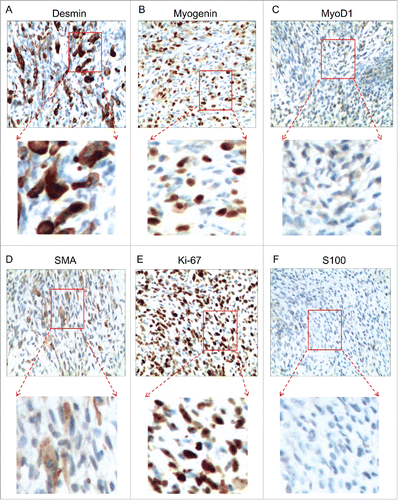
Histologic sections from tumor showed spindle cell arranged in diffuse sheets. The cells have high nucleocytoplasmic ratio and prominent nucleoli. Mitotic figures were high. Multinucleated tumor giant cells were present (). Immunohistochemical examination of the tumor specimen showed that the neoplasm was RMS, as the tumor cells were diffusely positive for Desmin, Myogenin, MyoD1 and SMA, while the tumor cells were negative for S-100 ().Citation5,Citation6 In addition, Vimentin and Ki-67 were highly expressed in the tumor tissue (). The rest examined markers, including CD34, CD31, CD68 and CD163 were positive, but cytokeratin (CK), ER and PR were negative.
Figure 1. HE staining of patient's tissue. A, Adjacent tissue. B, Photomicrograph showing eccentric nuclei, prominent nucleoli and occasional giant cells.
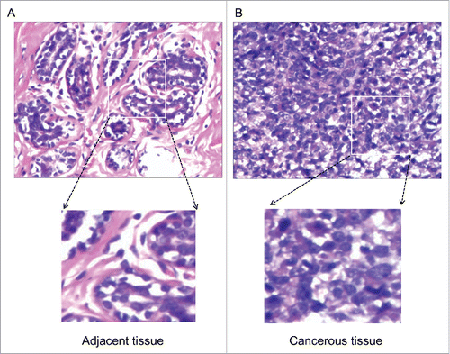
Figure 2. Immunohistochemical staining. Immunohistochemical staining showed positivity for Desmin, Myogenin, MyoD1, SMA, Ki-67, while negativity for S100.
Treatment and outcome
The breast surgery team was immediately involved. Segmental mastectomy was performed at first but mastectomy of the left breast was performed after diagnosis of RMS. Postoperatively, the patient received 6 rounds of combined chemotherapy with pirarubicin (THP), ifosfamide (IFO) and dacarbazine (DTIC). The patient continued to be in complete response at the last follow-up done 23 months post chemotherapy on March 27, 2017.
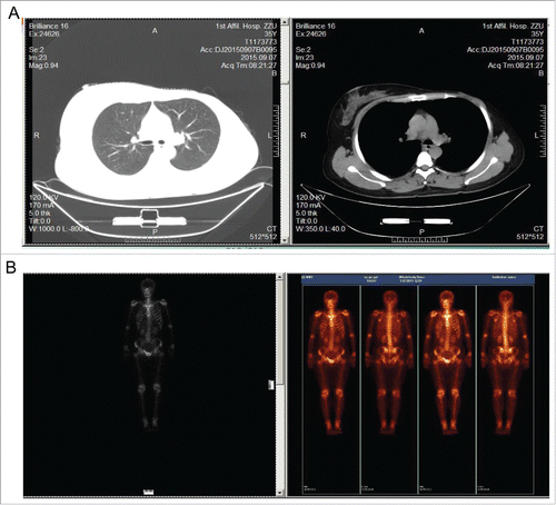
Metastatic work up, such as computed tomography (CT) of the abdomen and thorax were normal. Bone scan showed no evidence of disease elsewhere ().
Figure 3. Computed tomography (CT) and bone scan of the patient. A, CT of the thorax showed no evidence of disease elsewhere. B, Bone scan were normal.
Discussion
Primary non-epithelial malignant tumors of the breast consist of breast lymphomas, primary breast sarcomas, phyllode tumors and therapy-related breast sarcomas.Citation7 Pure non-epithelial primary malignant neoplasms of female breast are extremely uncommon, accounting for less than 1% of all breast malignancies.Citation8 Primary RMS of the breast in adults is extremely rare.Citation9
RMS predominantly presents in children aged less than 7 y old (4–8%).Citation10,Citation11 Though it seldom originates in the breast (0.2%), RMS affects diverse anatomic regions.Citation12,Citation13 The diagnosis of breast RMS is prone to be delayed. This is because benign diseases, for example, fibroadenoma and mastopathy take up the majority of breast mass, and doctors did not take immediate therapy when they deal with these cases. Although noninvasive examination, for example, ultrasonography and mammography are effective to locate the mass, they cannot distinguish RMS and were recommended on teenage women with breast mass (> 2 cm).Citation12 The cells composing of RMS are fusiform or polygonal or epithelioid, thus histological examination should distinguish it from other malignancies, including invasive lobular carcinoma, leukemia, malignant lymphoma and small cell carcinoma. It relies mainly on pathologic, immunohistochemical, and molecular biologic studies to diagnose RMS. Based on morphology of the present case, a list of differential diagnosis including RMS, malignant melanoma, and epithelioid sarcoma were included and immunohistochemistry was suggested for confirmatory diagnosis. The tumor cells were diffusely and positive for Desmin, Vimentin and SMA. Myogenin was focally positive. The tumor cells were immuno-negative for CK and S100. Only entrapped benign ductal epithelial cells showed CK positivity. Thus, we excluded the possibilities of malignant melanoma (S100−) andepithelioid sarcoma (CK−). However, tumor cells were positive for Desmin, Vimentin and Myogenin. Hence, a final diagnosis of RMS was established.
The therapy for primary breast sarcomas includes a multidisciplinary approach and surgery is the main treatment.Citation14 Wide local excision with negative margins is adequate. If there is local recurrence, then salvage mastectomy should be done. Axillary dissection is not required, as primary breast sarcomas do not present usually with axillary lymphadenopathy; it is required in cases of palpable lymphadenopathy, carcinosarcoma or liposarcoma. Role of adjuvant therapy in breast sarcomas is controversial as there is no prospective randomized trial. The role of radiotherapy is also controversial in the management of RMS.Citation15 Although it was reported rhabdomyosarcoma is sensitive to radiotherapy, and tumor measuring > 5 cm in the greatest dimension was suggested to undergo radiation therapy,Citation16 radiotherapy may also increase metastatic potential in alveolar rhabdomyosarcoma.Citation17 Results from an ongoing multi-institutional trial to validate the role of radiotherapy are awaited. In our case, mastectomy was done, and adjuvant chemotherapy was given. Although radiotherapy had been recommended by radiation oncologist, the patient refused radiotherapy after considering her age, negative surgical margin, and radiation therapy-induced side-effects. To rule out possibility of secondary sarcoma of breast, the patient was thoroughly checked and relevant investigations were done. However, no other primary tumor was detected. Besides, patient had no previous history of radiotherapy or chemotherapy especially for primary breast carcinoma. Hence, diagnosis of primary sarcoma of breast was established. Primary sarcoma of the breast is rare and primary RMS is extremely uncommon.
In conclusion, RMS of breast is a rare malignancy and mainly strikes children and adolescents. However, it can attack young women. Immunohistochemical staining is useful to make the right diagnosis. Oncologists should take immediate and active treatment once the diagnosis was established.
Disclosure of conflict of interest
No potential conflicts of interest were disclosed.
Acknowledgments
This work was supported by the National Natural Science Foundation of China (81372816).
References
- Donaldson SS. Rhabdomyosarcoma: contemporary status and future directions. The Lucy Wortham James Clinical Research Award. Archives of surgery. 1989;124:1015-20
- Binokay F, Soyupak SK, Inal M, Celiktas M, Akgul E, Aksungur E. Primary and metastatic rhabdomyosarcoma in the breast: report of two pediatric cases. European journal of radiology. 2003;48:282-4. doi:10.1016/S0720-048X(03)00041-X. PMID:14652147
- Pollard SG, Marks PV, Temple LN, Thompson HH. Breast sarcoma. A clinicopathologic review of 25 cases. Cancer. 1990;66:941-4
- Esnaola NF, Rubin BP, Baldini EH, Vasudevan N, Demetri GD, Fletcher CD, Singer S. Response to chemotherapy and predictors of survival in adult rhabdomyosarcoma. Annals of surgery. 2001;234:215-23. doi:10.1097/00000658-200108000-00012. PMID:11505068
- Pappo AS, Shapiro DN, Crist WM, Maurer HM. Biology and therapy of pediatric rhabdomyosarcoma. Journal of clinical oncology : official journal of the American Society of Clinical Oncology. 1995;13:2123-39. doi:10.1200/JCO.1995.13.8.2123. PMID:7636557
- Dias P, Chen B, Dilday B, Palmer H, Hosoi H, Singh S, Wu C, Li X, Thompson J, Parham D, et al. Strong immunostaining for myogenin in rhabdomyosarcoma is significantly associated with tumors of the alveolar subclass. The American journal of pathology. 2000;156:399-408. doi:10.1016/S0002-9440(10)64743-8. PMID:10666368
- Chugh R, Baker L. Nonepithelial malignancies of the breast. Oncology. 2004;18:665-73; discussion 73–6. PMID:15209192
- Kyriazis AP, Kyriazis AA. Primary rhabdomyosarcoma of the female breast: report of a case and review of the literature. Archives of pathology & laboratory medicine. 1998;122:747-9
- Audino AN, Setty BA, Yeager ND. Rhabdomyosarcoma of the Breast in Adolescent and Young Adult (AYA) Women. Journal of pediatric hematology/oncology. 2017;39:62-6. doi:10.1097/MPH.0000000000000710. PMID:27879537
- Pritchard J. Evidence of "stage shift" in IRS studies? Journal of clinical oncology : official journal of the American Society of Clinical Oncology. 1990;8:1768-9. doi:10.1200/JCO.1990.8.10.1768. PMID:2213111
- Crist WM, Garnsey L, Beltangady MS, Gehan E, Ruymann F, Webber B, Hays DM, Wharam M, Maurer HM. Prognosis in children with rhabdomyosarcoma: a report of the intergroup rhabdomyosarcoma studies I and II. Intergroup Rhabdomyosarcoma Committee. Journal of clinical oncology : official journal of the American Society of Clinical Oncology. 1990;8:443-52
- Hays DM, Donaldson SS, Shimada H, Crist WM, Newton WA, Jr., Andrassy RJ, Wiener E, Green J, Triche T, Maurer HM. Primary and metastatic rhabdomyosarcoma in the breast: neoplasms of adolescent females, a report from the Intergroup Rhabdomyosarcoma Study. Medical and pediatric oncology. 1997;29:181-9. doi:10.1002/(SICI)1096-911X(199709)29:3<181::AID-MPO4>3.0.CO;2-9. PMID:9212842
- Iniesta SL, Cereceda MT, Adams CS, Menor CE. Breast relapse after metastatic alveolar rhabdomyosarcoma: Is it an incurable entity? Indian journal of medical and paediatric oncology : official journal of Indian Society of Medical & Paediatric Oncology. 2016;37:119-21. doi:10.4103/0971-5851.180139
- Bhosale SJ, Kshirsagar AY, Sulhyan SR, Sulhyan SR. Rhabdomyosarcoma of the breast – a rare malignancy. The American journal of case reports. 2013;14:250-2. doi:10.12659/AJCR.883976. PMID:23869249
- Tiong SS, Dickie C, Haas RL, O'Sullivan B. The role of radiotherapy in the management of localized soft tissue sarcomas. Cancer biology & medicine. 2016;13:373-83. doi:10.20892/j.issn.2095-3941.2016.0028
- Zagars GK, Ballo MT, Pisters PW, Pollock RE, Patel SR, Benjamin RS, Evans HL. Prognostic factors for patients with localized soft-tissue sarcoma treated with conservation surgery and radiation therapy: an analysis of 1225 patients. Cancer. 2003;97:2530-43. doi:10.1002/cncr.11365. PMID:12733153
- Woods GM, Bondra K, Chronowski C, Leasure J, Singh M, Hensley L, Cripe TP, Chakravarti A, Houghton P. Radiation therapy may increase metastatic potential in alveolar rhabdomyosarcoma. Pediatric blood & cancer. 2015;62:1550-4. doi:10.1002/pbc.25516