
Article title: GLS2 is transcriptionally regulated by p73 and contributes to neuronal differentiation
Authors: Tania Velletri, Francesco Romeo, Paola Tucci, Angelo Peschiaroli, Margherita Annicchiarico-Petruzzelli, Maria Niklison-Chirou, Ivano Amelio, Richard Knight, Tak Mak, Gerry Melino, and Massimiliano Agostini
Journal: Cell Cycle
Bibliometrics: Volume 12, Issue 22, Pages 3564–3573
DOI: 10.4161/cc.26771
The E appeared incorrectly in print and online. The correct is provided on the next page.
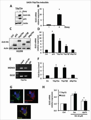
Figure 1. TAp73 drives the expression of GLS2. (A) SAOS-2-TAp73α inducible cell lines were treated with Doxycyclin (Doxy) for 24 h in order to overexpress the human TAp73α protein, and endogenous levels of GLS2 were assessed by real-time PCR (B). Induction of TAp73α led to a significant (P < 0.05) increase of GLS2 expression, as evaluated by real-time PCR. (C) H1299 cells were transfected with the indicated plasmids and expression of GLS2 was evaluated by real-time PCR as in (D). (E) TAp73 binds to the promoter of GLS2 as shown by ChIP. (F) TAp73 activates the GLS2 promoter as evaluated by luciferase activity. Co-transfection of a Renilla luciferase control plasmid was used to normalize the transfection efficiency. (G) Exogenous GLS2 localize in the mitochondria. SH-SY5Y were transfected with FLAG-GLS2 expressing vector and after 24 h stained with MitoTracker® Red CMXRos and antibody against FLAG epitope as described in “Materials and Methods”. A representative micrograph is shown. Magnification 40×. (H) TAp73 regulates GLS2 expression during neuronal terminal differentiation of neuroblastoma cells. SH-SY5Y cells were treated with 10 μM retinoic acid in order to induce differentiation. Retinoic acid (RA) treatment induces the expression of GLS2. Inhibition of TAp73 expression induced by retinoic acid prevents the upregulation of GLS2. Real-time PCR data are normalized to the housekeeping gene GAPDH and relative to control (Ctrl) Data represent mean ± s.d. of 3 different experiments. *P < 0.05.
The authors apologize for any inconvenience caused.