
ABSTRACT
Although Hmgn5 is involved in the regulation of cellular proliferation and differentiation, its physiological function during decidualization is still unknown. Here we showed that Hmgn5 was highly expressed in the decidual cells. Silencing of Hmgn5 expression by specific siRNA reduced the proliferation of uterine stromal cells and expression of Ccnd3 and Cdk4 in the absence or presence of estrogen and progesterone, whereas overexpression of Hmgn5 exhibited the opposite effects. Simultaneously, Hmgn5 might induce the expression of Prl8a2 and Prl3c1 which were 2 well-known differentiation markers for decidualization. In the uterine stromal cells, cAMP analog 8-Br-cAMP and progesterone could up-regulate the expression of Hmgn5, but the up-regulation was impeded by H89 and RU486, respectively. Attenuation of Hmgn5 expression could block the differentiation of uterine stromal cells in response to cAMP and progesterone. Further studies found that regulation of cAMP and progesterone on Hmgn5 expression was mediated by Hoxa10. During in vitro decidualization, knockdown of Hmgn5 could abrogate Hoxa10-induced upregulation of Prl8a2 and Prl3c1, while overexpression of Hmgn5 reversed the inhibitory effects of Hoxa10 siRNA on the expression of Prl8a2 and Prl3c1. In the stromal cells undergoing decidualization, Hmgn5 might act downstream of Hoxa10 to regulate the expression of Cox-2, Vegf and Mmp2. Collectively, Hmgn5 may play an important role during mouse decidualization.
Introduction
Uterine decidualization is essential for the establishment and maintenance of successful pregnancy and begins at the time of blastocyst attachment to the uterine epithelium on day 4.5 of pregnancy in mice. With the initiation of attachment, uterine stromal cells undergo extensive proliferation and subsequent differentiation into decidual cells with polyploidy, a process known as decidualization.Citation1,2 Decidualization primarily directed by ovarian estrogen and progesterone also requires the participation of various molecules.Citation1,2 Targeted gene deleting experiment has demonstrated that homeobox A10 (Hoxa10) is critical for decidualization.Citation3 Female deficiency of Hoxa10 led to a severe defect in decidualization, primarily due to reduced stromal cell responsiveness to progesterone.Citation3,4 But the underlying mechanism by which Hoxa10 regulates decidualization remains poorly understood.
High mobility group nucleosomal binding domain 5 (Hmgn5), also known as nucleosomal binding protein 1 (NSBP1), was a novel discovered member of Hmgn family which was characterized by the presence of 3 distinct functional domains: a bipartite nuclear localization signal, a conserved nucleosomal binding domain and an acidic C-terminal chromatin regulatory domain.Citation5-7 It could bind specifically to the nucleosome core particle, reduce the compacting of chromatin fiber and affect transcription.Citation5,6,8 Accumulating data have evidenced that dysregulation of Hmgn5 altered the expression of various genes.Citation8-10 In Rcho-1 cells which serve as an in vitro model for studying trophoblast cell differentiation, either up- or down-regulation of Hmgn5 led to the change of giant cell differentiation markers,Citation11 suggesting the role of Hmgn5 in cellular differentiation. This notion was further evidenced by the finding that overexpression of Hmgn5 induced the differentiation of mouse embryonic stem cells.Citation12 Simultaneously, Hmgn5 was also involved in regulating the proliferation and apoptosis of cancer cells.Citation13-16 But the effects of Hmgn5 on the proliferation and differentiation of uterine stromal cells during decidualization were still unknown. According to our (unpublished) microarray data, Hmgn5 was highly expressed in day 8 decidua and deciduoma under artificial decidualization compared with the uninjected uterine horn. On the basis of these observations, we hypothesize that Hmgn5 may be important for mouse decidualization. In this study, we found that elevated expression of Hmgn5 was observed in the decidua and decidualizing stromal cells. It could promote the proliferation and differentiation of uterine stromal cells. Further study found that Hmgn5 might act downstream of Hoxa10 to regulate the differentiation of uterine stromal cells in response to cAMP and progesterone.
Results
Hmgn5 mRNA expression during early pregnancy
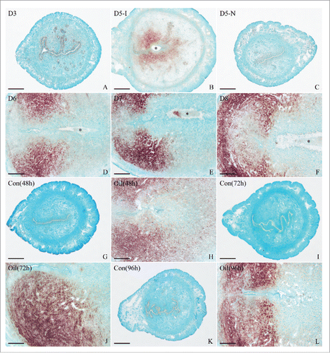
To evaluate the physiological relevance of Hmgn5 in early pregnancy events, we used in situ hybridization to examine the spatiotemporal expression pattern of Hmgn5 in mouse uterus. The results showed that there was no visible Hmgn5 mRNA signal on day 1 of pregnancy (Fig. S1B). From days 2 to 4 of pregnancy, Hmgn5 mRNA was mainly localized in the luminal and glandular epithelia (, Fig. S1C, D). On day 5 of pregnancy, Hmgn5 mRNA signal was obviously observed in the stromal cells surrounding the implanting blastocyst at implantation sites, but barely seen at inter-implantation sites (). From days 6 to 8 of pregnancy, Hmgn5 mRNA was highly expressed in the mesometrial decidua (). In addition, a specific Hmgn5 mRNA signal was only found in the embryo on day 7 of pregnancy (). However, when Hmgn5 antisense probe was replaced by Hmgn5 sense probe, there was no corresponding signal in the uterus on day 7 of pregnancy (Fig. S1A).
Figure 1. In situ hybridization of Hmgn5 expression in mouse uteri. A-F, Hmgn5 expression on days 3 (A), 5 (B, C), 6 (D), 7 (E) and 8 (F) of pregnancy. (G-L), Hmgn5 expression under artificial decidualization. 5-I, implantation site of day 5 of pregnancy; 5-N, inter-implantation site of day 5 of pregnancy; Con, uninjected uterine horn of control; Oil, oil-induced decidualization. Asterisks indicate embryo. Bar = 60 μm.
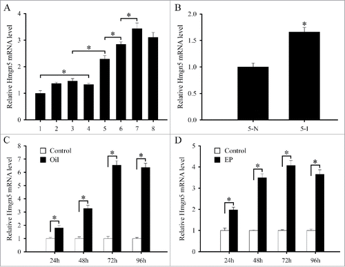
To quantify Hmgn5 mRNA expression, real-time PCR was performed. Accompanied by the progression of pregnancy, Hmgn5 expression was gradually elevated and reached a peak on day 7 and 8 of pregnancy (). Compared with the inter-implantation sites, a significantly high level of Hmgn5 expression was found at implantation sites on day 5 of pregnancy ().
Figure 2. Real-time PCR analysis of Hmgn5 expression in mouse uteri. (A) Hmgn5 expression on days 1–8 of pregnancy. (B) Hmgn5 expression at the implantation sites (5-I) and inter-implantation sites (5-N) on day 5 of pregnancy. (C) Hmgn5 expression under artificial decidualization. (D) Hmgn5 expression during in vitro decidualization. EP, estrogen plus progesterone. Data are shown mean ± SEM. Asterisks denote significance (P < 0.05).
Hmgn5 mRNA expression during decidualization
To better understand the basis of Hmgn5 potential involvement in decidualization, we employed an artificially induced decidualization model and further analyzed the expression of Hmgn5 in deciduoma. The results found that Hmgn5 expression was visualized in the decidualizing stromal cells after intraluminal oil infusion, while no visible signal was found in the uninjected control uterus (). Consistent with the above results, further quantitative analyses of Hmgn5 by real-time PCR revealed that an increased expression was also observed in the uteri undergoing artificially stimulated decidualization ().
We next employed mouse primary uterine stromal cells in vitro decidualization model and investigated the expression of Hmgn5. After stromal cells were induced to decidualization with both estrogen and progesterone, Hmgn5 expression was remarkably enhanced from 24 to 96 h of culture ().
Effects of Hmgn5 on stromal cell proliferation and differentiation during decidualization
Because decidualization requires successful progression of stromal cell proliferation and differentiation,Citation1,2 we first analyzed the effects of Hmgn5 on cell proliferation. After three different Hmgn5 siRNA duplexes were transfected into the uterine stromal cells, Hmgn5 siRNA 2 was the most effective one among them although Hmgn5 siRNA 1, 2 and 3 could strikingly suppress the expression of Hmgn5 (Fig. S2A). Therefore, Hmgn5 siRNA 2 was chosen for the following experiments. Knockdown of Hmgn5 with specific siRNA could reduce the proliferation of stromal cells in the absence or presence of estrogen and progesterone (). In contrast, overexpression of Hmgn5 could enhance the expression of Hmgn5 in the stromal cells and proliferation of stromal cells (, Fig. S2B). To understand the molecular basis for the proliferative role of Hmgn5, we subsequently analyzed the influences of Hmgn5 on the expression of cyclin A1 (Ccna1), Ccnb1, Ccnb2, Ccnd1, Ccnd3, Ccne1, cyclin-dependent kinase 1 (Cdk1), Cdk2, Cdk4 and Cdk6. The results showed that attenuation of Hmgn5 expression by siRNA efficiently suppressed the expression of Ccnd3 and Cdk4 in the absence or presence of estrogen and progesterone, whereas overexpression of Hmgn5 exhibited the opposite effects (). Hmgn5 did not significantly affect the expression of Ccna1, Ccnb1, Ccnb2, Ccnd1, Ccne1, Cdk1, Cdk2 and Cdk6 in the stromal cells which were untreated or treated with estrogen and progesterone (Fig. S2C-F).
Figure 3. Effects of Hmgn5 on the proliferation of uterine stromal cells. (A) Effects of Hmgn5 siRNA on the proliferation of uterine stromal cells. After transfection with Hmgn5 siRNA, stromal cells were analyzed by MTS assay in the absence or presence of estrogen and progesterone. (B) Effects of Hmgn5 overexpression on the proliferation of uterine stromal cells. After transfection with Hmgn5 overexpression plasmid, stromal cells were analyzed by MTS assay in the absence or presence of estrogen and progesterone. (C) Effects of Hmgn5 siRNA on the expression of Ccnd3 in the stromal cells. (D) Effects of Hmgn5 siRNA on the expression of Cdk4 in the stromal cells. (E) Effects of Hmgn5 overexpression on the expression of Ccnd3 in the stromal cells. (F) Effects of Hmgn5 overexpression on the expression of Cdk4 in the stromal cells. NC, control siRNA; siHmgn5, Hmgn5 siRNA; Con, empty pcDNA3.1 vector; Hmgn5, Hmgn5 overexpression plasmid.
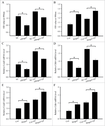
To further unveil the role of Hmgn5 in the differentiation of uterine stromal cells, we next analyzed its effects on the expression of prolactin family 8, subfamily a, member 2 (Prl8a2) and prolactin family 3, subfamily c, member 1 (Prl3c1), which were 2 well-known markers of uterine stromal cells differentiation during decidualization.Citation17 The result showed that treatment of stromal cells with Hmgn5 siRNA resulted in a marked reduction in the expression of Prl8a2 and Prl3c1 in the absence or presence of estrogen and progesterone (). Conversely, overexpression of Hmgn5 in the stromal cells caused a dramatic induction of Prl8a2 and Prl3c1 ().
Figure 4. Effects of Hmgn5 on the differentiation of uterine stromal cells. (A and B) Effects of Hmgn5 siRNA on the expression of Prl8a2 and Prl3c1. After transfection with Hmgn5 siRNA, the expression of Prl8a2 and Prl3c1 was determined by real-time PCR in the absence or presence of estrogen and progesterone. (C and D) Effects of Hmgn5 overexpression on the expression of Prl8a2 and Prl3c1. After transfection with Hmgn5 overexpression plasmid, the expression of Prl8a2 and Prl3c1 was determined by real-time PCR in the absence or presence of estrogen and progesterone.

Hmgn5 mediates the effects of Hoxa10 on the differentiation of uterine stromal cells
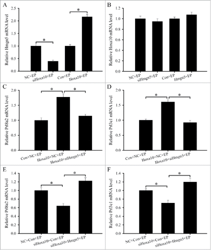
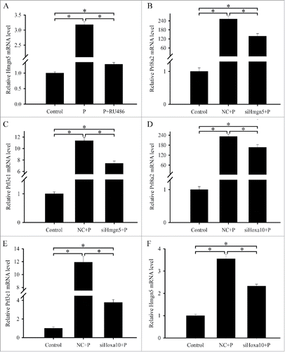
Hoxa10 was a major regulator of decidualization and its deficiency led to the aberrant Hmgn5 expression in the decidual bed with defective decidualization.Citation3,18,19 During in vitro decidualization, silencing of Hoxa10 with specific siRNA could decrease the expression of Hmgn5, while overexpression of Hoxa10 augmented its expression (). Neither inhibition nor overexpression of Hmgn5 affected the expression of Hoxa10 in the stromal cells treated with estrogen and progesterone (). Together these data imply that Hmgn5 may be a downstream target of Hoxa10 during decidualization. We next asked whether Hmgn5 could mediate the effects of Hoxa10 on the differentiation of uterine stromal cells. After the uterine stromal cells were co-transfected with Hoxa10 overexpression plasmid and Hmgn5 siRNA and then subjected to in vitro decidualization, we monitored the expression of differentiation markers Prl8a2 and Prl3c1 and found that attenuation of Hmgn5 expression could prevent the Hoxa10-induced up-regulation of Prl8a2 and Prl3c1 (). On the contrary, overexpression of Hmgn5 could improve the expression of Prl8a2 and Prl3c1 in the stromal cells which were transfected with Hoxa10 siRNA and then subjected to in vitro decidualization ().
Figure 5. Hmgn5 mediates the effects of Hoxa10 on the differentiation of uterine stromal cells. (A) Effects of Hoxa10 siRNA or overexpression on the expression of Hmgn5. (B) Effects of Hmgn5 siRNA or overexpression on the expression of Hoxa10. (C and D) Hmgn5 siRNA abrogated the effects of Hoxa10 overexpression on the expression of Prl8a2 and Prl3c1. After co-transfection with Hoxa10 overexpression plasmid and Hmgn5 siRNA, the expression of Prl8a2 and Prl3c1 was determined by real-time PCR in the presence of estrogen and progesterone. (E and F) Overexpression of Hmgn5 improved the effects of Hoxa10 siRNA on the expression of Prl8a2 and Prl3c1. siHoxa10, Hoxa10 siRNA; Hoxa10, Hoxa10 overexpression plasmid.
Hmgn5 acts downstream of Hoxa10 to mediate the effects of cAMP on the differentiation of uterine stromal cells
It has previously established that the process of decidualization was accompanied by elevated intracellular cAMP levels which could initiate the differentiation of uterine stromal cells prominently via protein kinase A (PKA) transduction pathway.Citation20 PKA inhibitor H89 could weaken the expression of Prl8a2 and Prl3c1 in the uterine stromal cells undergoing decidualization (). Meanwhile, Hmgn5 expression was also inhibited by H89 during in vitro decidualization (). These results collectively suggest that cAMP-PKA signal may be involved in regulating the expression of Hmgn5. To elucidate the regulation by cAMP of Hmgn5 expression, we treated the uterine stromal cells with cAMP analog 8-bromoadenosine-cAMP (8-Br-cAMP, 500 μM) and analyzed the expression of Hmgn5. The result found that a gradual increase in the level of Hmgn5 mRNA was observed between 1 to 12 h, reached a peak at 12 h and then slightly declined at 24 h (). This induction was abrogated by a pretreatment of H89 (). We next examined whether siRNA-mediated attenuation of Hmgn5 expression could impact on the cAMP-induced differentiation of stromal cells. Administration of 8-Br-cAMP to uterine stromal cells treated with Hmgn5 siRNA caused a robust decline in the expression of Prl8a2 and Prl3c1 ().
Figure 6. Hmgn5 acts downstream of Hoxa10 to mediate the effects of cAMP on the differentiation of uterine stromal cells. (A–C) Effects of H89 on the expression of Prl8a2, Prl3c1 and Hmgn5 during in vitro decidualization. (D) Hmgn5 expression in the uterine stromal cells treated with 8-Br-cAMP for 1, 3, 6, 12 and 24 h. (E) Hmgn5 expression after uterine stromal cells were treated with 8-Br-cAMP, or both 8-Br-cAMP and H89. (F and G) Hmgn5 mediated the effects of cAMP on the expression of Prl8a2 and Prl3c1. After transfection with Hmgn5 siRNA and addition of 8-Br-cAMP, the expression of Prl8a2 and Prl3c1 was determined by real-time PCR. (H) Effects of cAMP on the expression of Hmgn5 via Hoxa10.
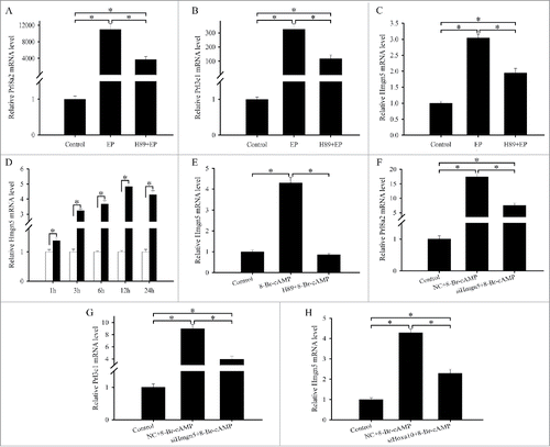
Our previous study have evidenced that Hoxa10 could mediate the effects of cAMP on the differentiation of stromal cells.Citation21 As stated above, Hmgn5 expression was regulated by Hoxa10 and cAMP. Thus we surmised that regulation of cAMP on the expression of Hmgn5 was mediated by Hoxa10. To verify the speculation, we transfected the stromal cells with Hoxa10 siRNA and then added 8-Br-cAMP and analyzed the expression of Hmgn5. The result showed that silencing of Hoxa10 could efficiently disturb the induction of 8-Br-cAMP on Hmgn5 expression ().
Hmgn5 mediates the effects of Hoxa10 on the expression of Cox-2, vegf and Mmp2
To reveal the underlying mechanism by which Hmgn5 governed the process of decidualization, we examined the regulation of Hmgn5 on the expression of cyclooxygenase-2 (Cox-2), vascular endothelial growth factor (Vegf), matrix metallopeptidase 2 (Mmp2) and Mmp9 genes. After transfection with Hmgn5 siRNA along with the addition of estrogen and progesterone, the expression of Cox-2, Vegf and Mmp2 were noticeably lessened compared with control (). In contrast, overexpression of Hmgn5 could up-regulate the expression of Cox-2, Vegf and Mmp2 during in vitro decidualization (). Interestingly, we did not observe any statistically significant difference in the expression of Mmp2 after transfection with Hmgn5 siRNA or overexpression plasmid, respectively ().
Figure 7. Hmgn5 mediates the effects Hoxa10 on the expression of Cox-2, Vegf and Mmp2 during in vitro decidualization. (A) Effects of Hmgn5 siRNA on the expression of Cox-2, Vegf, Mmp2 and Mmp9. (B) Effects of Hmgn5 overexpression on the expression of Cox-2, Vegf, Mmp2 and Mmp9. (C) Effects of Hoxa10 siRNA on the expression of Cox-2, Vegf and Mmp2. (D) Effects of Hoxa10 overexpression on the expression of Cox-2, Vegf and Mmp2. (E) The expression of Cox-2, Vegf and Mmp2 after stromal cells were co-transfected with Hoxa10 overexpression plasmid and Hmgn5 siRNA. (F) The expression of Cox-2, Vegf and Mmp2 after stromal cells were co-transfected with Hoxa10 siRNA and Hmgn5 overexpression plasmid.
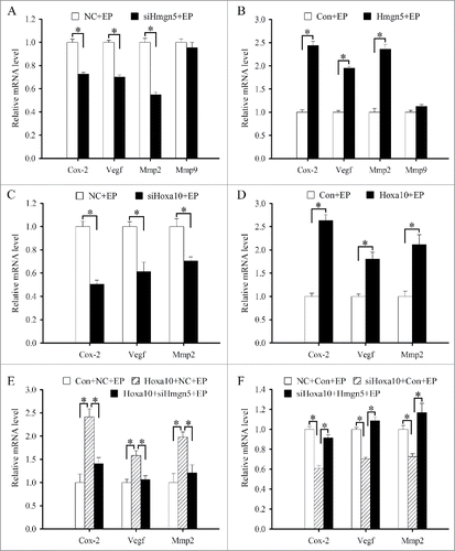
Because Hoxa10 could modulate the expression of Hmgn5, we also tested the regulation of Hoxa10 on the expression of Cox-2, Vegf and Mmp2 during in vitro decidualization. The results found that inhibition of Hoxa10 expression by siRNA diminished the expression of Cox-2, Vegf and Mmp2, whereas overexpression of Hoxa10 had the opposite effects on the expression of Cox-2, Vegf and Mmp2 (). We next determined whether Hmgn5 might mediate the effects of Hoxa10 on the expression of Cox-2, Vegf and Mmp2 in the stromal cells which were subjected to in vitro decidualization in the presence of estrogen and progesterone. After co-transfection with Hoxa10 overexpression plasmid and Hmgn5 siRNA, the expression of Cox-2, Vegf and Mmp2 were evidently reduced compared to transfection with Hoxa10 overexpression plasmid alone (). Conversely, overexpression of Hmgn5 reversed the inhibitory effects of Hoxa10 siRNA on the expression of Cox-2, Vegf and Mmp2 ().
Steroid hormonal regulation of Hmgn5 expression
To address the influence of estrogen and progesterone on uterine Hmgn5 expression, we employed ovariectomized mouse model injected with estrogen, progesterone, or estrogen plus progesterone. In the uteri of ovariectomized mice receiving sesame oil as a control, Hmgn5 mRNA signal was weakly seen in the luminal and glandular epithelium (). Estrogen treatment enhanced the expression of Hmgn5 in the uterine luminal epithelium at 24 h, but elicited no remarkable effects on the glandular epithelium (). Progesterone treatment caused a heightened expression of Hmgn5 in the luminal and glandular epithelium as well as in the uterine stromal cells (). After co-treatment with estrogen and progesterone, Hmgn5 expression was restricted to the luminal and glandular epithelium, but lower compared with progesterone treatment alone (), implying that estrogen seems to have an antagonistic effect on progesterone upregulation of Hmgn5 expression. Real-time PCR analysis confirmed the notion that co-treatment with estrogen and progesterone led to a reduction of Hmgn5 expression compared with progesterone treatment, although estrogen and/or progesterone stimulated the expression of Hmgn5 in the uteri of ovariectomized mice ().
Figure 8. In situ hybridization of Hmgn5 expression after ovariectomized mice were treated with sesame oil (Control), estrogen, progesterone or a combination of estrogen and progesterone. E, estrogen; P, progesterone. Bar = 60 μm.
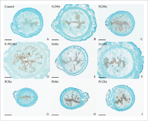
Figure 9. Hormonal regulation of Hmgn5 expression. (A) Real-time PCR analysis of Hmgn5 expression after ovariectomized mice were treated with sesame oil, estrogen, progesterone or a combination of estrogen and progesterone for 24 h. (B) Real-time PCR analysis of Hmgn5 expression in ovariectomized mouse uteri after injection of estrogen for 1, 3, 6, 12 and 24 h. (C) Hmgn5 expression in the uterine epithelial cells treated with estrogen or both estrogen and ICI 182,780. (D) Real-time PCR analysis of Hmgn5 expression in ovariectomized mouse uteri after injection of progesterone for 1, 3, 6, 12 and 24 h. (E) Hmgn5 expression in the uterine epithelial cells treated with progesterone or both progesterone and RU486. (F) Hmgn5 expression in uterine stromal cells treated with progesterone for 1, 3, 6, 12 and 24 h.
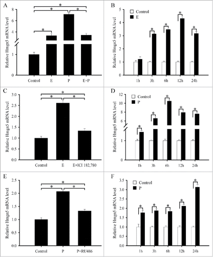
Based on above observations, we subsequently examined the expression of Hmgn5 at different time courses after estrogen or progesterone treatment. A high level of Hmgn5 mRNA signal was mainly detected in the uterine luminal and glandular epithelium at 3 and 6 h after estrogen treatment, and in the luminal epithelium at 12 h (). By real-time PCR analysis, the levels of Hmgn5 mRNA showed an increase after estrogen injection, with peak levels at 12 h followed by a decline at 24 h (). To further ascertain the regulation of estrogen on Hmgn5 expression, we treated the uterine epithelial cells with estrogen in vitro and found that Hmgn5 expression was distinctly up-regulated at 12 h (). Then we checked whether the induction of Hmgn5 expression by estrogen was depended on the estrogen receptor (ER). The results found that ER antagonist ICI 182,780 could reverse stimulatory effects of estrogen on Hmgn5 expression (), suggesting ER requirement for this induction. Progesterone could induce the accumulation of Hmgn5 mRNA in the uterine luminal and glandular epithelium from 3 to 24 h (). Interestingly, Hmgn5 mRNA signal was noted in the stromal cells at 12 and 24 h after ovariectomized mice were treated by progesterone (). Real-time PCR analysis revealed that progesterone injection resulted in a rise in uterine Hmgn5 mRNA levels that peaked at 6 h, then decreased at 12 h and sustained through 24 h (). In the in vitro cultured uterine epithelial cells, progesterone could raise the expression of Hmgn5 at 6 h (). But the up-regulation of Hmgn5 expression by progesterone was impeded by progesterone receptor (PR) antagonist RU486 (). In the meantime, administration of progesterone to the uterine stromal cells led to a notable increase in the expression of Hmgn5, which reached the highest level at 24 h, whereas RU486 markedly reduced the progesterone-dependent activation of Hmgn5 stromal expression ().
Figure 10. Hmgn5 acts downstream of Hoxa10 to mediate the effects of progesterone on the differentiation of uterine stromal cells. (A) Hmgn5 expression after uterine stromal cells were treated with progesterone or both progesterone and RU486. (B and C) Hmgn5 mediated the effects of progesterone on the expression of Prl8a2 and Prl3c1. After transfection with Hmgn5 siRNA and addition of progesterone, the expression of Prl8a2 and Prl3c1 was determined by real-time PCR. (D and E) Hoxa10 mediated the effects of progesterone on the expression of Prl8a2 and Prl3c1. (F) Effects of progesterone on the expression of Hmgn5 via Hoxa10.
Hmgn5 acts downstream of Hoxa10 to mediate the effects of progesterone on the differentiation of uterine stromal cells
Since progesterone is essential for the differentiation of uterine stromal cells during decidualization,Citation2,22 we next examined whether attenuation of Hmgn5 expression could impact on progesterone-mediated stromal differentiation. After transfection with Hmgn5 siRNA, the induction of Prl8a2 and Prl3c1 by progesterone was dramatically blocked (). As described above, Hmgn5 was a downstream target of Hoxa10 which was also regulated by progesterone.Citation4 Further analysis displayed that siRNA-mediated downregulation of Hoxa10 could reverse the stimulatory effects of progesterone on the differentiation of uterine stromal cells as evidenced by the reduced expression of Prl8a2 and Prl3c1 (). We next asked whether Hoxa10 might mediate the regulation of progesterone on Hmgn5 expression. To address this, we administrated progesterone to stromal cells transfected with Hoxa10 siRNA and tested the expression of Hmgn5. The result found that progesterone-induced expression of Hmgn5 was remarkably alleviated by Hoxa10 siRNA ().
Discussion
Although Hmgn5 is involved in the regulation of cellular proliferation and differentiation, its physiological role in the process of decidualization is still unknown. In this study, we found that Hmgn5 were highly expressed in the decidual cells, implying that Hmgn5 may be important for decidualization. Stromal cell proliferation is the first step of decidualization. Overexpression of Hmgn5 enhanced the proliferation of uterine stromal cells in the absence or presence of estrogen and progesterone, whereas inhibition of Hmgn5 with specific siRNA exhibited the opposite effects. Further study found that Hmgn5 could modulate the expression of Ccnd3 which was a well-known regulator of mammalian cell proliferation, and its function was dependent on the presence of Cdk4 or Cdk6.Citation1,23 In the uterine stromal cells, Hmgn5 could direct the expression of Cdk4, but did not change the expression of Cdk6, suggesting that Hmgn5 regulates stromal cells proliferation via targeting Ccnd3 and Cdk4. Following this marked proliferation, stromal cells begin to differentiate into decidual cells which are characterized by their pavement morphology and the elevated expression of Prl8a2 and Prl3c1 that are 2 well-known markers of uterine stromal cells differentiation during decidualization.Citation2,17,24 The present results revealed that Hmgn5 could stimulate the expression of differentiation markers Prl8a2 and Prl3c1 in the uterine stromal cells which were untreated or treated with estrogen and progesterone. Collectively, these results demonstrate that Hmgn5 plays an important role during decidualization.
It has been well established that progesterone is required for decidualization and its function is mediated through activation of the intracellular PR.Citation1,2,22 In the uterine stromal cells, progesterone could induce the expression of Hmgn5. Moreover, the induction was impeded by PR antagonist RU486, suggesting PR requirement for this induction. Attenuation of Hmgn5 expression by siRNA greatly diminished the progesterone-induced stromal differentiation which coincided with a gradual increase in intracellular cAMP level during human decidualization.Citation25 Simultaneously, uterine concentration of cAMP also elevates following blastocyst- and artificially induced decidualization in the mouse.Citation26,27 Previous studies have proved that cAMP is necessary for decidualization in human and mice.Citation20,28,29 As a ubiquitous second messenger molecule, cAMP can prominently activate the PKA signal transduction pathway.Citation20 PKA inhibitor H89 could restrain the differentiation of uterine stromal cells during in vitro decidualization as indicated by the drastically reduced expression of Prl8a2 and Prl3c1. In the stromal cells, cAMP analog 8-Br-cAMP induced the expression of Hmgn5, but this induction was abrogated by H89. Treatment of uterine stromal cells with Hmgn5 siRNA efficiently blocked the effects of cAMP on the expression of differentiation markers Prl8a2 and Prl3c1. Taken together, these results evidence that Hmgn5 is a critical downstream target of progesterone and cAMP in regulating the differentiation of uterine stromal cells. Further analysis revealed that regulation of progesterone and cAMP on Hmgn5 was mediated by Hoxa10.
It is generally accepted that Hoxa10 is of great importance to uterine decidualization. Ablation of Hoxa10 in mice led to severely compromised decidualization.Citation3 In Hoxa10-deficient mice, Hmgn5 was aberrantly expressed in the decidual bed with defective decidualization.Citation18,19 Likewise, overexpression of Hoxa10 could enhance the expression of Hmgn5 during in vitro decidualization, while silencing of Hoxa10 with specific siRNA reduced its expression, implying that Hmgn5 may be a downstream regulator of Hoxa10 during decidualization. This notion was supported by the present evidence that overexpression of Hmgn5 could improve the expression of Prl8a2 and Prl3c1 in the Hoxa10 siRNA-tranfected stromal cells, whereas attenuation of Hmgn5 expression by siRNA could prevent the Hoxa10-induced upregulation of Prl8a2 and Prl3c1, further reinforcing the effects of Hmgn5 on the differentiation of uterine stromal cells during decidualization.
Angiogenesis is a hallmark event during decidualization and is profoundly influenced by Vegf.Citation1,30 In the clear cell renal cell carcinoma (ccRCC) cells, knockdown of Hmgn5 could down-regulate the expression of Vegf.Citation13 Consistent with this finding, the present study provided evidence that Hmgn5 also modulated the expression of Vegf during in vitro decidualization. Furthermore, there was observation to suggest that Cox-2 also participated in uterine angiogenesis during decidualization because one cause of decidualization failure in Cox-2 deficient mice was deregulated vascular events.Citation1,30 In the uterine stromal cells, up- and down-regulation of Hmgn5 altered the expression of Cox-2 in the presence of estrogen and progesterone. Overall, these results offer convincing proof that Hmgn5 may direct uterine angiogenesis by affecting the expression of Cox-2 and Vegf genes during decidualization. Meanwhile, decidualization is involved in the extensive remodeling of uterine extracellular matrix which is regulated by Mmps.Citation1 It has previously been reported that silencing of Hmgn5 with specific siRNA decreased the expression of Mmp9 in osteosarcoma cells, breast or bladder cancer cells, the expression of Mmp2 in meningioma cells and the activity of Mmp2 and Mmp9 in ccRCC cells.Citation13-16,31 The present result evidenced that Hmgn5 could adjust the expression of Mmp2 in the uterine stromal cells undergoing decidualization, but did not change the expression of Mmp9. These data suggest a role of Hmgn5 in the remodeling of uterine extracellular matrix during decidualization. Further studies found that Hmgn5 could mediate the effects of Hoxa10 on the expression of Cox-2, Vegf and Mmp2 during in vitro decidualization.
In summary, Hmgn5 may play an important role during mouse decidualization and act downstream of Hoxa10 to regulate the differentiation of uterine stromal cells in response to cAMP and progesterone, and the expression of Cox-2, Vegf and Mmp2 during in vitro decidualization (Fig. S3).
Materials and methods
Animals
Matured mice (Kunming white strain) were caged in a controlled environment with a cycle of 14L:10D. All animal procedures were approved by the Institutional Animal Care and Use Committee of Jilin University. To confirm reproducibility of results, at least 3 mice per group were used in each stage or treatment in this study.
Pregnancy and pseudopregnancy
Adult female mice were mated with fertile or vasectomized males of the same strain to induce pregnancy or pseudopregnancy by co-caging, respectively (day 1 = day of vaginal plug). On days 1–4, pregnancy was confirmed by recovering embryos from the oviducts or uterus. The implantation sites on day 5 were identified by intravenous injection of 0.1 ml of 1% Chicago blue (Sigma, St. Louis, MO) in 0.85% sodium chloride.
Artificial induced decidualization
Artificial decidualization was induced by intraluminally infusing 25 µl of sesame oil into one uterine horn on day 4 of pseudopregnancy, while the contralateral uninjected horn served as a control. The mice were killed to collect uteri at 24, 48, 72 or 96 h after artificial induced decidualization. Decidualization was confirmed by weighing the uterine horn and histological examination of uterine sections.
Steroid hormonal treatments
Mature female mice were ovariectomized and, after 2 weeks, given a single injection of estradiol-17β (100 ng/mouse), progesterone (2 mg/mouse) or a combination of the same doses of estradiol-17β and progesterone, respectively. All steroids were dissolved in sesame oil and injected subcutaneously. Control mice received the vehicle only (sesame oil, 0.1 ml/mouse). Mice were sacrificed at different time to collect uteri after the hormonal injections.
Uterine epithelial and stromal cells from day 4 of pregnancy were isolated and cultured as previously described.Citation24 Cultured epithelial and stromal cells were also treated with 1 μM of progesterone or 0.1 nM of estradiol-17β, respectively. For further studies, cells were pretreated with RU486 (1 μM) for 2 h or ICI 182,780 (100 nM) before the addition of progesterone or estrogen, respectively. Steroids and antagonists were dissolved in ethanol. Controls received the vehicle only.
In situ hybridization
Total RNA from the mouse uteri was reverse-transcribed and amplified with Hmgn5 primers. Hmgn5 forward primer 5′- GATGGGAAATGCAAAGAGGA and reverse primer 5′- TCTTCTGCCTCCACCTTGTT were designed according to Mus musculus high mobility group nucleosomal binding domain 5 gene (Genbank accession number NM_016710). The amplified fragment (327 bp) of Hmgn5 was cloned into pGEM-T plasmid (pGEM-T Vector System 1, Promega, Madison, WI) and verified by sequencing. Hmgn5-containing plasmid was amplified with the primers for T7 and SP6 to prepare templates for labeling. Digoxigenin (DIG)-labeled antisense and sense cRNA probes were transcribed in vitro using a DIG RNA labeling kit (Roche Diagnostics GmbH, Mannheim, Germany).
Frozen sections (10 μm) were mounted on 3-aminopropyltriethoxy-silane (Sigma)-coated slides and fixed in 4% paraformaldehyde solution in PBS. Hybridization was performed as described previously.Citation24 Sections were counterstained with 1% methyl green. The positive signal was visualized as a dark brown color. The sense probe was also hybridized and served as a negative control. There was no detectable signal from sense probes.
Real-time PCR
Total RNAs from mouse uteri or cultured cells were isolated using TRIPURE reagent according to the manufacturer's instructions (Roche) and reverse-transcribed into cDNA using M-MLV reverse-transcriptase (Promega). Reverse transcriptase was performed at 42°C for 60 min with 2 μg total RNA in 25 μl volume. For real-time PCR, cDNA was amplified using FS Universal SYBR Green Real Master (Roche) on BIO-RAD CFX96™ Real Time Detection System. The conditions used for real-time PCR were as follows: 95°C for 3 min, followed by 40 cycles of 95°C for 15 s and 60°C for 1 min. All reactions were run in triplicate. The result was analyzed using CFX Manager Software. After analysis using the 2-ΔΔCt method, data were normalized to Gapdh expression. Primer sequences for real-time PCR were listed in .
In vitro decidualization
Mouse in vitro decidualization was performed as previously described with modification.Citation24 Briefly, uterine stromal cells were induced for in vitro decidualization with fresh medium supplemented with progesterone (1 μM) and estradiol-17β (10 nM) in DMEM-F12 with 2% charcoal-treated FBS (Biological Industries Ltd., Kibbutz Beit Hemeek, Israel).
Plasmid construction and transfection
Full-length Hmgn5 and Hoxa10 cDNA fragments were amplified by PCR from mouse uterus using the following primers: Hmgn5 (5′- GATATC (EcoRV) CGGAGAGCTGCAACAATGCCC and 5′- CTCGAG (XhoI) TTAGACAATACTCAGAGGCTC) and Hoxa10 (5′- GAATTC (EcoRI) ATGTCAGCCAGAAAGGG and 5′- GATATC (EcoRV) GGAAGCGAAAAGACGTTGT). The amplified products were purified and cloned into pGEM-T vector. The pGEM-T-Hmgn5, pGEM-T-Hoxa10 and pcDNA3.1 vector were cut by EcoRV/XhoI or EcoRI/EcoRV (TaKaRa, Dalian, China) at 37°C for 1 h, and then the fragments were ligated into pcDNA3.1 with T4 ligase (Promega) at 4°C overnight to construct pcDNA3.1-Hmgn5 and pcDNA3.1-Hoxa10, respectively. An empty pcDNA3.1 expression vector was served as control.
Transfection of uterine stromal cells was performed according to the manufacturer's protocol for lipofectamine 2000 (Invitrogen). After transfection with control plasmid (empty pcDNA3.1 vector) or overexpression plasmids, stromal cells were collected or induced for in vitro decidualization for 48 h.
RNA interference
The small-interfering RNA (siRNA) duplexes for targeting Hmgn5 and Hoxa10 as well as a scrambled sequence (control siRNA duplex, negative control) were designed and synthesized by GenePharma. The sequences were shown as follows: 5′- CACCGGAGUUGAAACCUAATT and 5′- UUAGGUUUCAACUCCGGUGTT (Hmgn5 siRNA 1); 5′- CCACAAAUGUCGUGG AAGATT and 5′- UCUUCCACGACAUUUGUGGTT (Hmgn5 siRNA 2); 5′- GACGAAUGCAA CAUGGAAATT and 5′- UUUCCAUGUUGCAUUCGUCTT (Hmgn5 siRNA 3); 5′- CCAAAUU AUCCCACAACAATT and 5′- UUGUUGUGGGAUAAUUUGGCG (Hoxa10 siRNA); 5′- UUCUCCGAACGUGUCACGUTT and 5′- ACGUGACACGUUCGGAGAATT (nonspecific scrambled siRNA, negative control). Transfections for siRNA were performed according to Lipofectamine 2000 protocol. After transfection with Hmgn5 siRNAs or Hoxa10 siRNAs, uterine stromal cells were collected or induced for in vitro decidualization for 48 h.
Cell proliferation
Proliferation assays were performed using MTS reagent (Promega) according to the manufacturer's directions. Uterine stromal cells were seeded at a density of 1×10⁵ /well in 96-well plates and cultured in the DMEM/F12 medium containing 2% heat-inactivated FBS. After transfection with Hmgn5 overexpression plasmid or siRNA, stromal cells were cultured or induced for in vitro decidualization for 48 h. Finally, 20 µl of MTS reagent was added to each well and incubated for 4h. Absorbance was measured at 490 nm using a 96-well plate reader. Every experiment was performed in triplicate.
Statistics
All the experiments were independently repeated at least 3 times. The significance of difference was analyzed by one-way ANOVA or Independent-Samples T Test using the SPSS software program (SPSS Inc., Chicago). The differences were considered significant at P < 0.05.
Table 1. Primers for real-time PCR.
Disclosure of potential conflict of interest
No potential conflicts of interest were disclosed.
KCCY_A_1220459_supplement.zip
Download Zip (3.8 MB)Funding
This work was financially supported by Special Funds for Scientific Research on Public Causes (201303119), National Natural Science Foundation of China (31472158 and 31372390) and College Student Innovation Experimental Program of Jilin University (2016).
References
- Dey SK, Lim H, Das SK, Reese J, Paria BC, Daikoku T, Wang H. Molecular cues to implantation. Endocr Rev 2004; 25:341-73; PMID:15180948; http://dx.doi.org/10.1210/er.2003-0020
- Zhang S, Lin H, Kong S, Wang S, Wang H, Wang H, Armant DR. Physiological and molecular determinants of embryo implantation. Mol Aspects Med 2013; 34:939-80; PMID:23290997; http://dx.doi.org/10.1016/j.mam.2012.12.011
- Benson GV, Lim H, Paria BC, Satokata I, Dey SK, Maas RL. Mechanisms of reduced fertility in Hoxa-10 mutant mice: uterine homeosis and loss of maternal Hoxa-10 expression. Development 1996; 122:2687-96; PMID:8787743
- Lim H, Ma L, Ma WG, Maas RL, Dey SK. Hoxa-10 regulates uterine stromal cell responsiveness to progesterone during implantation and decidualization in the mouse. Mol Endocrinol 1999; 13:1005-17; PMID:10379898; http://dx.doi.org/10.1210/mend.13.6.0284
- Furusawa T, Cherukuri S. Developmental function of HMGN proteins. Biochim Biophys Acta 2010; 1799:69-73; PMID:20123069; http://dx.doi.org/10.1016/j.bbagrm.2009.11.011
- Rochman M, Malicet C, Bustin M. HMGN5/NSBP1: a new member of the HMGN protein family that affects chromatin structure and function. Biochim Biophys Acta 2010; 1799:86-92; PMID:20123071; http://dx.doi.org/10.1016/j.bbagrm.2009.09.012
- González-Romero R, Eirín-López JM, Ausió J. Evolution of high mobility group nucleosome-binding proteins and its implications for vertebrate chromatin specialization. Mol Biol Evol 2015; 32:121-31; http://dx.doi.org/10.1093/molbev/msu280
- Rochman M, Postnikov Y, Correll S, Malicet C, Wincovitch S, Karpova TS, McNally JG, Wu X, Bubunenko NA, Grigoryev S, Bustin M. The interaction of NSBP1/HMGN5 with nucleosomes in euchromatin counteracts linker histone-mediated chromatin compaction and modulates transcription. Mol Cell 2009; 35:642-56; PMID:19748358; http://dx.doi.org/10.1016/j.molcel.2009.07.002
- Malicet C, Rochman M, Postnikov Y, Bustin M. Distinct properties of human HMGN5 reveal a rapidly evolving but functionally conserved nucleosome binding protein. Mol Cell Biol 2011; 31:2742-55; PMID:21518955; http://dx.doi.org/10.1128/MCB.05216-11
- Ciappio ED, Krausz KW, Rochman M, Furusawa T, Bonzo JA, Tessarollo L, Gonzalez FJ, Bustin M. Metabolomics reveals a role for the chromatin-binding protein HMGN5 in glutathione metabolism. PLoS One 2014; 9:e84583; PMID:24392144; http://dx.doi.org/10.1371/journal.pone.0084583
- Shirakawa H, Rochman M, Furusawa T, Kuehn MR, Horigome S, Haketa K, Sugita Y, Inada T, Komai M, Bustin M. The nucleosomal binding protein NSBP1 is highly expressed in the placenta and modulates the expression of differentiation markers in placental Rcho-1 cells. J Cell Biochem 2009; 106:651-8; PMID:19160411; http://dx.doi.org/10.1002/jcb.22046
- Pritsker M, Ford NR, Jenq HT, Lemischka IR. Genomewide gain-of-function genetic screen identifies functionally active genes in mouse embryonic stem cells. Proc Natl Acad Sci U S A 2006; 103:6946-51; PMID:16621925; http://dx.doi.org/10.1073/pnas.0509861103
- Ji SQ, Yao L, Zhang XY, Li XS, Zhou LQ. Knockdown of the nucleosome binding protein 1 inhibits the growth and invasion of clear cell renal cell carcinoma cells in vitro and in vivo. J Exp Clin Cancer Res 2012; 31:22; PMID:22420896; http://dx.doi.org/10.1186/1756-9966-31-22
- Zhou X, Yuan B, Yuan W, Wang C, Gao R, Wang J. The expression and clinical significance of high mobility group nucleosome binding domain 5 in human osteosarcoma. Tumour Biol 2014; 35:6539-47; PMID:24687550; http://dx.doi.org/10.1007/s13277-014-1825-0
- He J, Liu C, Wang B, Li N, Zuo G, Gao D. HMGN5 blockade by siRNA enhances apoptosis, suppresses invasion and increases chemosensitivity to temozolomide in meningiomas. Int J Oncol 2015; 47:1503-11; PMID:26315299
- Weng M, Song F, Chen J, Wu J, Qin J, Jin T, Xu J. The high-mobility group nucleosome-binding domain 5 is highly expressed in breast cancer and promotes the proliferation and invasion of breast cancer cells. Tumour Biol 2015; 36:959-66; PMID:25315189; http://dx.doi.org/10.1007/s13277-014-2715-1
- Clementi C, Tripurani SK, Large MJ, Edson MA, Creighton CJ, Hawkins SM, Kovanci E, Kaartinen V, Lydon JP, Pangas SA, DeMayo FJ, Matzuk MM. Activin-like kinase 2 functions in peri-implantation uterine signaling in mice and humans. PLoS Genet 2013; 9:e1003863; PMID:24244176; http://dx.doi.org/10.1371/journal.pgen.1003863
- Ma X, Gao F, Rusie A, Hemingway J, Ostmann AB, Sroga JM, Jegga AG, Das SK. Decidual cell polyploidization necessitates mitochondrial activity. PLoS One 2011; 6:e26774; PMID:22046353; http://dx.doi.org/10.1371/journal.pone.0026774
- Sroga JM, Gao F, Ma X, Das SK. Overexpression of cyclin D3 improves decidualization defects in Hoxa-10(−/−) mice. Endocrinology 2012; 153:5575-86; PMID:23008516; http://dx.doi.org/10.1210/en.2012-1528
- Gellersen B, Brosens J. Cyclic AMP and progesterone receptor cross-talk in human endometrium: a decidualizing affair. J Endocrinol 2003; 178:357-72; PMID:12967329; http://dx.doi.org/10.1677/joe.0.1780357
- Li DD, Guo CH, Yue L, Duan CC, Yang ZQ, Cao H, Guo B, Yue ZP. Expression, regulation and function of Hmgn3 during decidualization in mice. Mol Cell Endocrinol 2015; 413:13-25; PMID:26112184; http://dx.doi.org/10.1016/j.mce.2015.05.038
- Li Q, Kannan A, Wang W, Demayo FJ, Taylor RN, Bagchi MK, Bagchi IC. Bone morphogenetic protein 2 functions via a conserved signaling pathway involving Wnt4 to regulate uterine decidualization in the mouse and the human. J Biol Chem 2007; 282:31725-32; PMID:17711857; http://dx.doi.org/10.1074/jbc.M704723200
- Tan J, Raja S, Davis MK, Tawfik O, Dey SK, Das SK. Evidence for coordinated interaction of cyclin D3 with p21 and cdk6 in directing the development of uterine stromal cell decidualization and polyploidy during implantation. Mech Dev 2002; 111:99-113; PMID:11804782; http://dx.doi.org/10.1016/S0925-4773(01)00614-1
- Li DD, Yang ZQ, Guo CH, Yue L, Duan CC, Cao H, Guo B, Yue ZP. Hmgn1 acts downstream of C/EBPβ to regulate the decidualization of uterine stromal cells in mice. Cell Cycle 2015; 14:3461-74; PMID:26566865; http://dx.doi.org/10.1080/15384101.2015.1093704
- Jones MC, Fusi L, Higham JH, Abdel-Hafiz H, Horwitz KB, Lam EW, Brosens JJ. Regulation of the SUMO pathway sensitizes differentiating human endometrial stromal cells to progesterone. Proc Natl Acad Sci U S A 2006; 103:16272-7; PMID:17053081; http://dx.doi.org/10.1073/pnas.0603002103
- Rankin JC, Ledford BE, Baggett B. Early involvement of cyclic nucleotides in the artificially stimulated decidual cell reaction in the mouse uterus. Biol Reprod 1977; 17:549-54; PMID:200287; http://dx.doi.org/10.1095/biolreprod17.4.549
- Huang Z, Wang TS, Qi QR, Zuo RJ, Liang XH, Zhao XY, Yang ZM. Progesterone regulates secretin expression in mouse uterus during early pregnancy. Reprod Sci 2014; 21:724-32; PMID:24336673; http://dx.doi.org/10.1177/1933719113512527
- Ruan YC, Guo JH, Liu X, Zhang R, Tsang LL, Dong JD, Chen H, Yu MK, Jiang X, Zhang XH, Fok KL, Chung YW, Huang H, Zhou WL, Chan HC. Activation of the epithelial Na+ channel triggers prostaglandin E2 release and production required for embryo implantation. Nat Med 2012; 18:1112-7; PMID:22729284; http://dx.doi.org/10.1038/nm.2771
- Deng WB, Liang XH, Liu JL, Yang ZM. Regulation and function of deiodinases during decidualization in female mice. Endocrinology 2014; 155:2704-17; PMID:24797630; http://dx.doi.org/10.1210/en.2014-1015
- Wang H, Dey SK. Roadmap to embryo implantation: clues from mouse models. Nat Rev Genet 2006; 7:185-99; PMID:16485018; http://dx.doi.org/10.1038/nrg1808
- Wahafu W, He ZS, Zhang XY, Zhang CJ, Yao K, Hao H, Song G, He Q, Li XS, Zhou LQ. The nucleosome binding protein NSBP1 is highly expressed in human bladder cancer and promotes the proliferation and invasion of bladder cancer cells. Tumour Biol 2011; 32:931-9; PMID:21695596; http://dx.doi.org/10.1007/s13277-011-0195-0