
 ?Mathematical formulae have been encoded as MathML and are displayed in this HTML version using MathJax in order to improve their display. Uncheck the box to turn MathJax off. This feature requires Javascript. Click on a formula to zoom.
ABSTRACT
Objective: Both females and the elderly have been identified as vulnerable populations with increased injury and mortality risk in multiple crash scenarios. Particularly in frontal impacts, older females show higher risk to the chest and thorax than their younger or male counterparts. Thoracic geometry plays a role in this increase, and this study aims to quantify key parts of that geometry in a way that can directly inform human body models that incorporate the concept of person age.
Methods: Computed tomography scans from 2 female subject groups aged 20–35 and 65–99 were selected from the International Center for Automotive Medicine scan database representing young and old female populations. A model of thoracic skeletal anatomy was built for each subject from independent parametric models of the spine, ribs, and sternum, along with further parametric models of those components’ spatial relationships. Parameter values between the 2 groups are directly compared, and average parameter values within each group are used to generate statistically average skeletal geometry for young and old females. In addition to the anatomic measures explicitly used in the parameterization scheme, key measures of rib cage depth and spine curvature are taken from both the underlying subject pool and from the resultant representative geometries.
Results: Statistically significant differences were seen between the young and old groups’ spine and rib anatomic components, with no significant differences in local sternal geometry found. Vertebral segments in older females had higher angles relative to their inferior neighbors, providing a quantification of the kyphotic curvature known to be associated with age. Ribs in older females had greater end-to-end span, greater aspect ratio, and reduced out-of-plane deviation, producing an elongated and overall flatter curvature that leads to distal rib ends extending further anteriorly in older individuals. Combined differences in spine curvature and rib geometry led to an 18-mm difference in anterior placement of the sternum between young and old subjects.
Conclusions: This study provides new geometric data regarding the variability in anthropometry of adult females with age and has utility in advancing the veracity of current human body models. A simplified scaffold representation of underlying 3-dimensional bones within the thorax is presented, and the reported young and old female parameter sets can be used to characterize the anatomic differences expected with age and to both validate and drive morphing algorithms for aged human body models. The modular approach taken allows model parameters to hold inherent and intuitive meaning, offering advantages over more generalized methods such as principal component analysis. Geometry can be assessed on a component level or a whole thorax level, and the parametric representation of thorax shape allows direct comparisons between the current study and other individuals or human body models.
Introduction
Motor vehicle crashes (MVCs) are a leading cause of injury and mortality worldwide, causing over 2 million injuries and 33,000 deaths each year in the United States (NHTSA Citation2014). In frontal crashes, thoracic injuries and rib fractures in particular are the most common form of severe injury, and sternal fractures occur in approximately 1 in 5 severely injured occupants (Hanna and Hershman Citation2009). Each of these injuries also shows significantly higher incidence rates in the elderly over younger occupants (Hanna and Hershman Citation2009; Lee et al. Citation2006; Ridella et al. Citation2012). Bose et al. (Citation2011) showed that the odds for a belt-restrained female driver to sustain severe injuries were 47% higher than those for a similarly restrained male driver involved in a comparable crash. In frontal sled tests with postmortem human subjects, elderly female subjects were found to sustain a higher number of rib fractures than field studies or equivalent sled tests with larger male counterparts would predict (Shaw et al. Citation2017).
Computational modeling offers unique opportunities to specifically address the safety concerns of at-risk or vulnerable populations by providing tools that better represent those populations and the risks they face. For example, increased thorax injury risk is seen in simulations that incorporate age-specific parameters into finite element models of the thorax (Antona-Makoshi et al. Citation2015; Schoell et al. Citation2015). In order to adequately model age as a human factor, quantitative descriptions of the morphometric changes that occur with age must be made available. A number of studies have done so using generalized procrustes analysis (GPA) and principal component analysis (PCA; Gayzik et al. Citation2008; Shi et al. Citation2014; Wang et al. Citation2016; Weaver, Schoell, and Stitzel Citation2014). However, the quantitative results from these studies (the principal components themselves) can be difficult to interpret directly, and most studies report on changes to the rib cage as a whole, leaving the specific changes to the different skeletal components in the rib cage underreported.
The purpose of this study is to develop a self-contained quantification of the overall structure of the thoracic skeleton that includes measurements of individual skeletal components and to provide exemplar geometries representing a young female and an old female population.
Methodology
Two female groups were selected for this study representing a young (18 to 35 years of age, 111 subjects) and an old (over 65 years of age, 101 subjects) population. All subjects had chest and abdomen computed tomography (CT) scans obtained for trauma purposes between 2002 and 2016, collected from the International Center for Automotive Medicine (ICAM) morphomics database. Subjects with extreme skeletal abnormalities (in the form of extreme scoliosis, injury, or skeletal fixation devices) and without the full chest wall within the scan window were excluded. All scans were taken in a supine position, and scan slice spacing ranged from 0.625 mm (75% of scans) to 5 mm (16% of scans). The mean ± SD age within each group was 27.9 ± 4.5 years and 76.4 ± 8.6 years. Weights within the young and old groups (76.5 ± 24.3 kg and 72.9 ± 17.5 kg) were not significantly different (P = .2), whereas the younger cohort (165.2 ± 7.5 cm) was taller than the older cohort (160.1 ± 6.6 cm).
The overall size and shape of the spine, the ribs, and the sternum were characterized via separate parametric models described below. A further 2 sets of parameters were then used to model the spatial relationship between the spine and ribs and the ribs and sternum.
Spine parameterization
A simplified geometry of the spine was chosen in order to provide a basis for the placement of ribs and the rest of the thoracic skeleton. Though the spine itself contains highly detailed and specialized geometry, we wish simply to describe its position and curvature in a quantitative fashion. This can be done by taking a curve through the center of the spinal canal as the desired shape to be modeled. In order to produce this geometrically, we can treat the spinal canal as being formed by a series of vertebral segments and then choose a finite set of parameters to represent each segment using
| • | VBSEGSz: The height of each vertebral segment. | ||||
| • | VBSEGα: The angle of each vertebral segment with respect to its inferior neighbor. | ||||
| • | L5pitch: The position and angle of the lowest vertebral segment with respect to the body. | ||||
These quantities were measured from the CT scans via 3D landmarks placed in the center of the spinal canal at each vertebral level. Figure A.1 (see online supplement) depicts those landmarks and the measurements of vertebral segment size and relative angle for an example L4 vertebra.
Rib parameterization
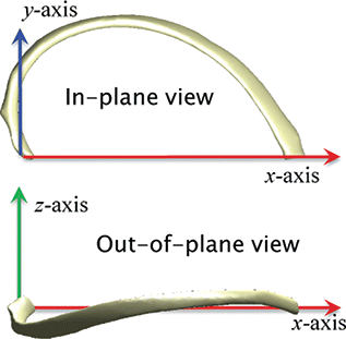
Overall rib shapes were characterized by modeling the centroidal path (i.e., the series of points at the center of consecutive cross-sections along the rib) via separate models of a rib’s in-plane shape and out-of-plane shape with respect to a local rib plane. The local rib plane consists of an x-axis starting at the rib’s proximal end (closest to the spine) and passing through its distal end point and a y-axis chosen to minimize the deviation from the x–y plane of a series of points along the rib’s central path as depicted in . The in-plane shape was modeled by a 6-parameter model from Holcombe et al. (Citation2016), which uses direct intrinsic geometric properties including a rib’s end-to-end span (Sx), aspect ratio (YPk), skewness (XPk), and inner angle at the proximal (vertebral) end of the rib in its local plane (φpia). Two additional parameters (Bd and Bp) are logarithmic spiral constants that modulate the local curvature of the rib in its distal and proximal regions, respectively. For full demonstration of the model parameter effects, the reader is directed to Holcombe et al. (Citation2016).
Figure 1. In-plane view and out-of-plane view of a single rib.
The out-of-plane deviation of each rib was modeled as a function of its overall rib arc length using a cubic Bezier curve with 2 free parameters. To ensure equivalent directions of deviation between left and right rib local coordinate systems, the z-deviation of right-sided ribs was reversed. Within this convention, the neck of the rib generally extends below the rib plane before passing back above the plane at around mid-shaft. The distal portion of the rib rises before eventually returning to the plane (at z = 0) at its distal end as shown in Figure A.2 (see online supplement). The mathematical bases for the Bezier curve are Bernstein polynomials of degree 3, 3ℓ(1 − ℓ)2 and 3ℓ2(1 − ℓ), where ℓ ranges from 0 to 1. A set of points along a rib deviating from the local plane can therefore be modeled using the regression coefficients corresponding to these two bases, ZA and ZB, as given in Equation Equation1(1)
(1) .
(1)
(1)
Rib position and orientation
The orientation of each rib with respect to a fixed body coordinate system in a scan was represented by 3 rotational angles. A rib’s pump-handle parameter (αPH) is specified as the angle between the rib local x-axis and the coronal plane. αLS is the angle between the rib local x-axis and the sagittal plane, and αBH (Margulies et al. Citation1989) is a rotation about the rib’s local x-axis after the prior rotations are performed. Positive αBH is defined as moving the lateral aspect of the rib superiorly regardless of rib side, such that an initial neutrally posed rib (on its correct side yet hanging directly inferiorly) can undergo successive rotations by αPH (up from the sagittal plane), αLS (away from the medial plane), and then by αBH (about the newly rotated x-axis, positive for left-sided ribs and negative for left-sided ribs), with the resulting rib being oriented correctly in the body habitus.
The proximal ends of ribs (i.e., the origins of each local rib plane) were then positioned in the body via simple parameters for their x-, y-, and z-coordinate offsets from the center of the spinal canal at their corresponding vertebral level. The precise location from which rib ends are offset was chosen to be the center of the spinal canal at the inferior aspect of the vertebral body. For example, the position of the proximal end of a given person’s sixth ribs might lie 12 mm lateral (RIBOFFx parameter) from the midline their T6 vertebra, 3 mm posterior (RIBOFFy) to the center of the spinal canal at T6, and 6 mm superior (RIBOFFz) to the inferior aspect of T6.
Sternum parameterization
The sternum was modeled by parameterizing the position of landmarks placed at each connecting notch between the sternum and segments of costal cartilage corresponding to ribs 1 through 7. These landmarks were first placed in the CT scan 3D space and then a local sternum plane was constructed such that
the origin sits at the sternal angle, specified as the mid-point of the 2 second rib sternal notches
the plane z-axis extends from the origin through the mid-point of the 2 seventh rib sternal notches
the lateral x-axis is perpendicular to the primary axis, pointing in the average left to right direction specified by each left–right pair of notch landmarks 1 through 7
Evaluated with respect to this plane, each notch location (for ribs 1–7, left and right) has a local x-, y-, and z-component, designated by STNOTCH[x|y|z]. Data for the left and right sides were averaged such that only one set of 7 rib levels is used (i.e., lateral symmetry is assumed), and the x-coordinate is specified as a positive lateral offset from the midline of the sternum. The landmarks and resulting sternum plane are illustrated in .
Figure 2. Sternum plane placement and orientation. The plane origin (largest sphere marker) is parameterized by its offset (STNM[y|z]Offset) from the midpoint between second rib distal ends (smallest sphere marker). The sternum plane pitch (STNMpitch) is measured against the vertical axis in the sagittal view.
![Figure 2. Sternum plane placement and orientation. The plane origin (largest sphere marker) is parameterized by its offset (STNM[y|z]Offset) from the midpoint between second rib distal ends (smallest sphere marker). The sternum plane pitch (STNMpitch) is measured against the vertical axis in the sagittal view.](/cms/asset/e1067e47-6250-49bd-99a2-cd1e7a9bb9bb/gcpi_a_1309526_f0002_oc.gif)
In order to place and orient the sternum appropriately in the body, parameters are included for the position of the sternum plane origin relative to the midpoint between the ends of the second ribs (STNM[y|z]Offset) in global Y (anterior) and Z (superior) directions. The inclination of the sternum is provided by sternum pitch angle (STNMpitch) given in degrees, with lower STNMpitch angles resulting in a more vertically oriented sternum.
Results
All parameters were measured directly from CT scans with average results per group reported below for each parametrically modeled body part or body part relationship. Where they exist, statistically significant differences between the average values for the young and old group at the P = .05 level are indicated.
Spine shape parameters
The relative angle of each vertebral body (from L4 through T1) to its inferior neighbor was measured from the underlying CT scans for all subjects (VBSEGα), along with each vertebral segment size (VBSEGSz), as illustrated in Figure A.1. Mean values from the young and the old groups are given in . Additionally, the absolute angle of the L5 vertebra in the sagittal plane (used to place this lowest vertebra at an appropriate orientation) was measured, with the L5 vertebra from old individuals (L5pitch = 23.2°) being tilted more anteriorly than the younger group (L5pitch = 15.3°, P < .001).
Table 1. Relative angle (VBSEGα, degrees) and segment size (VBSEGSz, mm) of each vertebral segment for young (Y) and old (O) age groupsa.
In lumbar and thoracic segments, it was seen that the relative angle between adjacent vertebrae was significantly larger in magnitude in the older population than in the younger population.
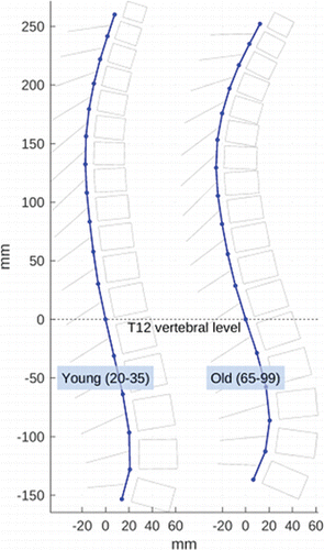
The results found for L5pitch and those in provide all that is needed to generate a curve composed of single-vertebra segments that runs along the center of the spinal canal. That shape is shown in for the young and old groups, clearly illustrating the effect of increasing spine kyphosis with age that is quantified by the differences in parameter values in . For ease of visualization, vertebral bodies and a line denoting the spinous process have been added.
Figure 3. Average group parameters used to rebuild average spine shapes of the young and old groups. Results reflect greater degrees of kyphosis with age.
Rib parameters
Using an optimization routine described in Holcombe et al. (Citation2016), each set of best-fitting in-plane rib parameters was found for all ribs in all subjects. Similarly, rib out-of-plane deviation parameters and rib angle parameters were measured from all subjects, and means for each group and from each parameter are given in Table B.1 (see online supplement). As shown in this table, there was a significant difference between young and old shape parameters at a majority of ribs for most parameters. Specifically, the parameters showing a consistent trend across all ribs include the rib end-to-end lengths (Sx), which were longer in the older population; rib aspect ratios (YPk), which were more extreme in the older population; rib out-of-plane deviation (ZA,ZB), which was reduced in the older population; and rib pump-handle angle (αPH), which was higher in the older population.
The relative position of the proximal (closest to the spine) end of each rib is provided in Table B.2 (see online supplement). Here it can be seen that upper ribs are positioned more laterally in the older group than in the younger group, and lower ribs are positioned more posteriorly and inferiorly relative to the spine in the older group than the younger group.
Sternum parameters
The placement with respect to the ends of the second ribs and the orientation of the local sternum plane were measured from all subjects. The mean parameter values from each group for the plane position are given in Table B.3 (see online supplement), with no significant differences seen between the young and old populations. Similarly, average positions of the sternal connection notch of ribs 1 through 7 relative to the local sternum plane are given in Table B.4 (see online supplement), with no statistically significant differences seen between the subject groups.
Collective results
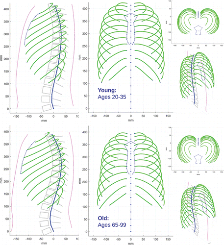
Using each of the parameterized geometries described above along with their average values from the young and the old subject groups, we can collect results into a parametric model of the thoracic skeleton and rib cage. Each individual piece of geometry is developed in an isolated fashion based on parameters defining that geometry, and the pieces are then combined by the additional parameters that define their spatial relationships. For example, the ribs as defined by shape parameters in Table B.1 (see online supplement) form a coherent model of isolated ribs, which can subsequently be combined with the structure for a spine by specifying their relationship (i.e., positional offsets RIBOFF[x, y, z] and orientation angles αPH, αLS, αBH) from that spine. This modular approach followed by the recombination of each piece of geometry from group parameter averages produces the overall thoracic skeleton geometries depicted in . Here, we see the accumulated effect of each of the differences in geometry parameters on overall thoracic shape.
Figure 4. Reconstructed skeletal geometry from average model parameters as measured from the young and old subject groups. Skin and vertebral body outlines included for visualization purposes only.
Discussion
In this study we have developed isolated models of thoracic skeleton components based on statistically average parameters from 2 subject groups and then combined those components to form an overall characterization of the skeleton of the thorax for comparison between the groups. Individual differences are seen at the component level by direct observation of component model parameters, and the resulting differences in overall thorax geometry are interrogated and discussed below.
Spine curvature
Results from show that the relative angles between adjacent vertebrae are, on average, significantly larger in magnitude in the old population compared to the younger group. These individual segment angle differences accumulate to produce the increased kyphotic spine curvature in the elderly geometry that is seen most clearly in and .
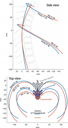
Figure 5. Reconstructed skeletal geometry from average model parameters as measured from the young and old subject groups. Both groups are centered at their T6 vertebra, and ribs 2 and 6 are shown along with the position of the sternum second rib notches. Vertebral body outlines included for visualization purposes only.
One way to validate the overall accuracy of spine geometry presented here—and to assess the ability of the accumulated spine model using average parameter values to represent spines that are actually present within the constituent population—is to use global measures of spine curvature that are not explicit model parameters. A standard clinical measure of spine curvature is the Cobb angle, which is derived from the inner angle in the sagittal plane subtended between the superior end-plate of the T4 vertebra and the inferior end-plate of the T9 vertebra. Taking end-plate surfaces as lying perpendicular to the spinal canal, this T4–T9 Cobb angle was measured from the CT scans in each population group and then measured from the two resulting geometries depicted in and . In the underlying population, older subjects (34.6° ± 10.2°) had significantly higher Cobb angles than the younger group (23.1° ± 6.4°, P < .001). Reconstructed geometries using average parameter values from the 2 subject groups offered excellent agreement to these population distributions, with Cobb angles for the old and young average geometries of 34.7° and 23.4°, respectively. These results also match well to a population of adults without disease or skeletal deformity measured by Goh (Citation2000), which had mean Cobb angles of 28.5° with standard deviation of 10.7°.
Recently, whole-spine geometry has been described by Sato et al. (Citation2016) for young male and female subjects in an automotive seated posture. In that study, the most prominent mode of variation reflected changes to the position along the spine of the posterior-most peak of the kyphotic region of the spine, and it was seen that more pronounced kyphosis corresponded to this peak occurring at a lower vertebral level. In the current study, the vertebral level found to extend most posteriorly ranged from T12 to T4, with the most common level being T7 (all vertebrae referenced at the inferior end plate level of its vertebral body), and with a standard deviation across the studied population of 1.38 vertebral body sizes. No significant differences in the level that extended furthest posteriorly were found between young and old populations (P > .05). Taking T4–T12 Cobb angle as a measure of overall kyphosis, a slight yet significant inverse relationship (P = .03, r2 = 0.02) between kyphosis and posterior-most vertebral level was seen, yet the direction of this relationship was reversed if T4–T9 Cobb angle was instead used to quantify kyphosis (P = .05, r2 = 0.017). These results suggest that the variability in posterior-most vertebral level reported by Sato et al. (Citation2016) is well supported, yet its association with kyphosis may not hold for larger sample sizes or may be dependent on the precise method used to evaluate kyphosis. Alternatively, the association may occur primarily as a function of subject posture, with the effect being seen in seated positions but not replicated in supine positions.
Rib shape and rib cage depth
Structural differences in the shape of the rib cage serve to alter its mechanical response to loading. One thoracic feature of interest is that of overall chest depth (i.e., the anterior–posterior depth of the rib cage and sternum). Increases in chest depth that come from superior rotation of the ribs are known to increase overall thoracic stiffness to anterior thoracic loading (Kent et al. Citation2005). Shi et al. (Citation2014) and Wang et al. (Citation2016) reported an increase in overall rib cage depth with age, and results from the current work illustrated in confirm this effect.
An alternative view comparing the two geometries is given in , which superimposes the young and old average geometry using the T6 vertebra as a mid-thorax reference location. Here it can be seen that the effect of spine kyphosis serves to shift the T2 vertebral bodies approximately 12 mm more anteriorly in the older group. At the distal ends of the second ribs, the older subject geometry is shifted 18 mm more anteriorly than the younger geometry, meaning that the combination of rib end-to-end length (Sx) and pump-handle angle (αPH) serve to push older second ribs 6 mm anteriorly independent of the changing spine shape. This trend is repeated at the sixth rib level, this time with a 14-mm anterior shift in rib distal end position for the older group.
In the current study, the choice for sternum position is specified by the relative location of the second rib notch to the distal ends of the second ribs. As highlighted by Table B.3 (see online supplement), none of the resulting offset parameters were seen to be significantly different between groups. Specifically, STNMyOffset placed the sternum marginally more anterior in younger rib cages than older ones by a distance of 1.8 mm. These results help to explain that the primary source of the observed depth increase of the chest in the elderly group comes from changes to the ribs and spine themselves, rather than from any relative shift of the sternum due to changes in costal cartilage. Similarly, no significant differences were seen in the local geometry parameters describing sternum shape (Table B.4; see online supplement). This result is consistent with Weaver, Schoell, Nguyen, et al. (Citation2014), who described sternal growth through childhood but found little to no change in shape in adults after skeletal maturity.
With the ribs and the spine primarily responsible for the increased rib cage depth, a comparison of the specific rib shape differences reported here between young and old females was made to previously available data from Shi et al. (Citation2014). Using the point cloud data from that study (generated using the average ages, heights, and body mass index from the 2 cohorts in the current study), the sixth rib was isolated and fitted to a local rib coordinate system. A visual comparison of the two pairs of ribs is given in Figure A.3 (see online supplement) and shows strong agreement between the studies. Specifically, ribs lengths and aspect ratios measured in ribs regenerated from Shi et al. (Citation2014) are within 0.25 standard deviations of the average population values from the current young and old cohorts. Though these specific rib bone shape changes were not previously commented on, the correspondence between geometric output offers replication of results provided by Shi et al. (Citation2014) and adds veracity both to the results of that study and to the specific age differences quantified here.
Accompanying these differences in the in-plane shapes of young and old ribs are changes in the out-of-plane deviation as represented by the reduced magnitudes in the ZA and ZB parameters seen in older ribs in Table B.1 (see online supplement). The maximum deviation of the sixth rib centroid above its fitted rib plane (as occurs in the region of the rib distal to the spine), was 8.7 ± 2.8 mm for the young ribs and 6.5 ± 2.5 mm for older ribs. When using the average parameter values for ZA and ZB to generate out-of-plane deviation from Equation Equation1(1)
(1) , comparable values of 8.8 mm (young) and 6.4 mm (old) were obtained. However, when considering the maximum deviation below the rib plane (as occurs proximal to the spine), the resulting deviation resulting from model parameters 9.4 and 6.7 mm were found to underestimate the average deviation seen within the constituent populations, which were 11.7 ± 3.5 mm and 8.9 ± 3.4 mm for the young and old sixth ribs, respectively. Though these results show that the overall trend (a reduction in out-of-plane deviation with age) is well modeled, they also suggest room for improving model accuracy, as discussed further below.
Discussion of methodology
This study has utilized a modular approach to the geometric modeling of thoracic anatomy. The focus has been on targeted models of individual anatomic components (ribs, spine, sternum) along with targeted models of those components’ relationships. This is in contrast to the more generalized technique of using GPA and PCA to quantify shape via a cloud of homologous landmarks placed across the thoracic skeleton. One potential drawback to PCA-based models is that their output coefficients can be difficult to interpret directly, and their physical meanings are often understood only after observation of their effects when applied to an underlying geometric structure. With the modular approach used here, however, each separate parametric model can be designed to include physically meaningful parameters such that their values can immediately provide insight. This is advantageous when trying to quantify trends or differences between demographic populations because parameter values within each population inherently provide the magnitude and direction of those geometric differences. A further advantage of the model and parameter set used here is that researchers can make direct comparisons of model parameter values even to individuals who were not part of the original study population. This utility allows individuals to be assessed in terms of their geometric normalcy (or abnormality) as described by the individual component parameters and without the need for processing to be completed on all body components.
One way of assessing the accuracy of modeled geometry is to compare the geometric features of any resulting geometry to those same features as measured from within an underlying population. This can be done using a PCA methodology by simply taking measurements (such as local rib, spine, or sternum measures or, indeed, aggregate measures of chest depths, spine shape, etc.) from the resultant geometry after it has been predicted by demographic factors. Another way to promote model accuracy—and one stressed in this study—is to incorporate as many of these same geometric measures as possible as direct parameters to any modular parametric model of geometry. This is reflected in the choices for rib shape model parameters to directly include rib size, aspect ratio, skewness, and orientation angles, along with choices for other anatomy such as vertebral segment size and relative angle to represent spine curvature. By explicitly making these choices for model parameters, we ensure that the most accurate estimate (at least to the accuracy of the chosen statistic, which in this study is a population group average) of such geometric properties is provided.
The results for out-of-plane deviation discussed above show that there is an inherent asymmetry in the deviation of ribs above and below the rib plane. The comparison provided to maximum deviation values from the constituent populations also suggests that this asymmetry is imperfectly modeled by the Bezier curve model of Equation Equation1(1)
(1) , most notably in the proximal regions of the rib. Though this definition serves to approximate the out-of-plane geometry using a compact definition (containing just two parameters), it also suffers from the fact that the specific parameters are not, themselves, inherent geometric properties. A model that directly incorporates, for example, values for the maximum deviation above and below the rib plane into its definition would better meet the desirable model properties discussed here and would in turn serve to improve the accuracy of the component model representing a rib’s out-of-plane deviation.
Limitations and applications
A major limitation of the parametric model of skeletal geometry presented here is that it includes highly simplified representations of human anatomy as 3D points and curves. This means that its results cannot be used directly as the basis for finite element human body geometry. The intention, however, is for the geometry presented here to be used firstly as a comparative tool in order to quantify the overall differences between young and old female geometry and, secondly, as potential morphing or kriging handles in order to manipulate the overall shape of previously digitized skeletal anatomy.
It should also be noted that all scans were performed in a supine posture. In order to fully understand the effect of these geometric changes on the final position of a seated occupant, a transformation from supine to seated posture would be required.
This study also does not address any interactions between anatomic component parameters and other demographic variables such as stature and weight, which might be expected to correlate with anatomical differences. Between the groups in this study, weights were not significantly different but stature was seen to differ significantly with the younger group on average 5 cm taller than the older group. This height difference might normally serve to extend distal rib ends anteriorly because height has previously been associated with overall rib size (Bellemare et al. Citation2006; Kindig and Kent Citation2013). However, it is seen that the greater spine curvature and elongated rib shape associated with age (Holcombe Citation2016) serve to overcome this height difference and produce greater forward extension of the upper spine and rib ends in the older (yet shorter) group.
In this study, we have used a modular approach to quantitatively describe thoracic anatomy and have shown its explanatory power for describing differences in the overall geometry of young and old females at both the individual component level and the level of a combined model of the overall thoracic skeleton. This work can be used as a reference for the building of, or morphing between, human body models representing a young and and old female demographic.
Supplemental Material
Download PDF (153 KB)Related Research Data
References
- Antona-Makoshi J, Yamamoto Y, Kato R, et al. Age-dependent factors affecting thoracic response: a finite element study focused on Japanese elderly occupants. Traffic Inj. Prev. 2015;16:S66–S74.
- Bellemare F, Fuamba T, Bourgeault A. Sexual dimorphism of human ribs. Respir. Physiol. Neurobiol. 2006;150:233–239.
- Bose D, Segui-Gomez M, Crandall JR. Vulnerability of female drivers involved in motor vehicle crashes: an analysis of US population at risk. Am. J. Public Health. 2011;101:2368–2373.
- Gayzik FS, Yu MM, Danelson KA, Slice DE, Stitzel JD. Quantification of age-related shape change of the human rib cage through geometric morphometrics. J. Biomech. 2008;41:1545–1554.
- Goh S. A comparison of three methods for measuring thoracic kyphosis: implications for clinical studies. Rheumatology. 2000;39:310–315.
- Hanna R, Hershman L. Evaluation of Thoracic Injuries Among Older Motor Vehicle Occupants. Washington, DC: NHTSA; 2009. DOT HS 811 101.
- Holcombe SA. The Development of Population-Wide Descriptions of Human Rib and Rib Cage Geometry [doctoral thesis]. Ann Arbor: University of Michigan; 2016.
- Holcombe SA, Wang SC, Grotberg JB. Modeling female and male rib geometry with logarithmic spirals. J. Biomech. 2016;49:2995–3003.
- Kent R, Lee S-HH, Darvish K, et al. Structural and material changes in the aging thorax and their role in crash protection for older occupants. Stapp Car Crash J. 2005;49:231–249.
- Kindig MW, Kent RW. Characterization of the centroidal geometry of human ribs. J. Biomech. Eng. 2013;111007–111007-9.
- Lee WY, Yee WY, Cameron PA, Bailey MJ. Road traffic injuries in the elderly. Emerg. Med. J. 2006;23:42–46.
- Margulies SS, Rodarte JR, Hoffman EA. Geometry and kinematics of dog ribs. J. Appl. Physiol. (1985). 1989;67:707–712.
- NHTSA. FARS encyclopedia. 2014. Available at: http://www-fars.nhtsa.dot.gov. Accessed July 20, 2016.
- Ridella SA, Rupp JD, Poland K. Age-related differences in AIS 3+ crash injury risk, types, causation and mechanisms. Paper presented at: IRCOBI Conference; 2012.
- Sato F, Odani M, Miyazaki Y, et al. Investigation of whole spine alignment patterns in automotive seated posture using upright open MRI systems. Paper presented at: IRCOBI Conference; 2016.
- Schoell SL, Weaver AA, Vavalle NA, Stitzel JD. Age- and sex-specific thorax finite element model development and simulation. Traffic Inj. Prev. 2015;16:S57–S65.
- Shaw G, Lessley D, Ash J, et al. Small female rib cage fracture in frontal sled tests. Traffic Inj. Prev. 2017;18:77–82.
- Shi X, Cao L, Reed MP, Rupp JD, Hoff CN, Hu J. A statistical human rib cage geometry model accounting for variations by age, sex, stature and body mass index. J. Biomech. 2014;47:2277–2285.
- Wang Y, Cao L, Bai Z, et al. A parametric ribcage geometry model accounting for variations among the adult population. J. Biomech. 2016;49(13):2791–2798.
- Weaver AA, Schoell SL, Nguyen CM, Lynch SK, Stitzel JD. Morphometric analysis of variation in the sternum with sex and age. J. Morphol. 2014;275:1284–1299.
- Weaver AA, Schoell SL, Stitzel JD. Morphometric analysis of variation in the ribs with age and sex. J. Anat. 2014;225:246–261.