ABSTRACT
Objectives: To quantify trunk muscle activation levels during whole body accelerations that simulate precrash events in multiple directions and to identify recruitment patterns for the development of active human body models.
Methods: Four subjects (1 female, 3 males) were accelerated at 0.55 g (net Δv = 4.0 m/s) in 8 directions while seated on a sled-mounted car seat to simulate a precrash pulse. Electromyographic (EMG) activity in 4 trunk muscles was measured using wire electrodes inserted into the left rectus abdominis, internal oblique, iliocostalis, and multifidus muscles at the L2–L3 level. Muscle activity evoked by the perturbations was normalized by each muscle's isometric maximum voluntary contraction (MVC) activity. Spatial tuning curves were plotted at 150, 300, and 600 ms after acceleration onset.
Results: EMG activity remained below 40% MVC for the three time points for most directions. At the 150- and 300 ms time points, the highest EMG amplitudes were observed during perturbations to the left (–90°) and left rearward (–135°). EMG activity diminished by 600 ms for the anterior muscles, but not for the posterior muscles.
Conclusions: These preliminary results suggest that trunk muscle activity may be directionally tuned at the acceleration level tested here. Although data from more subjects are needed, these preliminary data support the development of modeled trunk muscle recruitment strategies in active human body models that predict occupant responses in precrash scenarios.
Introduction
Numerical human body models with active muscles are promising tools for assessing vehicle safety systems (Iwamoto and Nakahira Citation2015; Meijer et al. Citation2013; Östh et al. Citation2015). To replicate the initial conditions of crash events, it is important for these models to predict occupant postural responses and restraint system interaction during vehicle precrash maneuvers. To achieve this goal, the models first need to be developed and validated using muscle recruitment schemes observed in volunteers. Prior work has shown that low back muscle activity increases during emergency braking (Ólafsdóttir et al. Citation2013); however, temporal and spatial data for trunk muscle activity during multidirectional precrash maneuvers are currently unavailable. Therefore, the objective of this study was to quantify trunk muscle activation levels during whole-body accelerations that simulate precrash events in multiple directions to identify recruitment patterns for the development of active human body models.
Methods
Four volunteers (1 female, 3 males, ages 23–56 years) gave their informed consent to participate, and the study was reviewed and approved by the University of British Columbia's Clinical Research Ethics Board. The volunteers were accelerated in eight directions (0°, ±45°, ±90°, ±135°, 180°) while seated on a sled-mounted car seat (2005 Volvo S40 driver's seat). An acceleration level of 0.55 g (net Δv = 4.0 m/s, Δt = 0.76 s) was chosen to simulate possible precrash vehicle maneuvers (typically below 1 g). The volunteers were seated with their hands on their lap and were restrained by a lap belt. The seatback angle was 22° and the feet were supported by footplates angled 55° from horizontal and longitudinally adjusted to form a 115° knee angle. Five sequential exposures in each of the eight directions were presented in four blocks, with each block containing two opposing directions. Block order and within-block direction order were randomized. Subjects experienced two practice trials, one in the forward (0°) and one in the rearward (180°) direction, before starting the experiment to familiarize them with the perturbation and to promote a habituated response. Subjects were unaware of the precise timing of each perturbation, which occurred unpredictably between 5 and 35 s after being instructed a trial was starting.
Electromyographic (EMG) activity in four trunk muscles was measured using indwelling wire electrodes inserted into the left rectus abdominis (RA), internal oblique (IO), iliocostalis (ILC), and multifidus (MU) muscles at the L2–L3 level. All EMG data were band-pass filtered at 140–1000 Hz (106th-order Butterworth) to remove motion artifacts and electrical noise, and then notch filtered to attenuate harmonics of 60 and 100 Hz within the bandpass region.
EMG data were normalized by each muscle's isometric maximum voluntary contraction (MVC) activity recorded during seated trunk flexion, extension, left lateral bending, or left rotation. EMG data from the MVC trials were filtered using a 50 Hz high-pass filter (second-order dual-pass Butterworth). During trunk flexion, lateral bending and rotation MVCs subjects sat on a stool and were constrained by a vertical seatback and a strap tightened around their upper torso. Trunk extension MVCs were performed on the car seat. Subjects were instructed to contract maximally for approximately 5 s during each MVC and received verbal encouragement during the contraction. The root mean square (RMS, 20 ms window) was calculated for all EMG data. Baseline activity was removed from both the MVC and perturbation data before normalizing the perturbation data by the MVC data for each muscle. The median normalized RMS activity across all volunteers for each muscle in each perturbation direction was extracted to determine the spatial tuning patterns at 150, 300, and 600 ms after acceleration onset. The 150 ms time point was chosen by visual inspection of the EMG time histories to approximate the instant of the peak initial EMG burst. The 300- and 600 ms time points were consequently selected to represent time points midway and late into the acceleration phase. ILC recordings from one –90° trial from a single subject were excluded due to signal artifact.
Results
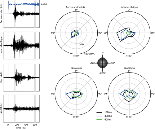
shows exemplar EMG data for a single trial of one subject, and the median spatial tuning curves for all subjects. Median EMG activity remained below 40% MVC for the 3 time points for most directions. At the 150- and 300 ms time points, left (–90°) and left rearward oblique (–135°) perturbations generally resulted in the highest EMG amplitudes. Median activity diminished by 600 ms for the anterior muscles, RA and IO, but not for the posterior muscles, ILC and MU; however, close inspection of the posterior muscle signals at 600 ms suggests that electrical noise may account for some of the signal at this time point.
Figure 1. Left: Exemplar filtered EMG time histories for left muscles from a single subject during a left rearward oblique acceleration (–135°). Dashed line shows t = 0 ms, and the gray lines indicate the 20 ms intervals that include the data contained in the RMS EMG extracted for the spatial tuning curves. Blue line shows the sled acceleration. Right: Median spatial tuning patterns of normalized (%MVC) muscle activation levels at 150 ms (black), 300 ms (blue), and 600 ms (green) after acceleration onset; 0° on the perimeter represents a forward acceleration.

Discussion
These preliminary results suggest that trunk muscle activity may be directionally tuned at the acceleration level tested here. An initial burst of activity at ∼150 ms was seen in most muscles in most directions, and amplitude varied between directions. Notably, all muscles had relatively high activity during the left and left rearward oblique (–90° and –135°) perturbations, suggesting a direction-dependent co-contraction of the trunk muscles. A similar, though smaller, co-contraction was seen in IO, ILC, and MU during acceleration to the right (90°). Preuss and Fung (Citation2008) also reported direction-specific recruitment patterns of the same trunk muscles for seated perturbations in eight directions. They found higher RA and IO activity in left forward oblique (–45°) perturbations compared to left rearward oblique (–135°) but similar patterns for MU. However, their lower perturbation kinematics (Δv = 0.45 m/s over 250 ms), lack of back support, and different event duration make a direct comparison to our data difficult.
Further work is needed to acquire data from additional subjects and to assess whether the preferred directions of activation of the muscles tested here are statistically significant. Moreover, electrical noise generated by the sled motors was present in the EMG signals, particularly in the ILC and MU muscles. Custom filtering methods were applied to attenuate this electrical noise, but these filters likely attenuated valid EMG signals in the 50 to 140 Hz range and thus may have affected the preliminary tuning curves presented here. Better isolation of the lower trunk from the sled will be implemented for future tests.
Although the volunteers did not know the exact onset of acceleration, they did get a warning 5–35 s prior to onset and the direction of the pending acceleration was evident. Anticipation of an imminent perturbation timing and direction has been shown to influence trunk muscle onset responses but not amplitudes (Milosevic et al. Citation2016). Nevertheless, it is possible that the subject's level of awareness influenced our results. More work is needed to evaluate the effects of subject awareness and perturbation acceleration levels on the temporal and spatial patterns of trunk muscle recruitment.
Despite these limitations, our initial findings provide a basis for estimating trunk muscle recruitment for human body models that simulate neuromuscular control in precrash conditions.
Additional information
Funding
References
- Iwamoto M, Nakahira Y. Development and validation of the Total HUman Model for Safety (THUMS) version 5 containing multiple 1D muscles for estimating occupant motions with muscle activation during side impacts. Stapp Car Crash J. 2015;59:53–90.
- Meijer R, Broos J, Elrofai H, de Bruijn E, Forbes P, Happee R. Modelling of bracing in a multi-body active human model. In: IRCOBI. Gothenburg, Sweden, 2013:576–587.
- Milosevic M, Shinya M, Masani K, Patel K, McConville KMV, Nakazawa K, Popovic MR. Anticipation of direction and time of perturbation modulates the onset latency of trunk muscle responses during sitting perturbations. J Electromyogr Kinesiol. 2016;26:94–101.
- Ólafsdóttir JM, Östh JKH, Davidsson J, Brolin KB. Passenger kinematics and muscle responses in autonomous braking events with standard and reversible pre‐tensioned restraints. In: IRCOBI. Gothenburg, Sweden, 2013:602–617.
- Östh J, Brolin K, Bråse D. A human body model with active muscles for simulation of pre-tensioned restraints in autonomous braking interventions. Traffic Inj Prev. 2015;16:304–313.
- Preuss R, Fung J. Musculature and biomechanics of the trunk in the maintenance of upright posture. J Electromyogr Kinesiol. 2008;18:815–28.
