
ABSTRACT
Strength is a primary and modifiable contributor to performance, injury risk, and rehabilitative success. The gold standard measure of strength is the isokinetic dynamometer, providing the clinician with the opportunity to manipulate contraction modality, range, and speed about a joint. However, criticism has highlighted a lack of functional relevance, with arbitrary selection of speed across the full range. To better understand function, biomechanical analysis of movement can inform a bespoke isokinetic data collection protocol. In this case, we use walking gait, a function goal for daily living and clinically assessed following knee replacement surgery for example. Concentric knee flexor and extensor demand was evident at an average angular velocity of 70°·s−1, whilst eccentric knee flexor and extensor demands were evident at speeds of 192°·s−1 and 165°·s−1, respectively. The efficacy of isokinetic dynamometry can be enhanced with bespoke data collection protocols, which better reflect the functional demands of the clinical movement objective.
Introduction
Strength is widely acknowledged as a fundamental element of human performance, and as a primary and modifiable risk factor for musculoskeletal injury. Strength is therefore a common outcome goal in sporting and clinical interventions. In a recent survey 90% of physiotherapists considered knee strength in their return to sport criteria, but only 6% of these practitioners measured knee strength using isokinetic dynamometry (Fausett et al., Citation2022). This is a poor reflection on what is considered to be the clinical gold standard for strength assessment and might reflect issues in accessibility, cost or expertise. Isokinetic dynamometry has also been criticized for not providing functional relevance or predicting injury (van Dyk et al., Citation2017). To make isokinetic strength testing more functional, function needs to be defined. Data collection often appears arbitrary with isokinetic protocols typically using a full range of motion at an arbitrarily selected speed (Coudeyre et al., Citation2016; Greig, Citation2006; van Dyk et al., Citation2017). More should be done to quantify the biomechanical characteristics of the functional goal, whether that be a return to walking gait post-surgery or a return to training or competition in elite sport, as examples. Understanding the functional demands of the rehabilitative goal and aligning these with muscle contraction type, speed and range of motion would facilitate the development of a bespoke isokinetic protocol.
As a case, we present the methodological approach to defining a bespoke isokinetic test for knee arthroscopy patients. More than 100,000 total knee replacement operations are performed in the UK per annum, with an estimated financial burden of £190 million (Chen et al., Citation2012). The primary rehabilitative target for a post-operative patient is often to restore normal walking gait. We therefore analysed walking gait for participants identified as a healthy, matched group (by age, sex and physical characteristics) for an ongoing knee arthroscopy study.
Materials and methods
Twenty-five healthy adults (8 M; 17 F, age: 57 ± 9 years; height: 1.66 ± 0.07 m; mass: 73 ± 16 kg; BMI: 26 ± 4) participated in this study. Inclusion criteria required that participants did not possess any medical issues which could influence gait, with no prior knee surgery. Institutional ethical approval was granted and written informed consent provided by participants prior to testing in accord with the Helsinki Declaration.
In a single testing session, each participant walked barefoot at a self-selected walking velocity (1.35 ± 0.14 m·s−1) along a 7 m walkway, completing five trials. A 10-camera motion capture system (Qualisys, Gothenburg, Sweden) operating at 200 Hz and two synchronized force platforms (Kistler, Winterthur, Switzerland) operating at 2,000 Hz captured kinematic and kinetic data, respectively. A six-degrees-of-freedom model (Langley et al., Citation2021) was used to track the position of the pelvis, thighs, shanks, and feet of participants. Marker trajectories were reconstructed, gap filled to a maximum of 10 frames, and labelled within Qualisys Track Manager (version 2018.1, Qualisys, Gothenburg, Sweden). Processed trials for each participant were exported as C3D files to Visual 3D (C-Motion Inc, Germantown, MD, USA). Kinematic and kinetic data were low pass filtered at 6 Hz and 24 Hz, respectively. Knee joint angles and moments were calculated using an XYZ Cardan sequence of rotations and Newton-Euler equations, facilitating calculation of derivatives in angular velocity and power. Initial contact and toe off were identified using a 10N threshold applied to the vertical ground reaction force data, and joint angles and moments were time normalized to 100% gait cycle duration. The gait cycle was then sub-divided into discrete temporal phases to align with methodological design choice in isokinetic data collection (contractile mode, speed, range), differentiating knee flexion and knee extension.
Results
summarizes the temporal pattern of variation in knee joint angle, moment and power throughout the gait cycle. subsequently presents the discrete temporal distribution of concentric (con) and eccentric (ecc) knee flexion (H) or extension (Q) during ground contact. Each discrete phase is annotated with the average knee joint angular range and average angular velocity.
Figure 1. The temporal pattern of sagittal plane knee angle, moment and power during the gait cycle. (Grey shaded band represents standard deviation about the mean).
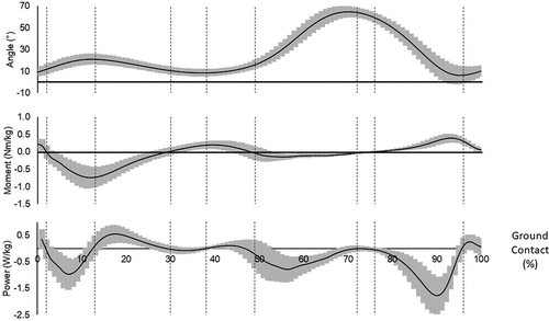
Figure 2. Identifying discrete challenges by modality, speed and range. (Grey shaded band represents standard deviation about the mean).
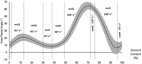
This analysis of healthy walking gait can be used to inform a bespoke isokinetic protocol described in .
Table 1. A summary of knee joint kinematics to inform isokinetic protocols.
Discussion
The aim of the present study was to evaluate “function” during walking gait, to inform musculoskeletal screening and rehabilitation in patients where normal walking gait is the functional goal, such as in cases of knee osteoarthritis or post-operatively for knee replacement. A bespoke isokinetic assessment or exercise intervention would include an assessment of concentric knee flexion and extension at ~70°·s−1. This concentric velocity is within the range of 30–180°·s−1 adopted in previous studies evaluating isokinetic muscle strengthening for knee osteoarthritis, as reviewed by Coudeyre et al. (Citation2016). However, the angular range at 6–65° would be challenging as the dynamometer needs to decelerate from the isokinetic speed, and thus data within the last 6° of knee extension are difficult to achieve. Isometric evaluation might be used to supplement the assessment where there is a clinical rationale to focus on the end range.
Whilst the concentric demands of walking gait are comparable with previously used isokinetic studies (Coudeyre et al., Citation2016), the eccentric demands for knee flexion and extension are at a far greater velocity than typically used in isokinetic evaluations. Eccentric knee extension is very rarely considered in isokinetic studies, despite its fundamental role in many daily activities including walking, helping to cushion the bending of the knee and providing joint stability (Arab et al., Citation2022). Jegu et al. (Citation2014) showed beneficial strength gains in patients with knee arthritis following eccentric knee extensor training in comparison to concentric, but the 30°·s−1 used by Jegu et al. is much lower than the 165°·s−1 observed during these walking trials.
The average eccentric knee flexor velocity was 192°·s−1, and whilst higher speeds up to 300°·s−1 have been used in some elite sporting populations (Greig, Citation2006) the tendency is to use slower, and seemingly arbitrary speeds. van Dyk et al. (Citation2017) reported that a “comprehensive” isokinetic protocol failed to predict injury risk in sport, but the eccentric knee flexor velocity at 60°·s−1 is considerably slower than the average velocity observed here during walking. Applying a more functionally relevant testing velocity might enhance the efficacy of isokinetic trials to predict injury and enhance rehabilitation. The previous discussion is based on an average of the repeated eccentric knee extensor phases (for example) during the gait cycle, and more specific attention could be paid to individual technical phases where clinically appropriate. Walking gait elicited an eccentric hamstring phase at an average of 259°·s−1, and another at 25°·s−1. The sparsely used fast:slow strength ratio (Rahnama et al., Citation2003) might identify athletes/patients at risk from rehabilitative progressions which migrate towards higher speed movements.
The data presented here should also inform bespoke data analysis, to enhance the prescription of rehabilitative or therapeutic interventions. Peak torque is the most commonly reported metric (Coudeyre et al., Citation2016), which reduces the whole data set to a singular peak and defines strength rather than weakness. Identifying where a patient is weak might better inform any subsequent intervention. The corresponding angle of peak torque might also have little functional relevance. If peak strength is achieved at >65° knee flexion it has no functional relevance to walking gait. The functional need is strength throughout this range of movement, at this speed, and analysis of peak torque does not reflect this. The standard clinical output generated by software packages also typically quantifies work, defined as the area under the strength curve, but strongly correlated with peak torque (Dvir, Citation2014). This would suggest redundancy in the frequently co-reported metrics of peak torque and work. A more useful metric might be the functional range, which defines the angular range across which strength can be maintained (Eustace et al., Citation2017). The functional range is independent of peak torque (Brown & Greig, Citation2022), and assessing strength through a range of motion might be more meaningful than previous studies which have considered the role of strength (Coudeyre et al., Citation2016) or range of motion (Campbell et al., Citation2023) in isolation with regard to their importance in osteoarthritis rehabilitation for example.
Isokinetic dynamometry might warrant its status as clinical gold standard if data collection and analysis can provide greater functional relevance. Raposo et al. (Citation2021) reported that strengthening exercise programmes are effective in knee osteoarthritis patients but provided no prescriptive detail of how to strengthen. Coudeyre et al. (Citation2016) reflected that weak methodology and heterogeneity in isokinetic protocols and outcome measures prohibited the development of guidelines for isokinetic muscle strengthening. Anecdotally, practitioners refer to a lack of time with patients/athletes, and given the implications for participant burden, more can be done to optimize isokinetic testing and evaluation. Clinical stakeholders have identified that optimal management of rehabilitation should be patient-centred and incorporate adequate equipment and specific training for clinicians (Dunphy et al., Citation2023).
The current study has a focus on walking gait as a rehabilitative goal, and future research should consider isokinetic data collection that is more functionally relevant to enhance the efficacy of clinical outcomes or predicting return to sport. Greater functional relevance can be established through biomechanical analysis of movement goals.
Acknowledgements
Our thanks to the participants who volunteered their time to participate in this study.
Disclosure statement
No potential conflict of interest was reported by the author(s).
Additional information
Funding
References
- Arab, F., Quddus, N., Khan, S. A., Alghadir, A. H., & Khan, M. (2022). Association of eccentric quadriceps torque with pain, physical function, and extension lag in women with grade ≤ II knee osteoarthritis. An observational study. Medicine, 101(31), e29923. https://doi.org/10.1097/MD.0000000000029923
- Brown, R., & Greig, M. (2022). The influence of isokinetic dynamometer configuration on eccentric hamstring strength metrics: Implications for testing and training. Research in Sports Medicine, 1–9. Ahead of Print. https://doi.org/10.1080/15438627.2022.2079988
- Campbell, T. M., Westby, M., Ghogomu, E. T., Fournier, J., Ghaedi, B. B., & Welch, V. (2023). Ahead of Print. Stretching, bracing, and devices for the treatment of osteoarthritis-associated joint contractures in nonoperated joints: A systematic review and meta-analysis. Sports Health. https://doi.org/10.1177/19417381221147281
- Chen, A., Gupte, C., Akhtar, K., Smith, P., & Cobb, J. (2012). The global economic cost of osteoarthritis: How the UK compares. Arthritis, 2012, 1–6. https://doi.org/10.1155/2012/698709
- Coudeyre, E., Jegu, A. G., Giustanini, M., Marrel, J. P., Edouard, P., & Pereira, B. (2016). Isokinetic muscle strengthening for knee osteoarthritis: A systematic review of randomized controlled trials with meta-analysis. Annals of Physical and Rehabilitation Medicine, 59(3), 207–215. https://doi.org/10.1016/j.rehab.2016.01.013
- Dunphy, E., Button, K., Murray, E., & Hamilton, F. L. (2023). Beyond guidelines: A qualitative clinical stakeholder study of optimal management of anterior cruciate ligament rehabilitation. Musculoskeletal Care, 21(1), 117–129. https://doi.org/10.1002/msc.1673
- Dvir, Z. (2014). Relevant, less relevant and irrelevant isokinetic strength test parameters: Some critical comments. Movement & Sport Sciences - Science & Motricité, 85(85), 15–21. https://doi.org/10.1051/sm/2013088
- Eustace, S. J., Page, R. M., & Greig, M. (2017). Contemporary approaches to isokinetic strength assessments in professional football players. Science and Medicine in Football, 1(3), 251–257. https://doi.org/10.1080/24733938.2017.1371851
- Fausett, W. A., Reid, D. A., & Larmer, P. J. (2022). Current perspectives of New Zealand physiotherapists on rehabilitation and return to sport following anterior cruciate ligament reconstruction: A survey. Physical Therapy in Sport, 53, 166–172. https://doi.org/10.1016/j.ptsp.2021.10.012
- Greig, M. (2006). The influence of soccer-specific fatigue on peak isokinetic torque production of the knee flexors and extensors. The American Journal of Sports Medicine, 36(7), 1403–1409. https://doi.org/10.1177/0363546508314413
- Jegu, A.-G., Pereira, B., Andant, N., & Coudeyre, E. (2014). Effect of eccentric isokinetic strengthening in the rehabilitation of patients with knee osteoarthritis: Isogo, a randomized trial. Trials, 15(1), 106. https://doi.org/10.1186/1745-6215-15-106
- Langley, B., Jones, A., Board, T., & Greig, M. (2021). Modified conventional gait model vs six degrees of freedom model: A comparison of lower limb kinematics and associated error. Gait and Posture, 89, 1–6. https://doi.org/10.1016/j.gaitpost.2021.06.016
- Rahnama, N., Reilly, T., Lees, A., & Graham-Smith, P. (2003). Muscle fatigue induced by exercise simulating the work rate of competitive soccer. Journal of Sports Sciences, 21(11), 933–942. https://doi.org/10.1080/0264041031000140428
- Raposo, F., Ramos, M., & Cruz, A. L. (2021). Effects of exercise on knee osteoarthritis: A systematic review. Musculoskeletal Care, 19(4), 399–435. https://doi.org/10.1002/msc.1538
- van Dyk, N., Bahr, R., Burnett, A. F., Whiteley, R., Bakken, A., Mosler, A., Farooq, A., & Witvrouw, E. (2017). A comprehensive strength testing protocol offers no clinical value in predicting risk of hamstring injury: A prospective cohort study of 413 professional football players. British Journal of Sports Medicine, 51(23), 1695–1702. https://doi.org/10.1136/bjsports-2017-097754