
Abstract
Based on epidemiological records of workers at Ni operations, regulatory guidelines commonly target specific Ni compounds for setting exposure limits. Thus, reliable methods of Ni speciation in airborne dust samples are required for effective monitoring of workplace exposure. Zatka sequential leaching has been routinely performed industry-wide since the 1990s for characterization of Ni in dust samples; however, limitations related to leaching kinetics have been identified, and optimization of the methodology is required to improve accuracy of data. In this study, Ni characterization of dust collected from a stainless steel operation was performed using Zatka sequential leaching (original and modified protocols) and quantitative mineralogy (QEMSCAN), a method novel to the field of industrial hygiene. Mineral analysis was also performed on bulk material collected from selected work areas at the plant. The results are compared with the objective of identifying opportunities to optimize the methods for characterizing dust that is unique to stainless steel manufacturing. The quantitative mineralogical analysis determined that the Ni dust is composed of oxidic Ni (chromite and trevorite, >80% of the Ni in most samples) and metallic Ni (Ni-Fe alloy), and the results were validated against chemical assays and alternate methods of mineral characterization. In contrast, the original Zatka method erroneously identified soluble Ni as a major Ni contributor, whereas the modified Zatka method identified sulfidic Ni. The mineralogy identified Ni-barren dust and grain sizes and liberation of individual Ni compounds as potential factors that can affect leaching selectivity. Clearly, for any sequential leaching method to be useful for these workplaces, they should be optimized by including reference materials that are representative of Ni substances present at stainless steel operations (chromite, trevorite, and Ni-Fe alloy). Improving methods of sequential leaching is important because the resolution of quantitative mineralogical techniques diminishes at <3 μm (respirable dust fraction). We recommend that quantitative mineralogy be performed in parallel with methods of sequential leaching to provide a robust system of characterization.
Introduction
Nickel (Ni) is an essential constituent in a number of high-demand products; however, exposure to certain forms and quantities can be toxic (USDHHS 2005). Although Ni can naturally be present at low levels in ambient environments, exposure to elevated levels occur more commonly at the occupational setting through inhalation of particulate matter (Institute 2008; Buxton et al. Citation2019). Adverse health effects associated with exposures at facilities that refined or processed sulfidic nickel ores have been documented from the early to mid-1900s when protective exposure measures were less stringent compared to present-day standards. A well-known example includes the increased incidents of respiratory cancers in workers employed at a nickel refinery in Clydach, Wales prior to 1930 (Hill Citation1939). Although these negative health effects were observed, lack of proper documentation and poor understanding of the types of Ni present within workplace airborne dust during the early production years have complicated interpretation of epidemiological records (Sivulka et al. Citation2014). In 1990, a seminal epidemiological study was published that shed some light on the main chemical forms of nickel associated with elevated respiratory cancer risks (Doll Citation1990)
Linking exposure data to specific health effects requires that the type of Ni present within the workplace is well defined because specific Ni compounds interact with biological tissues in different ways. Decades of epidemiological data demonstrates that exposure to different compounds of Ni can have variable impacts on health ranging from mild forms of contact dermatitis to severe chronic respiratory diseases (Doll Citation1990; USDHHS Citation2005; Buxton et al. Citation2019). Due to this variation, regulatory bodies, such as the American Conference of Governmental Industrial Hygienists (ACGIH®), recommend enforcing threshold limit values (TLV®) for individual Ni compounds in the workplace environment. The biologically relevant inhalable Ni species targeted for TLVs include (ACGIH Citation2001; U.S. Department of Health and Human Services Citation2005; Nickel Institute Citation2008):
Soluble Ni: includes Ni chlorides and other similar anions such as sulfosalts, carbonates, hydroxides, and carboxylate;
Variably Soluble: Ni sulfides and subsulphides; and
Poorly soluble to insoluble: Ni oxides and elemental nickel;
Although Ni uptake can occur through different routes (skin, oral, and inhalation), the most common and only route of relevance with regard to respiratory effects at the workplace is inhalation (Buxton et al. Citation2019). Upon inhalation there is greater opportunity for Ni to deposit and accumulate in the sinus cavities and lungs when it is in a poorly soluble form (Doll Citation1990; Oller et al. Citation2014). The risk of developing lung cancer is higher in animal studies with exposure to poorly soluble Ni compared to soluble Ni, although in the presence of both, soluble Ni may act as a tumor promoter due to its influence in increasing cell proliferation (Oller Citation2002; Goodman et al. Citation2009). The overall movement of Ni throughout the body and risk of developing disease is also dependent on the particle size distribution of the source material. Ultrafine particulates (< 2.5 μm) have a greater ability to penetrate and infiltrate the lymph and circulatory system compared to more coarse particulates (Goodman et al. Citation2011; Oller et al. Citation2014). This can affect the distribution of inhaled particulates to various organs in the body and where particles are deposited within the respiratory tract. In addition, finer particulates have greater surface area, increasing the rate of solubility, dissolution, and suspension within a liquid, including body fluids (Davidson et al. Citation2005).
In order to properly enforce and regulate the recommended limits and understand the associated epidemiology, a thorough system of characterization of particulate matter at the workplace is required; this should include size and chemical analysis of particulates collected with size selective personal samplers (ECS Citation1993; ISO Citation1995). To quantify concentrations of Ni in aerosols, the International Organization for Standardization recommends analysis by inductively coupled plasma with either atomic emission (ICP–OES) or mass spectrometry (ICP–MS) in combination with the digestion method appropriate for the filter substrate used to collect the aerosols (e.g., ISO 15202 and ISO 30011 for determining metals and metalloids in dust collected on specific sampling substrates (ISO Citation2004, Citation2010, Citation2020; Ashley Citation2015; International Organization for Standardization (ISO ) Citation2020).
When occupational exposure limit values (OELVs) refer to specific Ni compounds, supplementary analysis is required. Established in the early 1990s for evaluation of dust at “sulfidic ore” Ni and Cu refineries, the Zatka method employs a series of wet-chemical extraction procedures to selectively leach Ni species into the four biologically relevant categories: water soluble, sulfidic, metallic, and oxidic (Zatka et al. Citation1992; Conard et al. Citation2008). Although modifications have been made over time to improve the leaching steps (Conard et al. Citation2008), the Zatka method has been routinely performed industry-wide for determining apportionment of Ni compounds in dust.
Although the Zatka method has considerable promise, common limitations of the method are mostly related to the kinetics of leaching where the Ni concentration, particle size, and liberation can impact the time required to leach the Ni at each step (Bacon and Davidson Citation2008; Conard et al. Citation2008). Multiple studies comparing the Zatka method to speciation methods such as X-ray Diffraction (XRD) and X-ray Absorption Near Edge Spectroscopy (XANES), suggest that sequential leaching can skew the proportions of species depending on the effectiveness of the leach (Andersen et al. Citation1998; Ajiboye et al. Citation2007; Tirez et al. Citation2011; Van Loon et al. Citation2015). Other limitations can include rather minimal sample throughput and the requirement that the analyst be extremely skilled with the method.
The Zatka method requires further development for assessment of Ni species at intermediate or secondary alloy operations, such as stainless steel manufacturing. Achieving accurate characterization results is important for determining toxicological effects associated with exposure and mitigating risk of developing disease (Oller et al. Citation2009). Furthermore, in many situations, detailed characterization at the phase level is required and cannot be achieved using the Zatka method (e.g., differentiation between Ni sulfide minerals such as pentlandite and millerite) and additional characterization methods may be required. Phase-level characterization is important at locations such as smelters and refineries where a complex variety of Ni sub-species are handled.
Quantitative mineralogy can be a useful additional method for speciating Ni and other metals of interest in the inhalable fractions of solid particulates. Instruments of quantitative mineralogical analysis consist of scanning electron microscope- (SEM-) based automated systems that produces particle maps (color coded by mineral) through the collection of rapidly acquired X-rays. Examples include Quantitative Evaluation of Materials by Scanning Electron Microscope (QEMSCAN), Mineral Liberation Analysis (MLA), and Tescan Integrated Mineral Analyzer (TIMA). These systems classify mineral species based on the elements present within a particle, and are compared to reference libraries of known phase spectra. The systems are capable of measuring thousands of particles over a relatively short period, increasing the population size of the dataset. The rapid phase determination is useful for measuring air samples with low concentrations of Ni by extending measurement time to achieve the appropriate counting statistics. Abundance, particle size, morphology, and association can be documented using quantitative mineralogy (Gottlieb et al. Citation2000).
Methods of quantitative mineralogy are not widely utilized in industrial hygiene exposure assessment studies, but data obtained from this type of analysis can provide the characterization details necessary to support monitoring programs that rely on quantification of different Ni compounds (Williamson et al. Citation2013; Hrstka et al. Citation2018). The data generated from quantitative mineralogy can also be used to calibrate sequential leaching steps in order to optimize the method for effective characterization of Ni that is specific to individual operations containing a unique set of compounds.
In the present study, characterization of Ni in 18 stationary aerosol samples, taken in parallel for each method at 6 work areas (Smelting, Argon Oxygen Decarburization (AOD) Converter, Oxy-cutting, Raw Materials, Hot Roll Mill, and Grinding), was performed using Zatka sequential leaching and QEMSCAN for airborne aerosol samples collected at a stainless steel production site for the purpose of comparing and assessing the efficacy of the methodologies. Loose materials collected from each location by floor sweepings, were also analyzed using QEMSCAN to improve the interpretation of the data and gain a concrete understanding the minerals present within the different operations. An apportionment of Ni among each category was determined and is compared between the QEMSCAN and Zatka methods.
Gravimetric analysis and subsequent metals analysis should include wall deposits that occur when using samplers with cassettes as indicated by MDHS 14/4 HSE 2014. In this study only the filter was analyzed as in the common methods for Ni speciation, and only the sampling substrate is analyzed because the suction filtration apparatus is designed to mount the sampled filter directly to perform the different Zatka leaching procedures. Furthermore, the QEMSCAN method requires a direct on filter analysis where wall deposits cannot be included. Another reason to exclude the wall deposits was the low mass collected where the uncertainty on weighing the cassettes with filter compared to filter only would have a significant impact.
Methodology
Choice of worksites and workers
To compare the characterization methods, location for sampling at a stainless steel manufacturing plant in the EU were chosen based on a general classification scheme of 11 work areas included in an epidemiological study of High Nickel Alloy producing plants in the United States (Arena et al. Citation1998, Citation1999). Samples were taken at the steel plant and the hot roll mill. Sampling was only performed when producing austenitic stainless steel grades. At each work area, stationary samples were taken in parallel. The key sampling areas at the operation included: Smelting, Argon Oxygen Decarburization (AOD) Converter, Oxy-cutting, Raw Materials, Hot Roll Mill, and Grinding.
Sampling
Multiple sets of stationary air samples and settled dust samples were collected for each location so that similar samples could be used for Ni characterization analysis, total metal analysis and mineralogical analysis.
The samplers and devices used in this study have been described extensively and include the plastic inhalable Institute of Occupational Medicine (IOM; SKC Ltd, UK) personal sampler with plastic cassettes, the respirable Plastic cyclone (Higgins-Dewell) sampler with plastic cassettes (Casella), and the Sioutas Personal Cascade Impactor Sampler (SKC Ltd Uk) to separate particles in the following aerodynamic particle diameter ranges: <0.25, 0.25–0.5, 0.5–1.0, 1.0–2.5, and 2.5–10 μm (Misra et al. Citation2002)
Conard et al. (Citation2008) used DM450 acrylic copolymer membranes in the leach filtration steps because of better resistance to chemicals than cellulose ester filters (Conard et al. Citation2008). Because the DM450 acrylic copolymer membrane filters were no longer available and Van Loon et al. (Citation2015) used 1.2 um Versapore filters for air sampling, samples for Zatka analysis were taken with 25 mm 0.8 μm Versapor Acrylic Copolymer Membrane filters (Van Loon et al. Citation2015). In parallel, for the total metals and mineralogical analysis, samples were taken with 25 mm − 0.8 μm pore MCE membrane filters (SKC part number 225-1930) and 25 mm − 5 μm pore GLA-5000 PVC membrane filters (SKC part number 225-5-25).
In the Sioutas Cascade Impactor, 25 mm − 0.5 μm pore PTFE (SKC part number 225-3708) and a 37 mm − 2 μm pore PTFE (SKC part number 225-1709) were used, respectively, as collection substrate for the four stages and as the backup filter.
Settled dust samples were taken by sweeping a surface within the working area until a sample recipient with a capacity of 250 cm³ was completely filled. Separate sub-samples, for total metals and QEMSCAN analysis, were taken from the bulk sample as described in ISO 14488 (ISO Citation2007).
Gravimetric analysis
Prior to and after each sampling all filters were weighed at the Laboratory for Chemical Analysis of the University of Ghent on a Mettler Toledo MT5 microbalance following ISO 15767 (Workplace atmospheres—Controlling and characterizing uncertainty in weighing collected aerosols, ISO 15767 2009). For each sampling site and per collection substrate, at least three field blanks and a series of five laboratory blanks were weighed. Respectively, the limit of detection and limit of quantification are 82 and 272 μg for MCE filters, 17 and 58 μg for PVC filters, 22 and 74 μg for Versapore filters, and 10 and 32 μg for PTFE filters.
Total metal analysis in aerosols
The sampled filters were digested and analyzed at the Laboratory for Chemical Analysis of the University of Ghent following ISO 15202-2 Annex G (ISO Citation2020). A Multiwave 5000 microwave reaction system (Anton Paar, Austria) was used for sample digestion and the IRIS Intrepid II (Thermo Scientific, Madison, WI, USA) ICP-OES for analysis. Analytes included Al, As, Ba, Be, Cd, Co, Cr, Cu, Fe, Mg, Mn, Mo, Ni, Pb, Sb, Sn, Ti, V, W, Zn, and Zr.
Zatka sequential leaching
Both the original and modified versions of the Zatka method were used for sequential leaching (methodology summarized in Luk et al. (Citation2000)). The modifications to the original Zatka method are intended to improve the extraction technique with the addition of centrifugation, replacement of hot plate digestions with microwave digestions, optimized extraction process, and modified leaching reagents. The method involves a four-step process to extract soluble Ni (0.1 M ammonium citrate solution for the original Zatka method and deionized H2O and centrifugation for the modified Zatka method), sulfidic Ni (H2O2—citrate mix followed by centrifugation), metallic (methanol-bromine followed by centrifugation), and oxidic (HF–HNO3 with microwave digestion). Concentrations of Ni at each step were determined at the Laboratory for Chemical Analysis of the University of Ghent using a Multiwave 5000 microwave reaction system for digestion of sub-samples (Anton Paar) and an iCAP Q (Thermo Scientific) ICP-MS for the Ni analysis. A detailed description of the original and modified Zatka methodology are included as a supplementary file.
Mineralogy
A section of each >2.5 μm filter containing a sample of dust was cut to fit a 30 mm diameter epoxy mount. The dust dispersed on the filters appeared to be mostly homogenous for the samples, and a portion of the filter was retained for future assay. A thin layer of carbon was applied to the samples prior to QEMSCAN measurement.
Each mounted filter was measured by QEMSCAN. The instrument used was a FEI 650 SEM (Brno, Czech Republic) equipped with a field emission gun (FEG), capable of detecting particles as fine as 3 μm. The beam conditions included an accelerating voltage of 25 kV, beam current of 10 nA, and spot size of ∼0.5 μm. The aerosol samples were measured using Field Image mode and a species identification protocol (SIP), specifically designed for air filters from this operation. A pixel spacing of 0.5 μm was used for the measurement at a 1,000 X-ray counts averaged over one acquisition point. To obtain the most robust measurements possible, both the filter and particulates were measured, and the filter background was digitally removed from the data set once the measurements were completed.
Conventionally, a flat, polished sample surface is required for QEMSCAN to prevent X-ray scatter and produce the most robust measurements. Because the filters cannot be polished flat, caution was taken during identification and quantification of the particulates.
For example, interference due to X-ray scatter from adjacent particles may produce overlaps during identification but were corrected through data reduction and processing schemes during SIP development.
The QEMSCAN results are only representative of particles on the surface of the filter and for particles that are > 2–3 μm. Particles that measured < 2–3 μm in diameter were not included in the results due to poor X-ray generating volume at this size.
In preparation for mineralogical analysis, the settled dust was mounted in epoxy resin and polished. No water was used for the sample preparation to avoid dissolving any sulfate minerals. All polishing was completed using oil-based lubricants. QEMSCAN measurements were completed using particle analysis mode (PMA) and using the same SIP developed for the dust samples.
Limited Electron Probe Microanalysis (EPMA), and inspection of particles using SEM-imaging equipped with energy dispersive spectroscopy (EDS), were subsequently performed on selected samples to confirm phase identification and concentrations of metals in each phase.
The EPMA was performed at XPS using a Cameca SX-100 (Paris, France). A focused beam (spot size of < 1 μm), beam current of 20 nA, and an accelerating voltage of 20 kV were used for the analysis.
Statistical analysis
Gravimetric and Zatka leaching results from the stationary samples were analyzed using Excel (Microsoft Inc., Redmond, WA) and SPSS Statistics (version 26, IBM Corp., Armonk, NY). The EXPLORE function was used to evaluate distributions of data sorted by sampling substrate type. The gravimetric dust concentrations (μg/m³) were found to be normally distributed (Shapiro–Wilk test).
Results
Gravimetric analysis and particle size distributions
Paired samples correlations () show that the sampling efficiency is positively correlated between all different sampling substrates. There is a statistically significant average difference in gravimetric result (sample efficiency) of the IOM sampler with MCE filters and polyacrylate (PA) and PVC filters (). On average, MCE collected 166 μg/m³ more dust than PA filters and 194 μg/m³ more than PVC filters. Although the PA filters sampled 47 μg/m³ less dust than PVC filters, the average difference was not statistically significant.
Table 1. Paired sample correlations.
Table 2. Statistical comparison of gravimetric analysis of filter substrates.
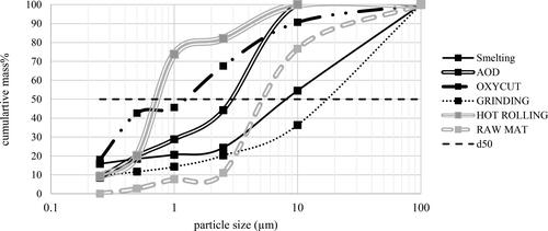
Based on the gravimetric results of the sampling substrates in the Sioutas Personal Cascade Impactor Sampler (0.25, 0.5, 1.0, 2.5, and 10 μm) and the AED50 of the inhalable sampler (100 μm), the particle size distribution was simulated for all sampled workplaces (% of the total mass) for the gravimetric results () and total Ni results (). The total mass from all Sioutas impactor substrates is subtracted from the inhalable sampling substrate mass to derive the 100 μm mass %. Smooth lines were drawn through the points for visual aid. For Ni, 50% of the mass collected in the work area smelting, raw material, and AOD, contained particles smaller than 10 μm. In the work area hot rolling the particles were smaller than 1 μm. Although > 50% of the Ni in Oxycutting is > 1 μm, the distribution is bimodal, and a significant amount of the Ni (∼43%) is carried in the particulates that are < 0.5 μm. At the work area smelting, only 45% the mass collected contained particles smaller than 10 μm.
Figure 1. Simulated particle size distributions for airborne dust in filters (mass%). Smooth lines are drawn through data points for visual aide.
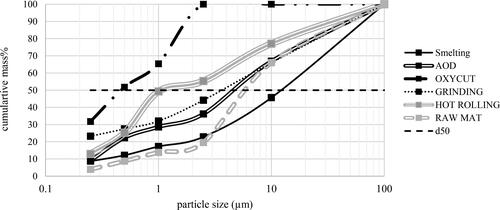
Figure 2. Simulated particle size distributions for Ni in airborne dust in filters (mass%). Smooth lines are drawn through data points for visual aide.
QEMSCAN
Settled dust: Overall dust
In the settled dust material collected from the source locations, the dominant non-Ni phases consist of Fe oxide minerals, chromite, Ca-Mg oxide/hydroxide minerals (possibly portlandite), and ambient silicate minerals (feldspar, Mg, and Al silicates). Trace to minor amounts of pyrite, sphalerite, Zn oxide or carbonate minerals, gypsum, and Al oxide minerals are also present.
Settled dust: Nickel compounds
The Ni minerals are composed of Fe-Cr-Ni oxide (chromite), Fe-Ni oxide (likely trevorite), Ni oxide, Fe-Ni metal, and Fe-Cr-Ni metal. EPMA performed on the chromite indicates that low levels of Ni are present in the chromite (average of ∼1%). Rare grains of Ni-Fe sulfide and Ni sulfate were detected. The Ni-Fe sulfide minerals contribute a negligible amount of the overall sample mass, consistent with the low bulk S concentrations (0.19–1.16 wt.%) determined through chemical assay. No soluble forms of Ni were detected.
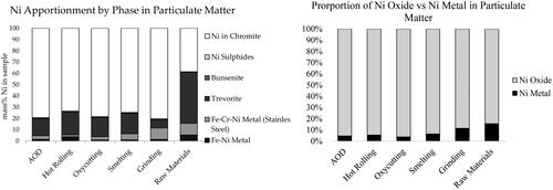
An apportionment of Ni (contributed by the individual Ni compounds detected) in the settled dust was calculated based on the amount of Ni present in each compound and the abundance of each compound in the sample (determined through a combination of QEMSCAN and EPMA analysis; ). A simplified apportionment containing the combined Ni in metals (Fe-Ni metal and Fe-Cr-Ni metal) and Ni in oxide minerals (Fe-Ni oxide and Ni in chromites) are also included in .
Figure 3. Ni Deportment in settled dust. Left: data presented as mass% of Ni in sample; right: data presented as concentration of Nin sample (wt%). A simplified deportment showing the proportion of Ni metal (Fe-Ni metal + Fe-Cr-Ni metal) to Ni oxide (Fe-Ni oxide + Ni in chromites) is included at the bottom of the figure.
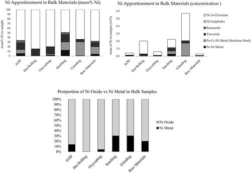
The majority of Ni is carried in chromite (ranging from 56% in AOD to 87% in Oxycutting). The remaining Ni is carried in Fe-Ni oxide minerals and Ni metals (Fe-Cr-Ni metal and Fe-Ni metal). The highest proportion of Ni contributed by metals is observed in the smelting and grinding samples. The Fe-Ni-Cr metal is coarse with shard-like textures, reaching particle sizes up to 500 μm.
The Fe-Ni-Cr metal is commonly associated with chromite, where the chromite forms rims around the metals. The Fe-Ni metal is slightly finer than the Fe-Ni-Cr metals, and commonly forms intergrowth textures with Fe-Ni oxide minerals (). shows images of selected Ni oxide particles found in the settled dust collected near sample AOD, along with associated EDS spectra.
Figure 4. QEMSCAN images of Ni metals present in settled dust. Images are cross-section of particles mounted in epoxy resin.
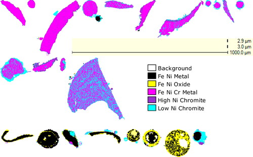
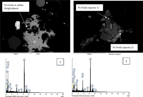
Figure 5. SEM images of Ni oxides enclosed in sulfate and silicate phases in the bulk sample MI8-414. Energy dispersive spectra (EDS) of points taken from Ni oxides are shown for validation of Ni compounds (elements such Ca, Si, and K are due to contributing volume from surrounding minerals).
Dust samples
The mineralogy of the dust samples resembles the source materials, with minor proportions contributed by Ni compound. In most samples, spinel (Fe-chromite and Ca-Mg spinel), Ca-Mg oxides/hydroxides (possibly portlandite or lime), Fe oxides (magnetite or hematite), a high-Ca, low-Mg silicate (Ca, Mg silicate), and ambient silicate minerals (mica, feldspar, other Mg silicate minerals) are the major phases present. The Oxycutting and Raw Materials Samples contain solid (Na-Ca)-Cl particulates and appear to have a Na- or Ca-Cl-rich residue that have accumulated on the surface of the filter.
Other minor to moderate phases include Ca-Mg sulfate minerals (can contain minor amounts of Fe), a Ca spinel with low Mg, graphite or carbon, Fe sulfide minerals, and Zn phases (Zn sulfide, sulfate, oxide, and possibly silicate minerals). Trace amounts of phases containing Pb, Cu, Mn, and W were also detected.
A Ni deportment in the dust samples was calculated () based on the types of Ni species present, and the Ni concentrations determined in the respective phases in the source materials. The dominant Ni species present in each sample are Ni-chromite and Fe-Ni oxide (likely trevorite). Together, trevorite and Ni-chromite (Ni oxide) carry > 85% of the Ni in the samples. The proportion of Ni contributed by each phase varies throughout the samples. Minor amounts of Ni are also contributed by Fe-Ni metal and Fe-Ni-Cr metal (Fe is the dominant metal). Where Fe-Ni metal is present it is intergrown with trevorite. Only rare grains of Ni sulfide were detected. Due to the low abundance and ultra-fine grain sizes of this phase, identification of the Ni sulfide is speculative. The Ni sulfides may contain sulfate or there could be overlapping signals from textures of Ni-bearing oxide and sulfate minerals. No discrete Ni sulfide was identified. The grain sizes of the Ni-bearing particulates range from 5 μm to 30 μm. The textures of the phases are typically bleb-like to irregular and may reflect the temperature of activity.
Figure 6. Ni apportionment in personal monitor filters (mass% of Ni in sample). A simplified deportment showing the proportion of Ni metal (Fe-Ni metal + Fe-Cr-Ni metal) to Ni oxide (Fe-Ni oxide + Ni in chromites) is included at the bottom of the figure.
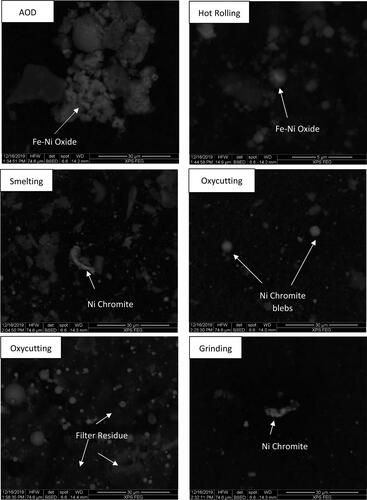
The calculated Ni apportionments are consistent with the Ni distributions determined for the settled dust; however, in the dust samples, less Ni is contributed by metallic Ni. This is likely due to the coarse grain sizes of the metallic Ni. To demonstrate this effect, the simplified Ni deportment (metallic Ni versus Ni oxide) in the settled dust was recalculated after digitally removing all particles > 20 μm (estimated average maximum particle size of Ni particles). The recalculated apportionment shows significantly less contribution from metallic Ni and is more similar to the apportionment determined for the particulate matter (). Minor variations between the oxide minerals in the apportionments suggest that either there is preference for some phases to become airborne over others (related to density and/or hardness of materials), or that there are some minor variations in mineral abundances between the different sampling periods at each location. Examples of the associations and grains sizes of the Ni-bearing particulates are shown in .
Figure 7. Comparison of simplified Ni deportment in particulate matter compared to the fine material (<10 µm) in the source materials. Note that digitally removing particles in the QEMSCAN data may not be fully representative of physical size class due to stereological bias in the mounted polished sections. Physical sizing is recommended prior to analysis to get the most accurate representation of size distributions. The graphs presented here are meant to show a general trend.
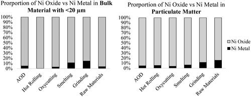
Figure 8. SEM photomicrographs of selected Ni-bearing particulates.
Sequential leaching of dust samples
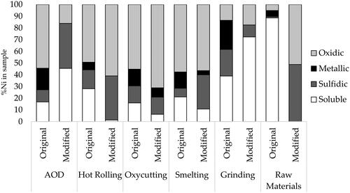
Analysis using the sequential leaching methods suggest that there are variable amounts of each soluble, sulfidic, metallic, and oxidic Ni; however, the apportionments of Ni species in the dust samples are not consistent between the original and modified Zatka leaching (). In a number of cases, the proportion of sulfidic Ni was higher using the modified leaching method (AOD, Hot Rolling, Smelting, and Raw Materials); however, in the Grinding sample, a higher proportion of sulfidic Ni is measured in the original Zatka method compared to the modified. In all samples, the proportion of metallic Ni is higher using the original Zatka method. The greatest consistency between results occurs in samples Oxycutting and Smelting, where oxidic Ni is the dominant species.
Figure 9. Ni apportionments determined by the original and modified Zatka sequential leaching (%Ni in sample).
Discussion
The Zatka method recovered significant quantities of Ni at the soluble and sulfidic leaching stages whereas the mineralogical analysis indicated that the majority of Ni is present in the oxide or metallic form with negligible amounts of soluble or sulfidic Ni. Although the Zatka method did not offer compositional information comparable to mineralogical characterization, from a health perspective, it is useful to understand solubility of a certain population of particulates. Further investigation to determine the specific causes of discrepancy between methods is required to understand why oxidic Ni behaves as soluble and sulfidic in certain samples. If the behavior of oxidic Ni during sequential leaching mimics the behavior in lungs or elsewhere in the body, the Zatka method may be considered an important measure of health risk rather than classification of composition. Possible explanations for the discrepancy between results are discussed.
Filter media and interference with leaching
The type of filter media used for collection of dust can be considered one possible source of discrepancy between the leaching methods and mineralogy.
As noted, the sampling efficiency of the IOM sampler may be different when operating in parallel with different sampling substrates. It is assumed that if a smaller mass is present on the filters, the particle size distribution of the aerosol samples will tend toward the smaller fraction which may have an impact on how the leaching solution will release certain fractions. When it is difficult to collect samples of high enough mass to perform fractionation procedures where the Ni concentrations are low, the statistical error associated with the lower number of Ni bearing particulates could increase.
Reagents and leaching time
The misidentification of sulfidic and soluble Ni determined by the Zatka methods may be related to leaching kinetics or reagents. With the exception of AOD and Raw Materials, soluble Ni was reported in higher proportions using the original Zatka method. The original method used an ammonium citrate leaching solution in place of deionized H2O, which may be too aggressive for samples containing no soluble forms of Ni, as also suggested previously by Oller et al. (Citation2009); however, the recovery of non-soluble Ni at the first stage of the modified Zatka method (leaching with only H2O) needs to be further investigated. Conard et al. (Citation2008) pointed out that when present in ultrafine or nano-size particles, elemental Ni may, at least partially, behave as soluble Ni during the leaching sequence. However, mineralogical analysis indicates that Ni metal is predominantly found in the coarser fraction at this plant.
There are a variety of non-Ni phases present which may impact the performance of leaching, including NaCl and Ca carbonate. Dissolution of some base-metals have been shown to increase in the presence of NaCl (Haudrechy et al. Citation1993; Park et al. Citation2007; Yazici and Deveci Citation2015; Castillo et al. Citation2019; Thubakgale et al. Citation2019). Activation of Ni in the presence of nonmetal salt compounds under the conditions used in this study should be investigated further to determine if it is promoting earlier release of Ni during the leaching sequence. In addition, Zatka et al. (Citation1992) reported that for dust collected on filters with a high natural alkalinity (glass fibres), soluble Ni was not efficiently recovered in the correct sequence. Although the observation reported by Zatka et al. (Citation1992) underestimated the proportion of soluble Ni, the result confirmed that leaching selectivity is affected by alkalinity, and the chemistry of the total dust may influence performance.
The complex mineralogy of dust samples highlights the need to optimize leaching procedures so that they are specific to a given operation. The majority of development work carried out for the Zatka method has focused on operations that process sulfide ores where the nature of the ore, host rock, and processing additives can influence the leaching kinetics. To determine the selectivity for each compound, Zatka et al. (Citation1992) used pure standards of Ni salts, pentlandite, Ni matte (assumed to be Ni3S2), and Ni metal to optimize the leaching conditions. During this development work, it was found that underestimation of Ni sulfide was common for samples with mass > 1 mg. An increase in residence time was required to attack the Ni sulfide in the higher mass samples (modified Zatka). However, in the samples studied here, Ni sulfide was falsely identified and is much more prominent in the modified Zatka method where leaching times for sulfide minerals are longer, suggesting that the longer leach time can have a significant impact in the absence of sulfidic Ni. It is also worth noting that alkalinity of Ni-barren dust at Ni sulfide processing operations may be different compared to samples collected from stainless steel operations due to different additives used for processing and the presence of mafic-ultramafic minerals that are commonly associated with sulfide ore. The types of phases present in the dust samples analyzed here are likely associated with activities performed by personnel, suggesting that the non-Ni bearing phases are also unique to these operations. Optimization of leaching conditions for samples containing high proportions of Ni in spinel and low proportions of Ni in sulfide minerals should be completed to improve chemical characterization methods for operations such as stainless steel processing facilities.
Liberation of Ni compounds
As discussed in numerous sequential leaching studies, the liberation of a particle can impact leaching kinetics (Zatka et al. Citation1992; Bacon and Davidson Citation2008; Conard et al. Citation2008). The mineralogical analysis of the settled dust that were prepared as cross-sections has shown that a variety of associations and grain/particle sizes amongst the different Ni phases are present. In particular, metallic Ni tends to be coarser, whereas the Ni-bearing chromite is finer grained and can be present as coatings on Ni metals. The particle images collected with the QEMSCAN show that chromite rimming can persist at particle sizes < 100 μm (inhalable). The effect of liberation is expected to be less prominent in respirable dust because most surfaces should be exposed at these fine particle sizes; however, particles in the inhalable fraction are coarser and therefore have greater opportunity to contain rimming or associations to other phases. Because the Ni in chromite is extracted at the most aggressive leaching stage (oxidic), any metallic Ni-containing chromite or oxide rims may not be effectively leached during the metallic stage and may therefore yield false results for the oxidic category. The liberation can also affect the QEMSCAN classification where only the surface of the dust is measured (as opposed to the settled dust prepared as cross-sections). In cases where fine particles of metallic Ni contain chromite rims, the electron beam may not penetrate the unexposed metallic Ni and the entire particle will be falsely identified as chromite. In order to identify characteristics that can influence the results of dust characterization analysis, samples of bulk source materials should be examined in parallel with dust samples.
The original Zatka method measured significant amounts of soluble Ni in a number of samples. Thorough inspection of the mineralogy of the source materials and dust indicated that Ni oxide (chromite and trevorite) is the major Ni carrier; however, in the source materials, the Ni oxide particles can be present as fine particles associated within Ca/K/Mg sulfate minerals, which may be soluble. The association with soluble minerals may contribute to leaching behavior.
Alternatively, the modeled QEMSCAN data for the settled dust showed that the Ni oxides and chromite may increase in the finer fractions, whereas the Ni metal was coarser. As suggested by Conard et al. Citation2008), particle size distribution can also impact leaching kinetics. Nickel oxide minerals that are present as fine and ultrafine particles may not require the same leaching times as coarser particles of the same compound. The textures of the material handled or processed at an operation should be characterized prior to speciation in order to identify limitations caused by mineral associations.
Despite the discrepancy between the sequential leaching and QEMSCAN, improving the methods to obtain accurate results can be of significant value The resolution of the QEMSCAN measurement begins to diminish at particle sizes < 3 μm, so it is important to establish a complementary speciation method that can provide reliable data for the respirable fraction dust. Sequential leaching is an accessible and inexpensive analysis compared to alternate methods of speciation, such as XANES, and warrants further development to optimize a method that is specific to the operation that contain a unique set of Ni compounds. To optimize the method for stainless steel manufacturing operations, reagent selectivity and leaching times should be adjusted using standards containing known contents of Ni materials that more accurately represent compounds present at the operation. If sequential leaching could be optimized for these operations, it can be performed in parallel with quantitative mineralogical analysis to provide a robust system of characterization of particulate matter of sizes.
Conclusion
The classification of Ni compounds using sequential leaching was not consistent with mineralogical analysis. Sequential leaching erroneously identified significant quantities of Ni in the dust as soluble and sulfidic. Although there is discrepancy, it was recognized that the solubility of a population of particulates may be a useful measure of health risk. In addition, optimizing the sequential leaching method specifically for stainless steel products would be beneficial in order to provide analysis complementary to the mineralogy, and improve characterization of respirable dust that contains particulates smaller than the resolving power of SEM-based instrumentation.
Comparison of the results and a concrete understanding of the mineralogy of the source materials has identified several points of opportunity for improving characterization of Ni at stainless steel operations or operations that process similar types of material.
The MCE and PVC filter membranes can interfere with the leaching reagents. Polycrylate filters are recommended for sequential leaching, but sampling methods need to be modified in order to acquire comparable sampling efficiencies.
Ni characterization by sequential leaching has only been optimized for samples present during processing of Ni sulfide ore. Selectivity for materials processed at stainless steel operations needs to be optimized by including reference materials representative of Ni compounds known to be present at these operations (e.g., chromite and Ni oxides).
The chemistry of ambient, non-Ni bearing dust needs to be evaluated in the event that it interferes with leaching reagents and alters selectivity. Non-Ni and accessory phases are rarely considered during evaluation of characterization methods. Mineralogical analysis will improve understanding of the ambient phases and their chemistry.
The type of Ni compound, size distribution, and degree of liberation are unique to each workplace. In the samples studied here, metallic Ni is coarser than oxidic Ni, thus oxidic Ni has greater opportunity to reside as dust. Rimming of Ni oxides (chromites) on metallic Ni is observed in some cases. Because particle size and liberation are known to impact leaching kinetics, mineralogical characterization of settled dust should be carried-out prior to sequential leaching to identify characteristics that can influence leaching performance.
Acknowledgments
We wish to thank the technical staff at XPS for their assistance with sample preparation for mineralogical analysis, Adriana Oller, Samuel Buxton, and Bruce Conard for their helpful review of the manuscript. The cost of this project was completely covered by the Nickel Institute, and no external funding was involved.
Data availability statement
The data that supports the findings of this study are available from the corresponding author upon reasonable request.
References
- Ajiboye B, Warner J, Dutton M. 2007. Speciation of nickel in aerosols: comparison of synchrotron XANES and sequential leaching. Saskatoon, Saskatchewan: Canadian Light Source Activity Report 2007.
- American Conference of Governmental Industrial Hygienists (ACGIH). 2001. Nickel and inorganic compounds, including nickel subsulfide. ACGIH TLV Chemical Substances 8th Edition Documentation, Cincinnati (OH).
- Andersen I, Berge SR, Resmann F. 1998. Speciation of airborne dust from a nickel refinery roasting operation. Analyst. 123(4):687–689. doi:https://doi.org/10.1039/a708031j
- Arena VC, Costantino JP, Sussman NB, Redmond CK. 1999. Issues and findings in the evaluation of occupational risk among women high nickel alloys workers. Am J Ind Med. 36(1):114–121. doi:https://doi.org/10.1002/(SICI)1097-0274(199907)36:1<114::AID-AJIM16>3.0.CO;2-V
- Arena VC, Sussman NB, Redmond CK, Costantino JP, Trauth JM. 1998 . Using alternative comparison populations to assess occupation-related mortality risk. Results for the high nickel alloys workers cohort. J Occup Environ Med. 40(10):907–916. doi:https://doi.org/10.1097/00043764-199810000-00012
- Ashley K. 2015. NIOSH manual of analytical methods 5th edition and harmonization of occupational exposure monitoring. Gefahrst Reinhalt Luft. 2015(1–2):7.
- Bacon JR, Davidson CM. 2008. Is there a future for sequential chemical extraction? Analyst. 133(1):25–46. doi:https://doi.org/10.1039/B711896A
- Buxton S, Garman E, Heim KE, Lyons-Darden T, Schlekat CE, Taylor MD, Oller AR. 2019. Concise review of nickel human health toxicology and ecotoxicology. Inorganics. 7(7):89. doi:https://doi.org/10.3390/inorganics7070089
- Castillo J, Sepúlveda R, Araya G, Guzmán D, Toro N, Pérez K, Rodríguez M, Navarra A. 2019. Leaching of white metal in a NaCl-H2SO4 system under environmental conditions. Minerals. 9(5):319. doi:https://doi.org/10.3390/min9050319
- Conard BR, Zelding N, Bradley GT. 2008. Speciation/fractionation of nickel in airborne particulate matter: improvements in the Zatka sequential leaching procedure. J Environ Monit. 10(4):532–540. doi:https://doi.org/10.1039/b714884d
- Davidson CI, Phalen RF, Solomon PA. 2005. Airborne particulate matter and human health: a review. Aerosol Sci Technol. 39(8):737–749. doi:https://doi.org/10.1080/02786820500191348
- Doll R. 1990. Report of the International Committee on nickel carcinogenesis in man. Scand J Work Environ Health. 16(1):1–82.
- European Committee for Standardization (ECS). 1993. Workplace atmospheres—size fraction definitions for measurement of airborne particles. EN 481, Brussels Belgium.
- Goodman JE, Prueitt RL, Dodge DG, Thakali S. 2009. Carcinogenicity assessment of water-soluble nickel compounds. Crit Rev Toxicol. 39(5):365–417. doi:https://doi.org/10.1080/10408440902762777
- Goodman JE, Prueitt RL, Thakali S, Oller AR. 2011. The nickel ion bioavailability model of the carcinogenic potential of nickel-containing substances in the lung. Crit Rev Toxicol. 41(2):142–174. doi:https://doi.org/10.3109/10408444.2010.531460
- Gottlieb P, Wilkie G, Sutherland D, Ho-Tun E, Suthers S, Perera K, Jenkins B, Spencer S, Butcher A, Rayner J. 2000. Using quantitative electron microscopy for process mineralogy applications. JOM. 52(4):24–25. doi:https://doi.org/10.1007/s11837-000-0126-9
- Haudrechy P, Foussereau J, Mantout B, Baroux B. 1993. Nickel release from 304 and 316 stainless steels in synthetic sweat. Comparison with nickel and nickel-plated metals. Consequences on allergic contact dermatitis. Corros Sci. 35(1–4):329–336. doi:https://doi.org/10.1016/0010-938X(93)90164-C
- Hill A. 1939. Statistical report to the Mond Nickel Company relating to the incidence of carcinoma of the respiratory system at the Clydach works. Wales: Mond Nickel Company.
- Hrstka T, Gottlieb P, Skala R, Breiter K, Motl D. 2018. Automated mineralogy and petrology-applications of TESCAN Integrated Mineral Analyzer (TIMA). J Geosci. 63(1):47–63. doi:https://doi.org/10.3190/jgeosci.250
- Health and Safety Executive (HSE). 2014. General methods for sampling and gravitmetric analysis of respirable, thoracic and inhalable aerosols. MDHS14/4. [accessed 2020 Nov.] https://www.hse.gov.uk/pubns/mdhs/pdfs/mdhs14-4.pdf.
- International Organization for Standardization (ISO). 1995. Air quality—particle size fraction definitions for health-related sampling, ISO 7708. Geneva (Switzerland): International Organization for Standardization.
- International Organization for Standardization (ISO) 2004. Workplace air—determination of metals and metalloids in airborne particulate matter by inductively coupled plasma atomic emission spectrometry—part 3: analysis, ISO 15202-3. Geneva (Switzerland): International Organization for Standardization.
- International Organization for Standardization (ISO). 2007. Particulate materials—sampling and sample splitting for the determination of particulate properties, ISO 14488. Geneva (Switzerland): International Organization for Standardization.
- International Organization for Standardization (ISO). 2010. Workplace air—determination of metals and metalloids in airborne particulate matter by inductively coupled plasma mass spectrometry ISO 30011. Geneva (Switzerland): Internation Organization for Standardization.
- International Organization for Standardization (ISO). 2020. Workplace air—determination of metals and metalloids in airborne particulate matter by inductively coupled plasma mass spectrometry: part 2: sample preparation ISO 15202-2. Geneva (Switzerland): International Organization for Standardization.
- Luk K, Grohse P, Gutknecht W. 2000. Development of a method for speciation of nickel. Research Triangle Institute, NC. 27709:49.
- Misra C, Singh M, Shen S, Sioutas C, Hall PA. 2002. Development and evaluation of a personal cascade impactor sampler (PCIS). J Aerosol Sci. 33(7):1027–1047. doi:https://doi.org/10.1016/S0021-8502(02)00055-1
- Nickel Institute. 2008. Safe use of nickel in the workplace: a guide for health maintenance of workers exposed to nickel, its compounds and alloys. Brussels (Belgium): Nickel Institute.
- Oller AR. 2002. Respiratory carcinogenicity assessment of soluble nickel compounds. Environ Health Perspect. 110(suppl 5):841–844. doi:https://doi.org/10.1289/ehp.02110s5841
- Oller AR, Cappellini D, Henderson RG, Bates HK. 2009. Comparison of nickel release in solutions used for the identification of water-soluble nickel exposures and in synthetic lung fluids. J Environ Monit. 11(4):823–829. doi:https://doi.org/10.1039/b820926j
- Oller AR, Oberdörster G, Seilkop SK. 2014. Derivation of PM10 size-selected human equivalent concentrations of inhaled nickel based on cancer and non-cancer effects on the respiratory tract. Inhal Toxicol. 26(9):559–578. doi:https://doi.org/10.3109/08958378.2014.932034
- Park K-H, Mohapatra D, Hong-In K, Xueyi G. 2007. Dissolution behavior of a complex Cu–Ni–Co–Fe matte in CuCl2–NaCl–HCl leaching medium. Sep Purif Technol. 56(3):303–310. doi:https://doi.org/10.1016/j.seppur.2007.02.013
- Sivulka DJ, Seilkop SK, Lascelles K, Conard BR, Jones SF, Collinson EC. 2014. Reconstruction of historical exposures at a Welsh nickel refinery (1953–2000). Ann Occup Hyg. 58(6):739–760.
- Thubakgale CK, Mbaya RK, Shongwe MB. 2019. Characteristics of leaching of nickel from a mafic overburden in sulfuric acid and sodium chloride medium at atmospheric pressure. JOM. 71(12):4616–4623. doi:https://doi.org/10.1007/s11837-019-03824-x
- Tirez K, Silversmit G, Vincze L, Servaes K, Vanhoof C, Mertens M, Bleux N, Berghmans P. 2011. Speciation and fractionation of nickel in airborne particulate matter: comparison between selective leaching and XAS spectroscopy. J Anal at Spectrom. 26(3):517–527. doi:https://doi.org/10.1039/C0JA00049C
- U.S. Department of Health and Human Services. 2005. Toxicological profile for nickel. Atlanta (GA): Agency for Toxic Substances and Disease Registry.
- Van Loon LL, Throssell C, Dutton MD. 2015. Comparison of nickel speciation in workplace aerosol samples using sequential extraction analysis and X-ray absorption near-edge structure spectroscopy. Environ Sci Process Impacts. 17(5):922–931. doi:https://doi.org/10.1039/c4em00603h
- Williamson BJ, Rollinson G, Pirrie D. 2013. Automated mineralogical analysis of PM10: new parameters for assessing PM toxicity. Environ Sci Technol. 47(11):5570–5577. doi:https://doi.org/10.1021/es305025e
- Workplace Atmospheres. 2009. Controlling and characterizing uncertainty in weighing collected aerosols, ISO 15767.
- Yazici E, Deveci H. 2015. Cupric chloride leaching (HCl–CuCl2–NaCl) of metals from waste printed circuit boards (WPCBs). Int J Miner Process. 134:89–96. doi:https://doi.org/10.1016/j.minpro.2014.10.012
- Zatka VJ, Warner JS, Maskery D. 1992. Chemical speciation of nickel in airborne dusts: analytical method and results of an interlaboratory test program. Environ Sci Technol. 26(1):138–144. doi:https://doi.org/10.1021/es00025a015
